View Article

  • Multifarious Lethal Effects and Systemic Toxicosis of Aluminium

  • 1 College of Pharmaceutical Sciences, Dayananda Sagar University, Bengaluru, Karnataka, India 
    2 Department of Pharmacology, Acharya and BM Reddy College of Pharmacy, Bengaluru, Karnataka
     

Abstract

Aluminium is the most abundant, bountiful and copious metal which is extensively scattered in our atmosphere. Aluminium is extensively used for commercial applications. Aside from this, aluminium too used in cooking household utensils, pharmacological agents including antacids where molecules get into the body of individual. Several studies of evidences for neurological intoxication of aluminium have reported, but exact intoxication of aluminium mechanism is not known. However, the investigation studies indicate and shows that the intoxication of aluminium give rise to disparity between the antioxidants and systemic indication of reactive oxygen species and ultimately leads to neuronal damage and necrosis of cell. Aluminium regarded as eminent and notable toxin, undergoes development of neuronal deterioration diseases, including Parkinsonism, Dementia, Amyotrophic lateral sclerosis (ALS), Alzheimer’s (AD), Gulf war syndrome. The circumstantial feasible pathogenesis and development of Aluminium toxicity of neurons was investigated and reviewed. This review outlines aluminium induced events like cell mediated toxicity, oxidative stress, apoptosis, glutamate toxicity, calcium homeostasis effects, effects on gene expressions, inflammatory events in the brain, glutamate toxicity, oxidative stress, Aluminium induced Neurofibrillary tangle (NFT) formation. Furthermore, this review provides a wide overview of systemic toxicosis like toxic ischemic stroke, anemia, Crohn’s disease, dementia, Alzheimer’s disease, myocarditis, cardiovascular effects, hematologic effects, gastrointestinal effects, neurologic effects, musculoskeletal effects, reproductive and developmental effects, hepato-renal and pancreatic effects, mammary gland and breast effects, breast cancer, oligospermia and infertility, diabetes mellitus, osteomalacia, inflammatory bowel diseases. Apart from these, we talk about various neurotoxins that are officially and frequently used for induction of neurotoxicity.

Keywords

Aluminium Chloride, Neurodegenerative, Neurotoxicity, Toxicosis, Intoxication, Alzheimer’s disease

Introduction

Most bountiful and rich metal is aluminium. To the individual body, it enters and approaches via nutriment, water, household utensils, drugs. Aluminium gathers and piles up in delicate and sensitive sites such as hippocampus sectors and frontal cortex sectors in the brain and examined as promising, prospective and definitive element to occurrence and development of deterioration of neuronal disorders like Alzheimer’s and Parkinson’s diseases. Aluminium intervened neurodegeneration resulted in Cerebral deterioration has been linked and associated with upraised amyloid ???? (A????) deposition, elevated Amyloid precursor protein (APP) in individuals of Alzheimer’s, impaired cholinergic projections, tau over expression, apoptotic neuronal damage and neuronal death, are also seen. Hence cerebral deterioration induced by the aluminium been used extensively for the examining the preclinical molecules against Alzheimer’s diseases (Nampoothiri et al., 2015) 3-30mg/day is the measured dietary intake of aluminium (Bharathi et al., 2008).  In tissues accumulation of unresolvable aluminium precipitates give rise to functional, and structural alterations and Neurological sequel of lethal aluminium subjection includes motor neuron degradation, encephalopathy, seizures, Parkinson’s disease and death. Further aluminium intervened cerebral deterioration give raise to cerebral cognitive disability, increased amyloid precursor protein (APP) (Lin et al., 2008; Walton et al., 2009). amyloid beta (Campbell et al., 2000; Kawahara et al., 2001), disabled cholinergic projections, neuronal cell damage and finally apoptotic death of neurons (Gulva et al., 1990; Ghiribi et al., 2001; Kawahara et al., 2003). Disintegration of homeostasis of metals includes calcium, magnesium, and iron leads to the aluminium toxicity and many biochemical alterations (Kawahara et al., 2011). Reactive oxygen species (ROS) Rendering and iron gathering exerted by aluminium give raise to genotoxicity in human cells of neurons and finally give raise to degeneration of neurons and neuronal death (Wu et al., 2012). Ultimately they cause neuronal cell death and large number of signalling cascades by undergoing oxidative stress from the induction of aluminium (Kumar et al., 2014). Finally, withdrawal of lethal metals from the individual body shows a functional and fundamental tool to circumvent the progression and development of many diseases, related to toxicity of metals (Fulgenzi et al., 2014).

Various chemical molecules with Aluminium are in large use in sevaral products related with human activities, this compounds are aluminium chloride, aluminium nitrate, aluminium hydroxide, aluminium phosphate,  aluminium potassium (potash alum), aluminium silicate, aluminium ammonium sulfate (ammonium alum) (Anon, 1982; Lewis, 2001) Other than this aluminium molecules are used in refining of crude oil, water treatment and purification , foils and cooking utensils manufacturing, glass, printing ink, ceramics, incandescent filaments, pottery, explosives, electric insulators, fire work, varnishes, paints, detergents, pesticides, cosmetics, vaccines, pharmaceuticals products. (Anon, 1982, 2008a; Malakoff, 2000; Lewis, 2001). This overview is a global view of systemic toxic effects of aluminium and its compounds, make up some aspects of subjection or exposure and updated  toxicosis in human body.

    1. Impact on oxidative stress induced by aluminium

Iron intervened lipid peroxidation is expedited by aluminium chloride which give rise and results notable disturbance in the reactive oxygen species production by elevating the iron concentration which is of redox active in the brain. Further elevation and rise of oxy-radicals & cellular homeostasis mislaying develop notable disturbance in the reactive oxygen species which is oxidative damage that give raise to cerebral deterioration (Amjad et al., 2015). Enzymes like haemoxygenase-1, superoxide dismutase etc. and malondialdehyde, carbonyls, nitrotyrosines, peroxynitriles, are the oxidative products liberated and released in the neurons (Markewberry et al., 1994). In glia, astrocytes and microglia, aluminium chloride status and its correlation to oxidative damage remain well detailed. Oxidative reactive oxygen species-antioxidant imbalance mainly characterized and categorised by elevated lipid peroxidation and a reduced level of antioxidant enzymes (Yokel et al., 1998) Against superoxide, super oxide dismutase function as a primary or frontline defence where the superoxide anion is transformed or converted and give rise to hydrogen peroxide and Oxygen, is a condition primarily under oxidative stress. It can furthermore detoxicate superoxide radicals to hydrogen peroxide, which is further converted to H2O by Catalase at the expense of reduced glutathione (GSH). In the reduced form glutathione is the great bountiful antioxidants intracellularly and is intricate and correlation in the uninterrupted forage of free radicals (Naidu et al., 2013).  Aluminium adapts the state of cellular redox by which enzymes are inhibited, correlated and intricate in defence of antioxidants which further results as inhibitors of free radicals process. In the regions of cerebrum, cerebral hemisphere and in the site of brain stem following aluminium subjection will be a notable and remarkable reduction in the activities of sodium oxide dismutase (Nehru et al., 2005). To the brain phospholipids which is negatively charged, aluminium get binds which contains polyunsaturated fatty acid and are striked simply by Oxygen (O2), Hydrogen peroxide (H2O2), and Hydroxide (OH) which are reactive oxygen species (ROS) (Verstraeten et al., 1997). In the fenton reaction aluminium energise and trigger iron initiated lipid peroxidation, which causes building of ROS and emergence of Fe3+. Al-O2 complex is formed by Al3+ in which superoxide is neutralized and further increases and elevate the O2 oxidative capacity (Exley et al., 2004). Schematic representation shows relationship among the enzymes of anti-oxidants, Al, ROS, and lipid peroxidation which is shown in Fig.1 (Customize from the exley research findings (Exley et al., 2004) (Halliwell and Gutteridge et al.,2007).

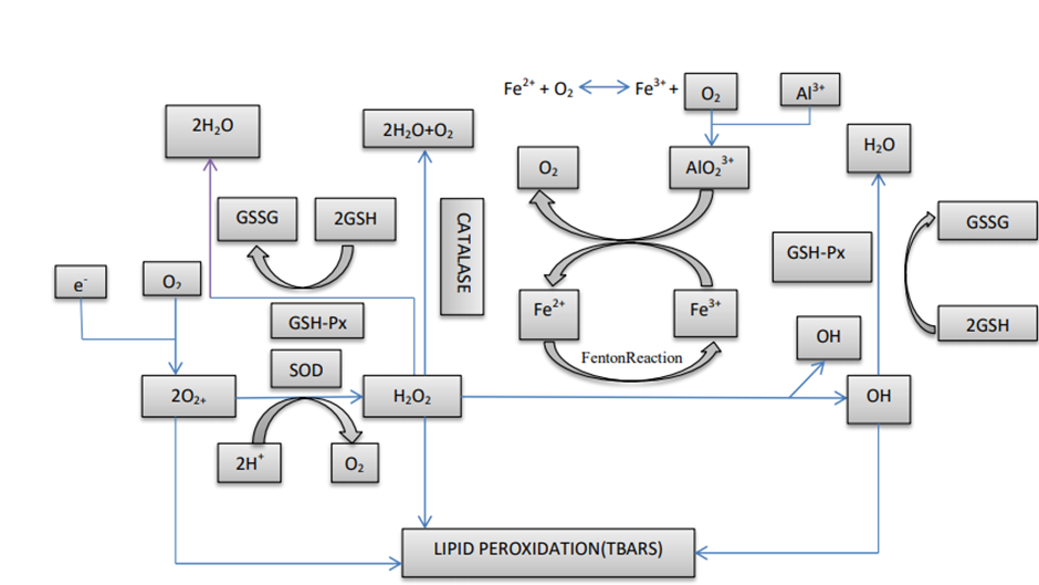
Fig.1. (coloured) Schematic imaging and representation of ROS, enzymes of anti-oxidant, aluminium and lipid peroxidation. TBARS-Thiobarbituric acid reactive substances, SOD—Superoxide dismutase, GPx-Glutathione peroxidase, GSSG—Oxidized glutathione.

1.2. Impact of aluminium as a Cholinotoxic agent

Acetylcholine is very much responsible for the cholinergic purpose or concern of the central nervous system (CNS). After release and let out from the presynaptic nerve terminals, acetylcholine is abruptly withdrawn and abolished by acetylcholine esterase(Ache) from synaptic cleft, which is a member to the family of carboxyl esterase’s and undergoes breakdown of acetylcholine into choline and acetate which is an inactive metabolite (Maheswari et al.,2014) Aluminium accumulates in the brain slowly undergoes aluminium complex formation with elevated and increased affinity for the enzyme anionic site which increases the reactive oxygen species and undergo liberation and generation of oxidative stress in the neuronal region (Halliwell et al., 2007) Ultimately, it shows and give raise to the  cholinergic dysfunction and indicates the aluminium cholinotoxic activity as a sequel of oxidative stress and damage.

1.3. Impact on Neurotoxicity induced by Aluminium

Aluminium is reported and details to influence a number of reactions in the central nervous system which is vital and it sequels various adverse side effects. Major and salient retort for growth of brain such as formation of neurotransmitter synthesis, axonal transport, inflammatory responses, transmission of synapsis and protein phosphorylation, protein Deterioration, inflammatory retort and gene expression. Al3+ getting on for negatively charged, ligands of oxygen donor where it has a greater affinity. Other class like organic and inorganic phosphates, deprotonated hydroxyl groups and carboxylate groups forms a powerful bond with Al3+. These feasible aluminium (Al3+) characteristic undergo secure, powerful affix with phosphate group of RNA and DNA, where topology of DNA is affecting and impacting the expression of gene essential for brain functioning. Moreover, Aluminium has powerful positive charge and small ionic radius in contrast to Ca2+, Zn2+, and Na+. Therefore, aluminium powerfully affix and attach to metal-binding amino acids like (tyrosine, arginine, histidine etc.). Ultimately, protein oligomerization induces and give raise to some important configurational difference that can restrict their deterioration and breakdown by proteases (Kawahara et al., 2011). Eventually aluminium undergoes damage and results death of neuronal cells and glial cells which is apoptosis. Moreover, it weakens the enzyme that intricate in the genesis of neurotransmitter and govern content of neurotransmitter. Aluminium also restricts receptors of neurotransmitter and Ca2+ channels which is voltage gated and further impedes synaptic transmission and ultimately leads to neurotoxicity, and weakens functions of the brain related learning and memory (Kawahara et al., 2011).

1.4. Impact on gene expression induced by aluminium

By redoing or altering the cerebral proteases expression and by operating the monoamine oxidase isotypes in the brain region, aluminium affects gene expression (Huh et al., 2015), induces to configurational changes and also further induce and give raise to topological DNA changes by binding to histone-DNA complex (Lukiw et al., 1978; Latha et al., 2002). The studies or research reveal that there is a raise in the level of glial cell marker, TNF-a and glial fibrillary acidic protein (GFAP) (Nedzvetsky et al., 2006). Furthermore, aluminium produce and induce the expressions of interleukin-1???? precursor, DAXX, phospholipase A2, NF-KB subunits. That intricate in the pro-apoptotic and pro-inflammatory signalling mechanisms (Alexandrov et al., 2005). The changed expressions of gene, gives induces the drop-in or lowered expressions of tubulin, Amyloid beta protein precursor, neurofilament and enolase which is neuron specific (Muma et al., 1996; Parhad et al., 1989)  mitochondrial cytochrome C oxidase receptors which is of reduced or lower number are induced by the aluminium and lowers the expressions of  brain derived neurotropic factor (BDNF) and finally neuron growth factors(Johnson et al., 2003). Protein binding intricate in the gene expressions are the expected and probable mechanism of altering the gene expressions. In the zinc finger domain Aluminium attach to the IIIA transcription factor and further inhibits its promoter binding (Hanas et al., 1996).

1.5. Impact on cell mediated excitotoxicity induced by aluminium.

Mitochondrial injury caused by the aluminium leads to triggering of overpriced highly reactive oxygen free radicals and hydroxyl free radicals and eventually cause hydrogen peroxide amass and gathering. Further the raise in amass of hydrogen peroxide intensify the influence of redox active iron further from relatively bound iron or by modulating the electron transport chain (Bondy et al., 1996). In addition, aluminium gives raise to oxidative damage through triggered Fenton reactions which is iron arbitrated, by enhancing the concentration of redox active iron. Also research manifests by aluminium superoxide dismutase is activated and further catalase is inhibited by aluminium (Bondy et al., 1996; yousef et al., 2004). Moreover, aluminium may enhance the excitotoxicity, which is glutamate induced and ultimately suggested and theorize that the outcome of neurotoxic of aluminium be arbitrated through an excitatory amino acid and glutamate (Nayak et al., 2001). neurodegenerative sequel of Aluminium and the probable mechanisms as related to excitotoxicity is outlined in Fig. 2.

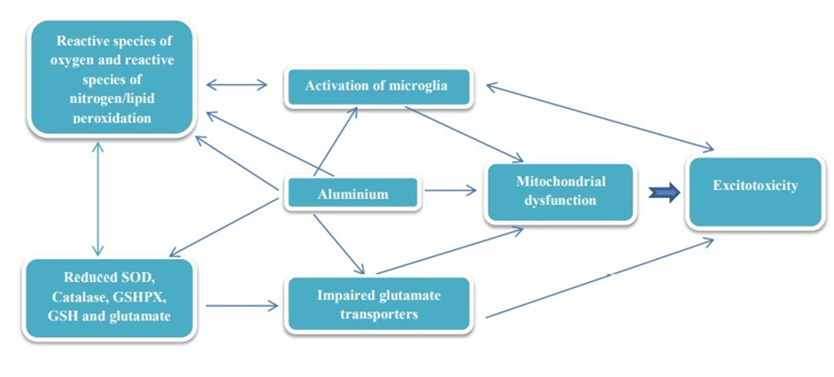
Fig.2. (coloured) Schematic representation and imaging of mechanisms of aluminium on neuroexictotoxicity

1.6. Impact on calcium homeostasis induced by aluminium.

Neurodegeneration and neuronal injury is induced by aluminium by interfering calcium homeostasis. Voltage dependent calcium channels are detained by aluminium and further obstruct the calmodulin dependent Ca2+/Mg2+ ATPase, in which very much accountable for protective activity in opposition to excitotoxicity (Exley et al., 1999). Furthermore, research in addition exhibit that there is a raise in levels of glutamate while gamma-aminobutyric acid(GABA) levels of brain were reduced, a circumstance that undergo maximizes excitotoxic damage experimental rats (Blaylock et al., 2012). Raising aluminium to metabolic changes similarly, in neuronal cytoplasam furtherance of the resting ca2+ and peak ca2+ levels, lowering of influx of ca2+, a modest inhibition of phosphatidylinositol 4,5-biphosphate (PIP2) hydrolysis by phosphatidylinositol-specific phospholipase-C (PI-PLC) in phosphoinositide signalling pathways, resulting in lowering inositol triphosphate (IP3) formation less protein kinase C (PKC) activation, and a obtuse rate of Ca2+ withdrawal from the cytoplasm. Analytical modifications in the Ca2+ homeostasis and Ca2+ signalling could occur from the continued gathering and amass of aluminium in neurons and eventually leading to the more considerable and damaged disruptions that influence ca2+ metabolism in neuronal disorder(Walton et al., 2012). By altering Ca2+ homeostasis neurodegenerative sequel of Aluminium and the probable pathways as related to excitotoxicity is outlined in Fig. 3.

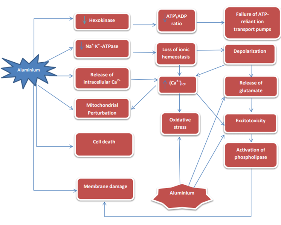
Fig.3.(Coloured) Schematic imaging of  pathways that the aluminium intercede or mediate neurodegenerative disease processing by modifying Ca2+ homeostasis. Ca2+ _ Calcium, Na+ _ Sodium,  K+- Potassium.

1.7. Impact on apoptosis induced by aluminium.

Mitochondria are where cytochrome C is liberated and released. This is induced by aluminium, lowering in Bcl-2 in twain or both endoplasmic reticulum and mitochondria, translocation of Bax into mitochondria, triggering of caspase-3 & activation and fragmentation of DNA (Ghiribi et al., 2001). Triggered cytochrome C from mitochondria gets binds to Apaf-1 and further  begin with apoptosis event which is induced by the aluminium (Savory et al., 2003) Caspase-9 is activated by the formed complex, that in succession operates the caspase-3 which is the effector caspase. Cytochrome C which eventually released and liberates, involve distinct pathways like, mitochondrial transition pore (MTP) opening, pro-apoptogenic Bax translocation of mitochondria which can shape the channel by itself, and Bax interaction with the voltage dependent anion channel (VDAC) to shape a larger channel which is cytochrome C permeable. Changes in mitochondria following cytotoxic stimuli is the considered primary event in the apoptosis (Vasudevaraju et al., 2008). Moreover, due to induction of aluminium the research shows that the incitement or triggering of SAPK/JNK (stress-activated protein kinase or c-jun N-terminal kinase) signal transduction pathway [39]. Ultimately due to the aluminium toxicity, apoptosis is found to be the common and broad mechanism of activity of the cell. Some attributed apoptotic properties like cell body shrinkage, chromatin and DNA fragmentation, and non-uniformed and irregularly shaped chromatin (Ghiribi et al., 2001; Maroney et al., 1998).

1.8. Impact on neurofibrillary tangles (NFTs) induced by aluminium.

Neurofibrillary tangles are found as a marker for neuronal diseases, such as Alzheimer’s diseases and Amyotrophic lateral sclerosis (ALS), and are the agglomeration of tau protein. Studies and research shows that abnormality induced by aluminium such as neurofilaments or tau in cytoskeletal, and further bring out the initiation of neurofibrillary tangles and degeneration of neurons of motor neurons in ALS.

(Kihira et al., 1993). In healthy neurons microtubules bounds tau proteins and is very much required and is vital to neuronal cytoskeleton formation and function. Al-ATP, glutamate receptor stimulated by glutamate and give raise to a maximized level of neuronal tau (Esclaire at al., 1997). The potentiality to individual self-gathering into structures of filamentous are the important feature of tau proteins that are the pathological signs of taupathies (Haase-Kohn et al., 2004).  Research studies show that the tau aggregates which is induced by the aluminium are not in the form of fibrillary but seems like amorphous form. Fibroblast toxicity could be produced by the induction of aluminium that expressed tau. Moreover, in these cell tau did not accumulate, but undergoes accumulation of neurofilaments in aluminium treated cells (Leterrier et al., 1992). Induction of aluminium in soluble form causes neurofilament monomers and tau. Further undergo results in the formation of accumulation in to non-fibrillar materials. Eventually Induction of aluminium to form neurofibrillary tangles and fibrillary bundles of neurofilaments (Leterrier et al., 1992; Troncoso et al., 1990)Ultimately aluminium induces and gives rise to tau hyperphosphorylation in which protein phosphatase-2A (PP2A) is inhibited actively in pyramidal cell. Further, caspases is operated by aluminium which shortens tau protein which is hyperphosphorylated, and amass it into granules. When joining the pile of gatherings appears as cytoplasmic pools. Elevated agglomeration of aluminium/shorten tau which is hyperphosphorylated may give raise to trigger polymerization. Polymerization of neurofibrillary tangles filaments within the aluminium/shorten tau which is hyperphosphorylated composite of the cytoplasmic pools (Walton et al., 2010). Formation of NFT by the effects of aluminium is outlined in Fig. 4

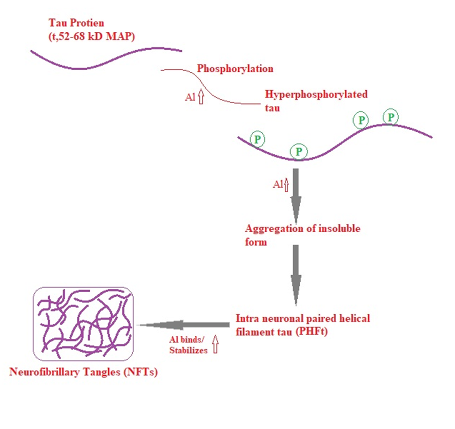
Fig.4. (coloured) Schematic representation of Aluminium induced NFT formation

2.   Systemic Toxicosis

2.1. Aluminium effects on pulmonary system.

Due to Aluminium exposure, there will be causation of pulmonary lesions in humans during the generation of aluminium products comprises, pulmonary granulomatosis, granulomatous pneumonia, pulmonary alveolar proteinosis, pulmonary fibrosis, and interstitial desquamative pneumonia (Chen et al., 1978; Herbert et al., 1982; de Vuyst et al., 1987). Due to exposure of aluminium there may be causation of asthma (Burge et al., 2000) Among the aluminium exposure and aluminium smelter workers reactive airways dysfunction syndrome was rarely reported (Wesdock et al., 2014) In person following incidental or volitional ingestion acute duration oral subjection to aluminium phosphide has been well outlined to seat pulmonary edema (Chopra et al., 1986; Khosla et al., 1988;) Rather than to aluminium exposure lethality was likely due to the emergence of highly lethal phosphine (Alter et al., 2001; Kamanyire et al., 2003; Moghadamnia et al., 2012) In experimental animals pulmonary lesions are infrequent and exposure of aluminium is not through aerosol vehicles.

2.2. Aluminium effects on cardiovascular system.

In case of aluminium exposure and aluminium phosphide poisoning, Toxic myocarditis, myocardial hypokinesia, left ventricular thrombosis and myocardial dysfunction were detailed (Hangouche et al., 2017).  As the delayed and detained complication of aluminium exposure and aluminium phosphide poisoning in the right middle cerebral artery due to thrombosis, ischemic stroke was detailed and reported (Abedini et al., 2014). Moreover, other aluminium compounds intoxication may not cause cardiovascular lesions. In embryonic chick heart cardiac teratogenesis was reported where also faults in ventricular myocardium as well as ventricular septation were detailed and reported (El Mazoudy et al., 2014) Further there was a notable and remarkable association between increase in maternal hair aluminium components and in offsprings risk or menace of total congenital heart weakness was seen. Eventually in subtypes such as septal defects, conotruncal defects and right ventricular outflow obstruction in female rat are seen (Wang et al., 2012). Ultimately due to aluminium toxicity cardiovascular effects are congenital heart defects, dysfunction of the myocardium, inflammation and cardiovascular thrombosis.

2.3. Aluminium effects on gastrointestinal system.

Aluminium intoxication and its ingestion orally may influence the intestinal microbiota, permeability and immune reaction which effect the conditions of inflammation locally (Vignal et al., 2016). Crohn’s diseases are genetically susceptible in individuals, aluminium relates to bring about and persistence of the chronic relapsing intestinal inflammation (Lerner et al., 2007). Excessive intestinal inflammation are characterized by inflammatory bowl diseases, comprise of diseases entities like crohn’s diseases and ulcerative colitis and experimental investigation in mice shows aluminium promoted intestinal inflammation, so that imply aluminium in the pathogenesis and development of inflammatory bowl dis78eases (de Chambrun et al., 2014) In transgenic mice, acute colitis and chronic colitis in which induced chemically are lacking interleukin 10 were aggravated by aluminium exposure orally, where aluminium increased the duration and force of intensity of intestinal inflammation and lowers the renewal of the intestinal epithelial mucosal cells (de chambrun et al., 2014) Eventually due to intoxication of aluminium intestinal barrier function was disabled and impaired beneath the basal circumstances; by aluminium and lipopolysaccharides there was a synergistic excite of expressions of  pro-inflammatory cytokine (de Chambrun et al., 2014) Furthermore in the mucosa of the small intestines of wistar rats aluminium chloride intoxication and exposure orally causes epithelial degeneration, goblet cell proliferation and lymphocyte infiltration (Buraimoh et al., 2012)

2.4. Aluminium effects on Musculoskeletal system.

Macrophagic myofasciitis (aluminic granuloma) is the major myopathy induced by aluminium intoxication and aluminium exposure associated and correlated with chronic fatigue syndrome and chronic arthromyalgia or myalgia (Exley et al., 2009; Gherardi et al., 2012; Rigolet et al., 2014). After intraperitoneal injection of aluminium lactate there will be take place of necrosis of  skeletal muscle which is transpire in the rats muscles of  diaphragm and abdominal regions adjacent to the peritoneum (Levine et al., 1992) Due to induction of dietary aluminium hydroxide supplementation Muscle fiber atrophy, with growth retardation, was detailed and outlined  in hypophosphatemia pigs (Haglin et al., 1994). Eventually due to aluminium subjection, contraction of smooth muscle which is give rise by k+ ion was impedes or inhibited (Nasu et al., 1998).  Osteoporosis, osteomalacia, rickets, exostosis, osteodystrophy and osteitis fibrosa are the bone diseases associated with Aluminium exposure (Sherrard et al., 1985; Chappard et al., 2016; Klein et al., 2019) Ultimately increase in aluminium levels in the bone results in long term exposure of aluminium orally (Ahn et al., 1995, Konishi et al., 1996)

2.5. Aluminium effects on reproductive system.

By Aluminium exposure Human reproduction may be affected negatively (Klein et al., 2014; Mouro et al., 2017) Studies shows that higher aluminium concentration had been reported in oligospermia patients than healthy individuals as human semen spermatozoa contain aluminium (Klein et al., 2014). Eventually, at human dietary level of aluminium and constant subjection of aluminium for 60days, the rat testes contain reduced levels of aluminium of 3.35 µg/g and associated with raise in oxidative damage, stress and inflammation, reduced daily sperm production, raise in abnormal spermatozoa, reduced sperm count and motility (Martinez et al., 2017). Ultimately bank voles (myodes glareolus) exhibits aluminium generate or bring about lesser standards and lower quantity of sperm than normal standard, but was not remarkably govern or influence in womens (Miska-Schramm et al., 2017). Aluminium effects on reproductive system is outlined in Fig. 5.

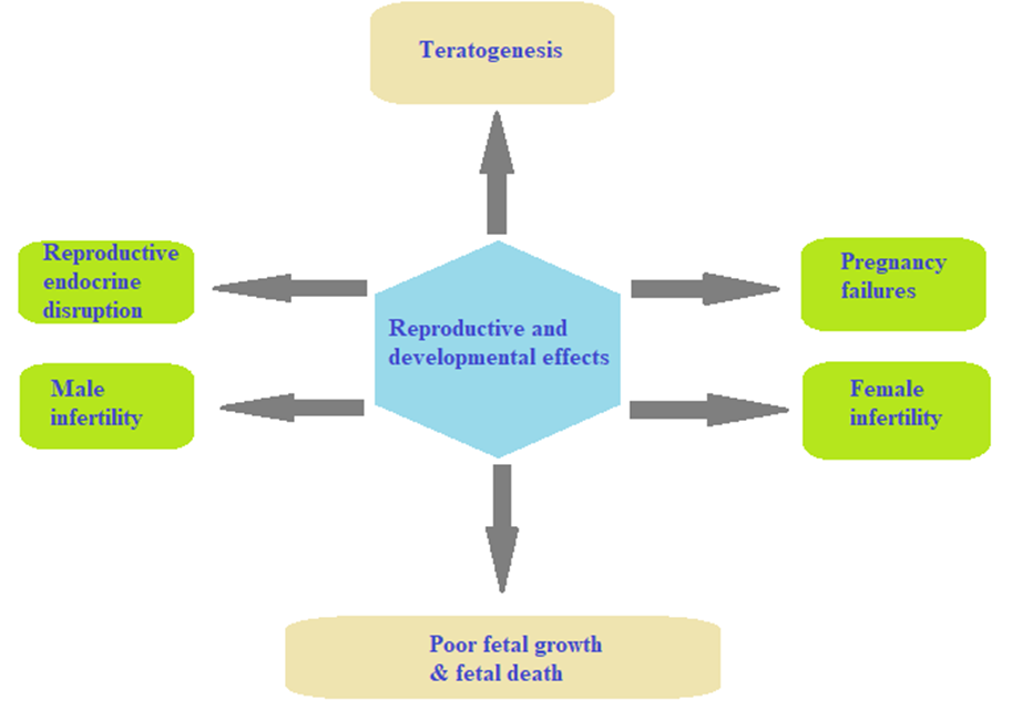
Fig.5. (coloured) Reproductive and developmental disorder in aluminium toxicity.

2.6. Aluminium effects on hepato-renal and pancreatic systems

Aluminium intoxication in the kidney and liver causes oxidative damage which leads to deterioration of the tissue and cell death or necrosis, and associated serum biochemical derangements (Nikolov et al., 2010; Mailloux et al., 2011; Bai et al., 2012). Eventually after aluminium chloride ingestion of 20mg/kg orally for 30days there was a notable and significant raise in the action of aspartate aminotransferase (AST), alkaline phosphatase (ALP), alanine aminotransferase (ALT), and bilirubin, as well as raise in serum urea and creatinine levels in the experimental animals (Abdel-Wahab et al., 2012). Studies shows aluminium phosphide pellets consuming was well outlined and reported to give raise to acute pancreatitis in one patient (Verma et al., 2007). After intermediate oral exposure (50 mg/kg for 28days) to aluminium chloride rats had moderate pancreatic islet necrosis, which was associated and correlated  with weaken fasting blood glucose and weaken tolerance of oral glucose (Igwenagu et al., 2017, 2019). Changes in metabolism due to hepatic and pancreatic lesions which results in hyperlipidaemia, hypercholesterolemia, hypertriglyceridemia, hyperglycaemia, hypoproteinemia (Omar et al., 2003; Kowalczyk et al., 2004; Turkez et al., 2011).

2.7. Aluminium effects on mammary gland or breast.

Aluminium intoxication may be involved in the causation and development of mammary gland conditions such as breast cancers and cyst (Darbre et al., 2016). In antiperspirant and other underarm cosmetics products, aluminium chlorohydrate may be an significant or major initiator of aluminium subjection (Pineau et al., 2014). In case control study (Linhart et al., 2017) The degree of aluminium levels in the tissues of breast were remarkably arise in breast cancer individuals than control (5.8 versus 3.8nmol/g). In which aluminium was notably associated with breast cancer incidence as the use of underarm cosmetics products containing aluminium. Patients of breast cancer had raise in levels of aluminium in tissues of breast than in blood serum (Darbre et al., 2013).  In cancer patients there were a raise in level of aluminium in nipple aspirates than healthy controls and raise in degree of fluid breast cyst than serum or milk (Darbre et al., 2011). Biological feature of epithelial cells of breast and carcinogenesis is contemplated an expected results which may influenced by the accumulating of aluminium in the tissues of the breast (Pineau et al., 2014). Eventually studies shows aluminium can induce injury of DNA in epithelial cells of human breast and later bring on cell proliferation (Darbre, Bakir et al., 2013; Darbre, Mannello et al., 2013). Ultimately by acting as a metalloestogen aluminium may increase the risk of breast cancer (Darbre et al., 2016).

  1. Chemicals used frequently to induce Alzheimer disease like symptoms in rodents.
    1. Aluminium

Heavy metals are promoter that through neurotoxicity it has been well set and accepted to knock the progress of the brain (Nallagouni et al., 2017).  It was identified and recognized that heavy metals which have been induced and give raise to elevated level of lethality is got connected with various conditions of diseases, which comprise cognitive disorder (Nallagouni et al., 2017). Nervous system is well deteriorated through heavy metals. Effect of aluminium on nervous system of all the heavy metals is well reported (Mujika et al., 2014; Chiroma et al., 2018; Exley et al., 2013). Aluminium is an omnipresent, ubiquitously dispersed industrial and environmental toxicant associated with anemia, osteomalacia, hepatic neurological disorders (Han et al., 2013). Patient suffering from Alzheimer’s diseases has been identified with high amount of aluminium which gives raise to effects of toxicological behaviour, encompass encephalopathy, anemia and bone disease. Eventually recent studies show, orally administered aluminium (300mg/kg) in the brain of rat was detailed to give raise oxidative stress, cholinergic deficit and Aβ & NFTs accumulation (Chiroma et al., 2018).

    1. Scopolamine

Scopolamine which is parasympatholytics and predominantly approved and accepted chemical in pilot studies to show deterioration of memory (Burbser et al., 2012) Scopolamine ingestion eventually give raise to deterioration in verbal recall, visual recognition memory, visuospatial recall psychomotor speed, visuospatial praxis and visuoperceptual processes (Flicker et al., 1990). This parasympatholytics has been well recognized to cause deterioration of ability to remember or recall. Furthermore it give raise to the excitation of glycogen synthase kinase-3 beta (GSK-3β), arborization of dendrites connected with alterations in CREC, amino-3-hydroxy-5-methyl-4-isoxazole propionic acid receptor (AMPA), Homer1, and inadequate spine maturation (Wu et al., 2013).

    1. Colchicine

Colchicine are promising potential drug, through cholinergic neuron dysfunction  induce dementia, further by impede cholinergic reversal or eroding cholinergic passage cascades (Kumar et al., 2006; Kumar et al., 2009). Hippocampal lesions could be induced by colchicine resulting in cognitive decline and  colchicine could cause toxicity of neurons and deterioration of memory by inhibiting cholinergic pathways, there by number of cholinergic neurons are reduced and in the same way decline cholinergic renewal in essence within the hippocampal region of brain (Evrard et al., 1998). Deterioration of memory was seen due to induction of colchicine perhaps an outcome of a decline in levels of serotonin, norepinephrine, dopamine within the area of caudate nucleus region, hippocampus region and entire cortex region (Ganguly et al., 2008). Moreover, generation of protein carbonyls following lipid peroxidation was caused by colchicine (Saini et al., 2012). Expression degree of cyclooxygenase 1 and 2 (COX-1 and COX-2) levels had also been found to raise by colchicine (Subbaramaiah et al., 2000) And generation of  ROS (Kumar et al., 2010) Further elevates glutamate/GABA ratio in the cortex (Yu et al., 1997).

    1. Streptozotocin

Streptozotocin found in the strain of Streptomyces achromogenes, a compound derived from glucosamine nitrosourea (Neha et al., 2014).  Streptozotocin used in anti-cancer treatment is another alkylating agent it has been shown to spoof or impersonate certain quality of nitrosourea, an anti-cancer agent further more cause deterioration of memory (Klinkenberg et al., 2010). Induction and give raise of neuronal toxicity and tau hyperphosphorylation is bring about by streptozotocin, which result in generation of Reactive oxygen species (ROS) and Reactive species of nitrogen (RNS) (Zhou et al., 2013). Studies shows that streptozotocin damage the enzyme glycolytic action in the brain region which ultimately outlined in the decreasing of concentration of creatine phosphate and ATP. These devastated  system of energy and decreased synthesis of acetyl CoA can impede cholinergic conductance (Neha et al., 2014).

    1. Ethanol

Persistent ingestion of alcohol is related with lots of  complications, which encompass deterioration of social skills, languages, hyperactivity, motor dysfunction and learning deficits (Spinetta et al., 2008). Pre-existing research has manifest that ingestion of ethanol elevates the productions of reactive oxygen species and sequel in lowering of antioxidants in the region of brain (Patil et al., 2015). Further research and investigation shows that ethanol could deteriorate cholinergic neurons and hippocampal neurons with resultant effects on disruption of learning and memory (Mailliard et al., 2004). Eventually elevated treatment of ethanol sequels in nitric oxide(NO) in excessive manner, which have been establish to devastate  memory recollection and learning skills, while further elevated dose of ethanol devastate the glutamatergic system and elevates GABAergic conveyance in the region of brain that relates with memory (Mailliard et al., 2004). Ultimately ethanol further increases the amount of adenosine, which may give rise to deterioration of memory (Cui et al., 2015).

    1. Excitotoxin (Ibotenic acid)

Ibotenic acid a strapping neurotoxicant that irritates signs and pathophysiology which is abnormal development similar to Alzheimer diseases (Clark et al., 2012; Karthick et al., 2016). In avoiding Alzheimer’s diseases pathology it is a functional model to admire drug efficiency. Furthermore it has found that intra hippocampal ingestion of ibotenic acid (5 µg/µl PBS) shows deterioration of recollection, learning and outlines raise in activity of AChE as well as increased MDA levels, there by induce toxicity of neuron (Rattan et al., 1992). Bilateral ICV injection of ibotenic acid may bring out Alzheimer diseases-like symptoms and pathology, ultimately research and certain investigation shows that ibotenic acid lowers the cholinergic neurons activity in rats (Kumar et al., 2011).

    1. Sodium azide(NaN3)

Sodium azide (NaN3) a mitochondrial toxin is a white crystalline solid embroil in the output or generation of explosion of lead azide (Neha et al., 2016). Sodium azide intake has give raise dysfunction of mitochondria and further critical enzymes of mitochondria, cytochrome oxidase is further inhibited (Wong-Riley et al., 1997). In the mitochondrial respiratory chain Cytochrome oxidase enzyme is critical one. Its involvement obstruct with mitochondrial complex-IV and declines the degree of  ATP, which induce or give raise to metabolic deterioration and reactive species of oxygen production (Bennett et al., 1996). NAN3 causes oxidative deterioration that results in breaking of the cell and necrosis which is death of the cell. In the cortical and hippocampal regions of NaN3 treated rats, progressive and continuous neuronal depletion and necrosis were well observed, thus shows the dominance of sodium azide. These studies and investigations affirm that intraperitoneal injection of sodium azide for 15days give raise and resulted in comparable level of dementia, and moreover ultimately inducing neurodegenerative disorder suggesting the involvement of NaN3 (Gao et al., 2018).

    1. Okadaic acid

Okadaic acid (OKA), is a marine microalgae product and major polyether toxin, that causes toxicity of diarrhetic shell fish (Kamat et al., 2010). It has been reported that the IC injection of OKA induce cognitive disorder in rats, Thus building it as approachable and worth model to investigate and screening anti-dementia drug (Kamat et al., 2011). Okadic acid has been established to be a prohibitive and tolerable antagonist of serine/threonine phosphatases 1 and 2A  in terms of mechanism of action (Cohen et al., 1990; Ishihara et al., 1989)  is connected with small term and protracted deterioration of memory in rats (Maidana et al., 2006) and undergo tau hyper phosphorylation and death of nerve cell in both invivo (He et al., 2001) and in vitro (Cagnoli et al., 1996) From the pre-existing research recognized that okadaic acid lowers basal synaptic transmission and impedes the synaptic plasticity formation (Koss et al., 2007). Further, Okadic acid has established to develop Ca2+ in a cells of hippocampal neurons by inotropic receptors of excitatory amino acid, Thus give raise to neuronal loss (Femandez et al., 1993) Moreover studies and research has signify that Okadic acid activates the generation of Reactive species of oxygen in the site of hippocampus in the region of brain and declines potency of mitochondria and activity, Finally results in deterioration of mitochondria in the  rats brain (Kamat et al., 2011).

CONCLUSION

There is a lot of studies and evidences that suggest aluminium in the development of circumstances that give raise to cognitive decline, it is greatly undertake that aluminium is well acknowledge toxin of neuron and that bring out deterioration of neuron and death of neuron. Aluminium is extremely plentiful and dispersed as industrial and atmospheric poison and is also contain in edibles, further involve in skeletal, neurological, haematological disorders. Aluminium get into the human form in higher amount it can enhance and undergo the emergence of reactive oxygen species, and triggers glial cells. Eventually reactive oxygen species generation and events of inflammation at last give raise to necrosis. Ultimately summarizes that neurotoxicity caused by aluminium in various way by enhancement of ROS production, modulating inhibition of DNA repair enzyme, and pathways alterations like NF-KB, JNK, DNA binding etc. All these episodes especially and finally leads to death of cell, genomic instability, and cognitive decline. More ever it is very much understandable that additional scientific approach and investigations are crucial against this type of episodes to find superior way to avert the Alzheimer induced cognitive disease progressions.

REFERENCES

  1. Abedini, M., Fatehi, F., Tabrizi, N., 2014. Ischemic stroke as a rare manifestation of aluminium phosphide poisoning: a case report. Acta Med Iran. 52, 947–949.
  2. Abdel-Wahab, W.M., 2012. AlCl3-induced toxicity and oxidative stress in liver of male rats: protection by melatonin. Life Sci J. 9, 1173–1182.
  3. Ahn, H.W., Fulton, B., Moxon, D., Jeffery, E.H., 1995. Interactive effects of fluoride and aluminum uptake and accumulation in bones of rabbits administered both agents in their drinking water. J Toxicol Environ Health. 44, 337–350.
  4. Alexandrov, P.N., Zhao, Y., Pogue, A.I., Tarr, M.A., Kruck, T.P., Percy, M.E., Cui, J.G., Lukiw, W.J., 2005. Synergistic effects of iron and aluminum on stress related gene expression in primary human neural cells. J. Alzheimer’s Dis. 8, 117– 127.
  5. Alter, P., Grimm, W., Maisch, B., 2001. Lethal heart failure caused by aluminium phosphide poisoning. Intensive Care Med 27, 327–331.
  6. Amjad, S., Umesalma, S., 2015. Protective effect of Centella asiatica against aluminium-induced neurotoxicity in cerebral cortex, striatum hypothalamus and hippocampus of rat brain histopathological, and biochemical approach. J. Mol. Biomark. Diagn. 6, 1–7.
  7. Anon., 1982. Drinking Water and Health. National Research Council, National Academy Press, Washington, DC 4, 155–201.
  8. Anon., 2008a. Aluminum and compounds. Hazardous Substances Data Bank. National Library of Medicine.
  9. Bai, C., Wang, F., Zhao, H., Li, Y., 2012. Effects of subchronic aluminum exposure on liver function in rats. J Northeast Agric Univ. 19, 62–65.
  10. Bennett, M.C., Mlady, G.W., Kwon, Y.H., Rose, G.M., 1996. Chronic in vivo sodium azide infusion induces selective and stable inhibition of cytochrome c oxidase. J Neurochem. 66, 2606–11.
  11. Blaylock, R.L., 2012. Aluminum induced immunoexcitotoxicity in neuro developmental and neurodegenerative disorders, Curr. Inorg. Chem. 2, 1–6.
  12. Bondy, S.C., Kirstein, S., 1996. The promotion of iron-induced generation of ROS in nerve tissue by aluminium, Mol. Chem. Neuropathol. 27,185–194.
  13. Bubser, M., Byun, N., Wood, M.R., Jones, C.K., 2012. Muscarinic receptor pharmacology and circuitry for the modulation of cognition. Handb Exp Pharmacol. 208, 121–66.
  14. Buraimoh, A.A., Ojo, S.A., 2012. Effects of aluminium chloride exposure on the histology of the small intestine of wistar rats. Int J Appl Sci Technol. 2, 123–132.
  15. Burge, P.S., Scott,J.A., McCoach, J., 2000. Occupational asthma caused by aluminium. Allergy. 55, 77–780.
  16. Cagnoli, C.M., Kharlamov, E., Atabay, C., Uz, T., Manev, H., 1996. Apoptosis induced in neuronal cultures by either the phosphatase inhibitor okadaic acid or the kinase inhibitor staurosporine is attenuated by isoquinolinesulfonamides H-7, H-8, and H9. J Mol Neurosci. 7, 65–76.
  17. Campbell, A., Kumar, A., La Rosa, F.G., Prasad, K.N., Bondy S.C., 2000.Aluminum increases levels of beta-amyloid and ubiquitin in neuroblastoma but not in glioma cells. Proc. Soc. Exp. Biol. Med. 223, 397–402.
  18. Chappard, D., Bizot, P., Mabilleau, G., Hubert, L., 2016. Aluminum and bone: review of new clinical circumstances associated with Al(3+) deposition in the calcified matrix of bone. Morphologie. 100, 95–105.
  19. Chen, W.J., Monnat Jr., R.J., Chen, M., Mottet, N.K., 1978. Aluminum induced pulmonary granulomatosis. Hum Pathol. 9, 705–711.
  20. Chiroma, S.M., Moklas, M.A.M., Taib, C.N.M., Baharuldin, M.T., Amon, Z., 2018. d-galactose and aluminium chloride induced rat model with cognitive impairments. Biomed Pharmacother. 103, 1602–8.
  21. Chopra, J.S., Kalra, O.P., Malik, V,S., Sharma, R., Chandna, A., 1986. Aluminum phosphide poisoning: A prospective study of 16 cases in one year. Postgrad Med J. 62, 1113–1115.
  22. Clark, D., Tuor, U.I., Thompson, R., Institoris, A., Kulynych, A., Zhang, X., 2012. Protection against recurrent stroke with resveratrol: endothelial protection. PLoS One.7.
  23. Cohen, P., Holmes, C.F., Tsukitani, Y., 1990. Okadaic acid: a new probe for the study of cellular regulation. Trends Biochem Sci. 15, 98–102.
  24. Cui, S.Q., Wang, Q., Zheng, Y., Xiao, B., Sun, H.W., Gu, X.L., 2015. Puerarin protects against damage to spatial learning and memory ability in mice with chronic alcohol poisoning. Braz J Med Biol Res. 48, 515–22.
  25. Cui, S.Q., Wang, Q., Zheng, Y., Xiao, B., Sun, H.W., Gu, X.L., 2015. Puerarin protects against damage to spatial learning and memory ability in mice with chronic alcohol poisoning. Braz J Med Biol Res. 48, 515–22.
  26. Darbre, P.D., Pugazhendhi, D., Mannello, F., 2011. Aluminium and Human breast diseases. J Inorg Biochem. 105, 1484–1488.
  27. Darbre, P.D., Bakir, A., Iskakova, E., 2013. Effect of aluminium on migratory and invasive properties of MCF-7 human breast cancer cells in culture. J Inorg Biochem. 128, 245–249.
  28. Darbre, P.D., Mannello, F., Exley, C., 2013. Aluminium and breast cancer: sources of exposure, tissue measurmenets and mechanisms of toxicological actions of breast of breast biology. J Inorg Biochem. 128, 257–261.
  29. Darbre, P.D., 2016. Aluminium and the human breast. Morphologie. 100, 65–74.
  30. de Chambrun, G.P., Body-Malapel, M., Frey-Wagner, I., Djouina, M., Deknuydt, F., Atrott, K., Esquerre, N., Altare, F., Neut, C., Arrieta, M.C., Kanneganti, T.D., Rogler, G., Colombel, J.F., Cortot, A., Desreumaux, P., Vignal, C., 2014. Aluminum enhances inflammation and decreases mucosal healing in experimental colitis in mice. Mucosal Immunol. 7, 589–601.
  31. de Vuyst, P., Dumortier, P., Schandené, L., Estenne, M., Verhest, A., Yernault, J.C., 1987. Sarcoid-like lung granulomatosis induced by aluminum dusts. Am Rev Respir Dis. 135, 493–497.
  32. El Mazoudy, R.H., Bekhet, G.A., 2016. In ovo toxico-teratological effects of aluminum on embryonic chick heart and vascularization. Environ Sci Pollut Res Int. 23, 21947–21956.
  33. Esclaire, F., Lesort, M., Blanchard, C., Hugon, J., 1997. Glutamate toxicity enhances tau gene expression in neuronal cultures. J. Neurosci. Res. 49, 309–318.
  34. Exley, C., 1999. A molecular mechanism of aluminium-induced Alzheimer’s disease. J. Inorg. Biochem. 76, 133–140.
  35. Exley, C., 2004. The pro-oxidant activity of aluminium. Free. Radic. Biol. Med. 36, 380–38.
  36. Exley, C., Swarbrick, L., Gherardi, R.K., Authier, F.J., 2009. A role for the body burden of aluminium in vaccine-associated macrophagic myofasciitis and chronic fatigue syndrome. Med Hypotheses. 72, 135–139.
  37. Exley, C., 2013. Human exposure to aluminium. Environ Sci Process Impacts. 15, 1807–16.
  38. Evrard, P.A., Ragusi, C., Boschi, G., Verbeeck, R.K., Scherrmann, J.M., 1998. Simultaneous microdialysis in brain and blood of the mouse: extracellular and intracellular brain colchicine disposition. Brain Res. 786, 122–7.
  39. Fernández, M.T., Zitko, V., Gascón, S., Torreblanca, A., Novelli, A., 1993. Neurotoxic effect of okadaic acid, a seafood-related toxin, on cultured cerebellar neurons. Ann N Y Acad Sci. 679, 260–9.
  40. Flicker, C., Serby, M., Ferris, S.H., 1990. Scopolamine effects on memory, language, visuospatial praxis and psychomotor speed. Psychopharmacology (Berl) 100, 243–50.
  41. Fulgenzi, A., Vietti, D., Ferrero, M.D., 2014. Aluminium involvement in neurotoxicity. Biomed. Res. Int. 1–5.
  42. Fu, H.J., Hu, Q.S., Lin, Z.N., Ren, T.L., Song, H., Cai, C.K., Dong, S.Z., 2003. Aluminum induced apoptosis in cultured cortical neurons and its effect on SAPK/JNK signal transduction pathway. Brain Res. 980, 11–23.
  43. Ganguly, R., Guha, D., 2008. Alteration of brain monoamines & EEG wave pattern in rat model of Alzheimer’s disease & protection by Moringa oleifera. Indian J Med Res. 2008. 128, 744–51.
  44. Gao, C., Chang, P., Yang, L., Wang, Y., Zhu, S., Shan, H., 2018. Neuroprotective effects of hydrogen sulfide on sodium azideinduced oxidative stress in PC12 cells. Int J Mol Med. 41, 242–50.
  45. Gherardi, R.K., Authier, F.J., 2012. Macrophagic myofaciitis: characterization and pathophysiology. Lupus. 21, 184–189.
  46. Ghiribi, O., Herman, M.M., Forbes, M.S., Dewitt, D.A., Savory J., 2001. GDNF protects against aluminum-induced apoptosis in rabbits by upregulating Bcl-2 and Bcl-XL and inhibiting mitochondrial Bax translocation. Neurobiol. Dis 8, 764–773.
  47. Gulya, K., Rakonczay, Z., asa, P.K., 1990. Cholinotoxic effects of aluminum in rat brain. J. Neurochem. 54, 1020–1026.
  48. Haase-Kohn, C., Stieler, J., Arendt, T., Holzer, M., 2004. Pseudophosphorylation of tau protein alters its ability for self-aggregation. J. Neurochem. 88, 1509– 1520.
  49. Haglin, L., Essén-Gustavsson, B., Lindholm, A., 1994. Hypophosphatemia induced by dietary aluminium hydroxide in growing pigs: effects on erythrocytes, myocardium, skeletal muscle and liver. Acta Vet Scand. 35, 263– 271.
  50. Halliwell, B.J., Gutteride, M., 2007. Free Radicals in Biology and Medicine, fourth ed. Oxford University Press New York.
  51. Hangouche, A.J.E., Fennich, H., Alaika. O., Dakka, T., Raisouni, Z., Oukerraj, L., Doghmi, N., Cherti, M., 2017. Reversible myocadial injury and intraventricular thrombus associated with aluminium phosphide poisoning. Case Rep Cardiol.
  52. Han, S., Lemire, J., Appanna, V.P., Auger, C., Castonguay, Z., Appanna, V.D., 2013. How aluminum, an intracellular ROS generator promotes hepatic and neurological diseases: the metabolic tale. Cell Biol Toxicol. 29, 75–84.
  53. Hanas, J.S., Gunn, C.G., 1996. Inhibition of transcription factor IIIA DNA interactions by xenobiotic metal ions. Nucleic Acids Res. 24, 924–930.
  54. He, J., Yamada, K., Zou, L.B., Nabeshima, T., 2001. Spatial memory deficit and neurodegeneration induced by the direct injection of okadaic acid into the hippocampus in rats. J Neural Transm (Vienna). 108, 1435–43.
  55. Herbert, A., Sterling, G., Abraham, J., Corrin, B., 1982. Desquamative interstitial pneumonia in an aluminum welder. Hum Pathol. 13, 694–699.
  56. Huh, J.W., Choi, M.M., Lee, J.H., Yang, S.J., Kim, M.J., Choi, J., Lee, K.H., Lee, J.E., Cho, S.W., 2005. Activation of monoamine oxidase isotypes by prolonged intake of aluminum in rat brain. J. Inorg. Biochem. 99, 2088–2091.
  57. Igwenagu, E., 2017. Pathologic effects of oral aluminium administration in non-diabetic and diabetic rats. MVSc dissertation, Department of Veterinary Pathology, University of Maiduguri, Maiduguri, Nigeria.
  58. Igwenagu, E., Igbokwe, I.O., Egbe-Nwiyi, T.N., 2019. Fasting hyperglycaemia, glucose intolerance and pancreatic islet necrosis in albino rats associated with subchronic oral aluminium chloride exposure. Comp Clin Pathol.
  59. Ishihara, H., Martin, B.L., Brautigan, D.L., Karaki, H., Ozaki, H., Kato, Y., 1989. Calyculin A and okadaic acid: inhibitors of protein phosphatase activity. Biochem Biophys Res Commun. 159, 871–7.
  60. Johnson, J., Sharma, R.P., 2003. Aluminum disrupts the pro-inflammatory cytokine/ neurotrophin balance in primary brain rotation-mediated aggregate cultures: possible role in neurodegeneration. Neurotoxicology. 24, 261–268.
  61. Kamat, P.K., Tota, S., Saxena, G., Shukla, R., Nath, C., 2010.  Okadaic acid (ICV) induced memory impairment in rats: a suitable experimental model to test anti-dementia activity. Brain Res. 1309, 66–74.
  62. Kamat, P.K., Tota, S., Shukla, R., Ali, S., Najmi, A.K., Nath, C., 2011. Mitochondrial dysfunction: a crucial event in okadaic acid (ICV) induced memory impairment and apoptotic cell death in rat brain. Pharmacol Biochem Behav.. 100, 311–9.
  63. Kamanyire, R., Murray, V., 2003. Occupational exposures to fumigants. J Toxicol Clin Toxicol. 41, 489–490.
  64. Karthick, C., Periyasamy, S., Jayachandran, K.S., Anusuyadevi, M., 2016. Intrahippocampal Administration of Ibotenic Acid Induced Cholinergic Dysfunction via NR2A/NR2B Expression: Implications of Resveratrol against Alzheimer Disease Pathophysiology. Front Mol Neurosci. 26.
  65. Kawahara, M., Kato, M., Kuroda, Y., 2001. Effects of aluminum on the neurotoxicity of primary cultured neurons and on the aggregation of b-amyloid protein. Brain Res. Bull. 55, 211–217.
  66. Kawahara, M., Kato-Negishi, M., Hosoda, R., Imamura, L., Tsuda, M., Kuroda Y., 2003. Brain-derived neurotrophic factor protects cultured rat hippocampal neurons from aluminum maltolate neurotoxicity. J. Inorg. Biochem. 97, 124–131.
  67. Kawahara, M., Kato-Negishi, M., 2011. Link between aluminum and the pathogenesis of Alzheimer’s disease: the integration of the aluminum and amyloid cascade hypotheses. Int. J. Alzheimer’s Dis. 2011, 1–17.
  68. Khosla, S.N., Nand, N., Khosla, P., 1988. Aluminum phosphide poisoning. J Trop Med Hyg. 91, 196–198.
  69. Kihira, T., Yoshida, S., Mitani, K., Yasui, M., Yase, Y., 1993. ALS in the Kii Peninsula of Japan, with special reference to neurofibrillary tangles and aluminium. Neuropathology 13, 125–136.
  70. Klein, GL., 2019. Aluminum toxicity to bone: A multisystem effect. Osteoporos Sarcopenia. 5, 2–5.
  71. Klein, J.P., Mold, M., Mery, L., Cottier, M., Exley, C., 2014. Aluminum content of human semen: implications for semen quality. Reprod Toxicol. 50, 43–48.
  72. Klinkenberg, I., Blokland, A., 2010. The validity of scopolamine as a pharmacological model for cognitive impairment: a review of animal behavioral studies. Neurosci Biobehav Rev. 34, 1307–50.
  73. Konishi, Y., Yagyu, K., Kinebuchi, H., Salto, N., Yamaguchi, T., Ohtsuki, Y., 1996. Chronic effect of aluminum ingestion on bone in calcium-deficient rats. Basic Clin Pharmacol Toxicol. 78, 429–434.
  74. Kowalczyk, E., Kopff, A., K?dziora, J., B?aszczyk, J., Kopff, M., Niedworok, J., Fija?kowski, P., 2004. Effect of long-term aluminium chloride intoxication on selected biochemical parameters and oxidative-antioxidative balance in experimental animals. Pol J Environ Stud. 13, 41–43.
  75. Kumar, A., Seghal, N., Padi, S.V., Naidu. P.S., 2006. Differential effects of cyclooxygenase inhibitors on intracerebroventricular colchicine-induced dysfunction and oxidative stress in rats. Eur J Pharmacol. 551, 58–66.
  76. Kumar, A., Dogra, S., Prakash, A., 2009. Neuroprotective effects of Centella asiatica against intracerebroventricular colchicine induced cognitive impairment and oxidative stress. Int J Alzheimers Dis.
  77. Kumar, A., Dogra, S., Prakash, A., 2010. Protective effect of naringin, a citrus flavonoid, against colchicine-induced cognitive dysfunction and oxidative damage in rats. J Med Food. 13, 976–84.
  78. Kumar, A., Prakash, A., Pahwa, D., 2011. Galantamine potentiates the protective effect of rofecoxib and caffeic acid against intrahippocampal Kainic acid-induced cognitive dysfunction in rat. Brain Res Bull. 85, 158–68.
  79. Kumar, V., Gill, K.D., 2014. Oxidative stress and mitochondrial dysfunction in aluminium neurotoxicity and its amelioration. a review, Neurotoxicology. 41, 154–166.
  80. Latha, K.S., Anitha, S., Rao, K.S., Viswamitra, M.A., 2002. olecular understanding of aluminum-induced topological changes in (CCG) 12 triplet repeats: relevance to neurological disorders, Biochem. Biophys. Acta 1588.56–64.
  81. Lerner, A., 2007. Aluminum is a potential envirnmental factor for Crohn’s disease induction: extended hypothsesis. Ann N Y Acad Sci. 329–345.
  82. Leterrier, J.F., Langui, D., Probst, A., Ulrich, J., 1992. A molecular mechanism for the induction of neurofilament bundling by aluminum ions, J. Neurochem. 58, 2060–2070.
  83. Levine, S., Saltzman, A., Drakontides, A.B.. 1992. Parenteral aluminum compounds produce a local toxic myopathy in rats: importance of the anion. Toxicol Pathol. 20, 405–415.
  84. Lewis, RJ., 2001. Hawley’s Condensed Chemical Dictionary, Wiley-Interscience. 39-46.
  85. Linhart, C., Talasz, H., Morandi, E.M., Exley, C., Lindner, H.H., Taucher, S., Egle, D., Hubalek, M., Concin, N., Ulmer, H., 2017. Use of underarm cosmetic products in relation to risk of breast cancer: a case-control study. EBioMedicine. 21, 78– 85.
  86. Lin, R., Chen, X., Li, W., Han, Y., Liu, P., Pi, R., 2008. Exposure to metal ions regulates mRNA levels of APP and BACE1 in PC12 cells: blockage by curcumin. Neurosci. Lett.  440, 344–347.
  87. Lukiw, W.J., Kruck, T.P., McLachlan, D.R., 1978.  Alterations in human linker histoneDNA binding in the presence of aluminum salts in vitro and in Alzheimer’s disease. Neurotoxicology. 8, 291–301.
  88. Maheswari, S.L., Murali, R.V., Balaji, R., 2014. Aluminium induced cholinotoxicity in zebra fish brain—a sequel of oxidative stress. Int. J. Adv. Res. 2, 322– 335.
  89. Maidana, M., Carlis, V., Galhardi, F.G., Yunes, J.S., Geracitano, L.A., Monserrat, J.M., 2006. Effects of microcystins over short- and long-term memory and oxidative stress generation in hippocampus of rats. Chem Biol Interact. 159, 223–34.
  90. Mailliard, W.S., Diamond, I., 2004. Recent advances in the neurobiology of alcoholism: the role of adenosine. Pharmacol Ther. 101, 39–46.
  91. Mailloux, R.J., Lemire, J., Appanna, V.D., 2011. Hepatic response to aluminum toxicity: dyslipidemia and liver diseases. Exp Cell Res. 317, 2231–2238.
  92. Malakoff, D., 2000. Aluminum is put on trial as a vaccine booster. Science. 288, 1323–1234.
  93. Markewberry, W.R., 1994. Oxidative stress hypothesis in Alzheimer’s disease. Free Radic. Biol. Med 23, 134–147.
  94. Maroney, A.C., Glicksman, M.A., Basma, A.N., Walton, K.M., Knight Jr., W., Murphy, C.A., Bartlett, B.A., Finn, J.P., Angeles, T., Matsuda, Y., Neff, N. T., Dionne, C.A., 1998. Motoneuron apoptosis is blocked by CEP-1347 (KT 7515), a novel inhibitor of the JNK signaling pathway. J. Neurosci. 18, 104–111.
  95. Martinez, C.S., Escobar, A.G., Uranga-Ocio, J.A., Precanba, F.M., Vassallo, D.V., Exley, C., Miguel, M., Wiggers, G.A., 2017. Aluminum exposure for 60 days at human dietary levels impairs spermatogenesis and sperm quality in rats. Reprod Toxicol. 73, 128–141.
  96. Miska-Schramm, A., Kapusta, A., Kruczek, M., 2017. The effect of aluminum exposure on reproductive ability of the bank vole (Myodes glareolus). Biol Trace Elem Res. 177, 97–106.
  97. Moghadamnia, A.A., 2012. An update on toxicology of aluminum phosphide. DARU J Pharm Sci. 20, 1–8.
  98. Mouro, V.G.S., Menezes, T.P., Lima, G.D.A., Domingues, R.R., Souza, A.C., Oiveira, J.A., Matta. S.L.P., Machado-Neves, M., 2017. How bad is aluminum exposure to reproductive parameters? Biol Trace Elem Res Sept 8.
  99. Mujika, J.I., Rezabal, E., Mercero, J.M., Ruipérez, F., Costa, D., Ugalde, J.M., 2014. Aluminium in biological environments: a computational approach. Comput Struct Biotechnol J.
  100. Muma, N.A., Singer, S.M., 1996. Aluminum-induced neuropathology: transient changes in microtubule-associated proteins, Neurotoxicol. Teratol 18, 679–690.
  101. Naidu, N.R., Bhat, S., Dsouza, U., 2013. Effect of long term administration of aluminium chloride on oxidative stress and acetylcholinesterase activity in rat brains. Int. J. Pharm. Biol. Sci. 3, 616–622.
  102. Nallagouni, C.S., Reddy, K.P., 2017. Aluminium and fluoride impacts cortex, hippocampus and dentate gyrus structure in rats: protective effect of resveratrol. Int J Appl Biol Pharm Technol. 8, 89–97.
  103. Nampoothiri, R., John, J., Kumar, N., Mudgal, J., Nampurath, G.K., Chamallamudi, M.R., 2015. Modulatory role of simvastatin against aluminium chloride-induced behavioural and biochemical changes in rats. Behav. Neurol 1–9.
  104. Nasu, T., Suzuki, N., 1998. Effect of aluminum ions on K+-induced contraction in ileal longitudinal smooth muscle. Comp Biochem Physiol C Pharmacol Toxicol Endocrinol. 120, 137–143.
  105. Nayak, P., Chatterjee, A.K., 2001. Effects of aluminium exposure on brain glutamate and GABA systems: an experimental study in rats, Food Chem. Toxicol. 39, 1285–1289.
  106. Nedzvetsky, S., Tuzcu, M., Yasar, A., Tikhomirov, A.A., Baydas, G., 2006. Effects of vitamin E against aluminum neurotoxicity in rats. Biochemistry Moscow 71, 239–244.
  107. Neha, R.K., Sodhi, R.K., Jaggi, A.S., 2014. Singh, N., Animal models of dementia and cognitive dysfunction. Life Sci. 109, 73– 86.
  108. Neha, A., Bindra, C.S., 2016. Jain UK. Investigations on Molecular Mechanism Involved in Neuroprotective Effect of Vitamin D against Sodium Azide Induced Alzheimer’s Disease in Rats. World Journal of Pharmaceutical Research. 511, 54– 72.
  109. Nehru, B., Anand, P., 2005. Oxidative damage following chronic aluminium exposure in adult and pup rat brains. J. Trace Elem. Med. Biol. 19, 203–208.
  110. Nikolov, I.G., Joki, N., Vicca, S., Patey, N., Auchere, D., Benchitrit, J., 2010. Tissue accumulation of lanthanum as compared to aluminum in rats with chronic renal failure: possible harmful effects after long-term exposure. Nephron Exp Nephrol. 115, 112–121.
  111. Omar, H.M., Khadiga, A.H., Abd-Elghaffar, S.K., Ahmed, E.A., 2003. Aluminium toxicity in rats: the role of tannic acid as antioxidant. Assiut Univ Bull Environ Res. 6, 1–14.
  112. Parhad, M., Krekoski, C.A., Mathew, A., Tran, P.M., 1989.  Neuronal gene expression in aluminum myelopathy, Cell. Mol. Neurobiol. 9, 123–13.
  113. Patil, S., Tawari, S., Mundhada, D., Nadeem, S., 2015. Protective effect of berberine, an isoquinoline alkaloid ameliorates ethanol-induced oxidative stress and memory dysfunction in rats. Pharmacol Biochem Behav. 136, 13–20.
  114. Pineau, A., Fauconneau, B., Sappino, A.P., Deloncle, R., Guillard, O., 2014. If exposure to aluminium in antiperspirants presents health risk, its content should be reduced. J Trace Elem Med Biol. 28, 147–150.
  115. Rattan, A.K., Tejwani, G.A., 1992. The neurotoxic actions of ibotenic acid on cholinergic and opioid peptidergic systems in the central nervous system of the rat. Brain Res. 571, 298–305.
  116. Rigolet, M., Aouizerate, J., Couette, M., Ragunathan-Thangarajah, N., Aoun-Sebaiti, M., Gherardi, R.K., Cadusseau, C., Authier, F.J., 2014. Clinical features in patients with long-lasting macrophagic myofasciitis. Front Neurol.
  117. Saini, N., Singh, D., Sandhir, R., 2012. Neuroprotective effects of Bacopa monnieri in experimental model of dementia. Neurochem Res. 37, 1928–37.
  118. Savory, J., Herman, M.M., Ghiribi, O., 2003. Intracellular mechanisms underlying aluminum-induced apoptosis in rabbit brain. J. Inorg. Biochem. 97, 151–154.
  119. Sherrard, D.J., Ott, S.M., Andress, D.L., 1985. Pseudohyperparathyroidism syndrome associated with aluminum intoxication in patients with renal failure. Am J Med. 79, 127–130.
  120. Spinetta, M.J., Woodlee, M.T., Feinberg, L.M., Stroud, C., Schallert, K., Cormack, L.K., 2008. Alcohol-induced retrograde memory impairment in rats: prevention by caffeine. Psychopharmacology (Berl). 201, 361–71.
  121. Subbaramaiah, K., Hart, J.C., Norton, L., Dannenberg, A.J., 2000. Microtubule-interfering agents stimulate the transcription of cyclooxygenase-2. Evidence for involvement of ERK1/2 AND p38 mitogen-activated protein kinase pathways. J Biol Chem. 275, 14838–45
  122. Troncoso, J.C., March, J.L., Haner, M., Aebi, U., 1990. Effect of aluminum and other multivalent cations on neurofilaments in vitro: an electron microscopic study. J. Struct. Biol. 103, 2–12.
  123. Türkez, H., Geyikoglu, F., Colak, S., 2011. The protective effect of boric acid in aluminum-induced hepatotoxicity and genotoxicity in rats. Turk J Biol. 35, 293–301.
  124. Vasudevaraju, P., Govindaraju, M., Palaniswamy, A.P., Sambamurti, K. Rao, K.S.J., 2008. Molecular toxicity of aluminium in relation to neurodegeneration. Ind. J. Med  Res. 128, 545–556.
  125. Verma, S.K., Ahmad, S., Shirazi, N., Barthwal, S.P., Khurana, D., Chugh, M., Gambhir, H.S., 2007. Acute pancreatitis: a lesser-known complication of aluminum phosphide poisoning. Hum Exp Toxicol. 26, 979–981.
  126. Verstraeten, S.V., Nogueira, L.V., Schreier, S., Oteiza, P.I., 1997. Effect of trivalent metal ions on phase separation and membrane lipid packing: role in lipid peroxidation, Arch. Biochem. Biophys. 338, 121–127.
  127. Vignal, C., Desreumaux, P., Body-Malapel, M., 2016. Gut: An underestimated target organ for aluminium. Morphologie. 100, 75–84.
  128. Walton, J.R., Wang, M.X., 2009. APP expression, distribution and accumulation are altered by aluminum in a rodent model for Alzheimer’s disease. J. Inorg. Biochem. 103, 1548–1554.
  129. Walton, J.R., 2010. Evidence for participation of aluminum in neurofibrillary tangle formation and growth in Alzheimer’s disease. J. Alzheimer’s Dis. 22, 65–77.
  130. Walton, J.R., 2012. Aluminium disruption of calcium homeostasis and signal transduction resembles change that occurs in aging and Alzheimer’s disease. J. Alzheimer’s Dis. 29, 255–273.
  131. Wang, N., She, Y., Zhu, Y., Zhao, H., Shao, B., Sun, H., Hu, C., Li, Y., 2012. Effects of subchronic aluminum exposure on the reproductive function of female rats. Biol Trace Elem Res. 145, 382–387.
  132. Wesdock, J,C., Arnold, I.M.F., 2014. Occupational and environmental health in the aluminum industry. J Occup Environ Med. 56, S5–11.
  133. Wong-Riley, M., Antuono, P., Ho, K.C., Egan, R., Hevner, R., Liebl, W., 1997. Cytochrome oxidase in Alzheimer’s disease: biochemical, histochemical, and immunohistochemical analyses of the visual and other systems. Vision research. 37, 3593– 3608.
  134. Wu, Y.Y., Wang, X., Tan, L., Liu, D., Liu, X.H., Wang, Q., 2013. Lithium attenuates scopolamine-induced memory deficits with inhibition of GSK-3β and preservation of postsynaptic components. J Alzheimers Dis 37, 515–27.
  135. Wu, Z., Du, Y., Xue, H., Wu, Y., Zhou, B., 2012. Aluminum induces neurodegeneration and its toxicity arises from increased iron accumulation and reactive oxygen species (ROS) production. Neurobiol. 33, 199.
  136. Yokel, R.A., O’Callaghan, J.P., 1998. An aluminium-induced increase in GFAP is attenuated by some chelators, Neurotoxicol. Teratol. 20, 55–60.
  137. Yousef, M.I., 2004. Aluminium-induced changes in hematobiochemical parameters, lipid peroxidation and enzyme activities of male rabbits: protective role of ascorbic acid. Toxicology 199, 47–57.
  138. Yu, Z., Cheng, G., Hu, B., 1997. Mechanism of colchicine impairment on learning and memory, and protective effect of CGP36742 in mice. Brain Res. 750, 53–8.
  139. Zhou, S., Yu, G., Chi, L., Zhu, J., Zhang, W., Zhang, Y., 2013. Neuroprotective effects of edaravone on cognitive deficit, oxidative stress and tau hyperphosphorylation induced by intracerebroventricular streptozotocin in rats. Neurotoxicology. 38, 136–45.

Reference

  1. Abedini, M., Fatehi, F., Tabrizi, N., 2014. Ischemic stroke as a rare manifestation of aluminium phosphide poisoning: a case report. Acta Med Iran. 52, 947–949.
  2. Abdel-Wahab, W.M., 2012. AlCl3-induced toxicity and oxidative stress in liver of male rats: protection by melatonin. Life Sci J. 9, 1173–1182.
  3. Ahn, H.W., Fulton, B., Moxon, D., Jeffery, E.H., 1995. Interactive effects of fluoride and aluminum uptake and accumulation in bones of rabbits administered both agents in their drinking water. J Toxicol Environ Health. 44, 337–350.
  4. Alexandrov, P.N., Zhao, Y., Pogue, A.I., Tarr, M.A., Kruck, T.P., Percy, M.E., Cui, J.G., Lukiw, W.J., 2005. Synergistic effects of iron and aluminum on stress related gene expression in primary human neural cells. J. Alzheimer’s Dis. 8, 117– 127.
  5. Alter, P., Grimm, W., Maisch, B., 2001. Lethal heart failure caused by aluminium phosphide poisoning. Intensive Care Med 27, 327–331.
  6. Amjad, S., Umesalma, S., 2015. Protective effect of Centella asiatica against aluminium-induced neurotoxicity in cerebral cortex, striatum hypothalamus and hippocampus of rat brain histopathological, and biochemical approach. J. Mol. Biomark. Diagn. 6, 1–7.
  7. Anon., 1982. Drinking Water and Health. National Research Council, National Academy Press, Washington, DC 4, 155–201.
  8. Anon., 2008a. Aluminum and compounds. Hazardous Substances Data Bank. National Library of Medicine.
  9. Bai, C., Wang, F., Zhao, H., Li, Y., 2012. Effects of subchronic aluminum exposure on liver function in rats. J Northeast Agric Univ. 19, 62–65.
  10. Bennett, M.C., Mlady, G.W., Kwon, Y.H., Rose, G.M., 1996. Chronic in vivo sodium azide infusion induces selective and stable inhibition of cytochrome c oxidase. J Neurochem. 66, 2606–11.
  11. Blaylock, R.L., 2012. Aluminum induced immunoexcitotoxicity in neuro developmental and neurodegenerative disorders, Curr. Inorg. Chem. 2, 1–6.
  12. Bondy, S.C., Kirstein, S., 1996. The promotion of iron-induced generation of ROS in nerve tissue by aluminium, Mol. Chem. Neuropathol. 27,185–194.
  13. Bubser, M., Byun, N., Wood, M.R., Jones, C.K., 2012. Muscarinic receptor pharmacology and circuitry for the modulation of cognition. Handb Exp Pharmacol. 208, 121–66.
  14. Buraimoh, A.A., Ojo, S.A., 2012. Effects of aluminium chloride exposure on the histology of the small intestine of wistar rats. Int J Appl Sci Technol. 2, 123–132.
  15. Burge, P.S., Scott,J.A., McCoach, J., 2000. Occupational asthma caused by aluminium. Allergy. 55, 77–780.
  16. Cagnoli, C.M., Kharlamov, E., Atabay, C., Uz, T., Manev, H., 1996. Apoptosis induced in neuronal cultures by either the phosphatase inhibitor okadaic acid or the kinase inhibitor staurosporine is attenuated by isoquinolinesulfonamides H-7, H-8, and H9. J Mol Neurosci. 7, 65–76.
  17. Campbell, A., Kumar, A., La Rosa, F.G., Prasad, K.N., Bondy S.C., 2000.Aluminum increases levels of beta-amyloid and ubiquitin in neuroblastoma but not in glioma cells. Proc. Soc. Exp. Biol. Med. 223, 397–402.
  18. Chappard, D., Bizot, P., Mabilleau, G., Hubert, L., 2016. Aluminum and bone: review of new clinical circumstances associated with Al(3+) deposition in the calcified matrix of bone. Morphologie. 100, 95–105.
  19. Chen, W.J., Monnat Jr., R.J., Chen, M., Mottet, N.K., 1978. Aluminum induced pulmonary granulomatosis. Hum Pathol. 9, 705–711.
  20. Chiroma, S.M., Moklas, M.A.M., Taib, C.N.M., Baharuldin, M.T., Amon, Z., 2018. d-galactose and aluminium chloride induced rat model with cognitive impairments. Biomed Pharmacother. 103, 1602–8.
  21. Chopra, J.S., Kalra, O.P., Malik, V,S., Sharma, R., Chandna, A., 1986. Aluminum phosphide poisoning: A prospective study of 16 cases in one year. Postgrad Med J. 62, 1113–1115.
  22. Clark, D., Tuor, U.I., Thompson, R., Institoris, A., Kulynych, A., Zhang, X., 2012. Protection against recurrent stroke with resveratrol: endothelial protection. PLoS One.7.
  23. Cohen, P., Holmes, C.F., Tsukitani, Y., 1990. Okadaic acid: a new probe for the study of cellular regulation. Trends Biochem Sci. 15, 98–102.
  24. Cui, S.Q., Wang, Q., Zheng, Y., Xiao, B., Sun, H.W., Gu, X.L., 2015. Puerarin protects against damage to spatial learning and memory ability in mice with chronic alcohol poisoning. Braz J Med Biol Res. 48, 515–22.
  25. Cui, S.Q., Wang, Q., Zheng, Y., Xiao, B., Sun, H.W., Gu, X.L., 2015. Puerarin protects against damage to spatial learning and memory ability in mice with chronic alcohol poisoning. Braz J Med Biol Res. 48, 515–22.
  26. Darbre, P.D., Pugazhendhi, D., Mannello, F., 2011. Aluminium and Human breast diseases. J Inorg Biochem. 105, 1484–1488.
  27. Darbre, P.D., Bakir, A., Iskakova, E., 2013. Effect of aluminium on migratory and invasive properties of MCF-7 human breast cancer cells in culture. J Inorg Biochem. 128, 245–249.
  28. Darbre, P.D., Mannello, F., Exley, C., 2013. Aluminium and breast cancer: sources of exposure, tissue measurmenets and mechanisms of toxicological actions of breast of breast biology. J Inorg Biochem. 128, 257–261.
  29. Darbre, P.D., 2016. Aluminium and the human breast. Morphologie. 100, 65–74.
  30. de Chambrun, G.P., Body-Malapel, M., Frey-Wagner, I., Djouina, M., Deknuydt, F., Atrott, K., Esquerre, N., Altare, F., Neut, C., Arrieta, M.C., Kanneganti, T.D., Rogler, G., Colombel, J.F., Cortot, A., Desreumaux, P., Vignal, C., 2014. Aluminum enhances inflammation and decreases mucosal healing in experimental colitis in mice. Mucosal Immunol. 7, 589–601.
  31. de Vuyst, P., Dumortier, P., Schandené, L., Estenne, M., Verhest, A., Yernault, J.C., 1987. Sarcoid-like lung granulomatosis induced by aluminum dusts. Am Rev Respir Dis. 135, 493–497.
  32. El Mazoudy, R.H., Bekhet, G.A., 2016. In ovo toxico-teratological effects of aluminum on embryonic chick heart and vascularization. Environ Sci Pollut Res Int. 23, 21947–21956.
  33. Esclaire, F., Lesort, M., Blanchard, C., Hugon, J., 1997. Glutamate toxicity enhances tau gene expression in neuronal cultures. J. Neurosci. Res. 49, 309–318.
  34. Exley, C., 1999. A molecular mechanism of aluminium-induced Alzheimer’s disease. J. Inorg. Biochem. 76, 133–140.
  35. Exley, C., 2004. The pro-oxidant activity of aluminium. Free. Radic. Biol. Med. 36, 380–38.
  36. Exley, C., Swarbrick, L., Gherardi, R.K., Authier, F.J., 2009. A role for the body burden of aluminium in vaccine-associated macrophagic myofasciitis and chronic fatigue syndrome. Med Hypotheses. 72, 135–139.
  37. Exley, C., 2013. Human exposure to aluminium. Environ Sci Process Impacts. 15, 1807–16.
  38. Evrard, P.A., Ragusi, C., Boschi, G., Verbeeck, R.K., Scherrmann, J.M., 1998. Simultaneous microdialysis in brain and blood of the mouse: extracellular and intracellular brain colchicine disposition. Brain Res. 786, 122–7.
  39. Fernández, M.T., Zitko, V., Gascón, S., Torreblanca, A., Novelli, A., 1993. Neurotoxic effect of okadaic acid, a seafood-related toxin, on cultured cerebellar neurons. Ann N Y Acad Sci. 679, 260–9.
  40. Flicker, C., Serby, M., Ferris, S.H., 1990. Scopolamine effects on memory, language, visuospatial praxis and psychomotor speed. Psychopharmacology (Berl) 100, 243–50.
  41. Fulgenzi, A., Vietti, D., Ferrero, M.D., 2014. Aluminium involvement in neurotoxicity. Biomed. Res. Int. 1–5.
  42. Fu, H.J., Hu, Q.S., Lin, Z.N., Ren, T.L., Song, H., Cai, C.K., Dong, S.Z., 2003. Aluminum induced apoptosis in cultured cortical neurons and its effect on SAPK/JNK signal transduction pathway. Brain Res. 980, 11–23.
  43. Ganguly, R., Guha, D., 2008. Alteration of brain monoamines & EEG wave pattern in rat model of Alzheimer’s disease & protection by Moringa oleifera. Indian J Med Res. 2008. 128, 744–51.
  44. Gao, C., Chang, P., Yang, L., Wang, Y., Zhu, S., Shan, H., 2018. Neuroprotective effects of hydrogen sulfide on sodium azideinduced oxidative stress in PC12 cells. Int J Mol Med. 41, 242–50.
  45. Gherardi, R.K., Authier, F.J., 2012. Macrophagic myofaciitis: characterization and pathophysiology. Lupus. 21, 184–189.
  46. Ghiribi, O., Herman, M.M., Forbes, M.S., Dewitt, D.A., Savory J., 2001. GDNF protects against aluminum-induced apoptosis in rabbits by upregulating Bcl-2 and Bcl-XL and inhibiting mitochondrial Bax translocation. Neurobiol. Dis 8, 764–773.
  47. Gulya, K., Rakonczay, Z., asa, P.K., 1990. Cholinotoxic effects of aluminum in rat brain. J. Neurochem. 54, 1020–1026.
  48. Haase-Kohn, C., Stieler, J., Arendt, T., Holzer, M., 2004. Pseudophosphorylation of tau protein alters its ability for self-aggregation. J. Neurochem. 88, 1509– 1520.
  49. Haglin, L., Essén-Gustavsson, B., Lindholm, A., 1994. Hypophosphatemia induced by dietary aluminium hydroxide in growing pigs: effects on erythrocytes, myocardium, skeletal muscle and liver. Acta Vet Scand. 35, 263– 271.
  50. Halliwell, B.J., Gutteride, M., 2007. Free Radicals in Biology and Medicine, fourth ed. Oxford University Press New York.
  51. Hangouche, A.J.E., Fennich, H., Alaika. O., Dakka, T., Raisouni, Z., Oukerraj, L., Doghmi, N., Cherti, M., 2017. Reversible myocadial injury and intraventricular thrombus associated with aluminium phosphide poisoning. Case Rep Cardiol.
  52. Han, S., Lemire, J., Appanna, V.P., Auger, C., Castonguay, Z., Appanna, V.D., 2013. How aluminum, an intracellular ROS generator promotes hepatic and neurological diseases: the metabolic tale. Cell Biol Toxicol. 29, 75–84.
  53. Hanas, J.S., Gunn, C.G., 1996. Inhibition of transcription factor IIIA DNA interactions by xenobiotic metal ions. Nucleic Acids Res. 24, 924–930.
  54. He, J., Yamada, K., Zou, L.B., Nabeshima, T., 2001. Spatial memory deficit and neurodegeneration induced by the direct injection of okadaic acid into the hippocampus in rats. J Neural Transm (Vienna). 108, 1435–43.
  55. Herbert, A., Sterling, G., Abraham, J., Corrin, B., 1982. Desquamative interstitial pneumonia in an aluminum welder. Hum Pathol. 13, 694–699.
  56. Huh, J.W., Choi, M.M., Lee, J.H., Yang, S.J., Kim, M.J., Choi, J., Lee, K.H., Lee, J.E., Cho, S.W., 2005. Activation of monoamine oxidase isotypes by prolonged intake of aluminum in rat brain. J. Inorg. Biochem. 99, 2088–2091.
  57. Igwenagu, E., 2017. Pathologic effects of oral aluminium administration in non-diabetic and diabetic rats. MVSc dissertation, Department of Veterinary Pathology, University of Maiduguri, Maiduguri, Nigeria.
  58. Igwenagu, E., Igbokwe, I.O., Egbe-Nwiyi, T.N., 2019. Fasting hyperglycaemia, glucose intolerance and pancreatic islet necrosis in albino rats associated with subchronic oral aluminium chloride exposure. Comp Clin Pathol.
  59. Ishihara, H., Martin, B.L., Brautigan, D.L., Karaki, H., Ozaki, H., Kato, Y., 1989. Calyculin A and okadaic acid: inhibitors of protein phosphatase activity. Biochem Biophys Res Commun. 159, 871–7.
  60. Johnson, J., Sharma, R.P., 2003. Aluminum disrupts the pro-inflammatory cytokine/ neurotrophin balance in primary brain rotation-mediated aggregate cultures: possible role in neurodegeneration. Neurotoxicology. 24, 261–268.
  61. Kamat, P.K., Tota, S., Saxena, G., Shukla, R., Nath, C., 2010.  Okadaic acid (ICV) induced memory impairment in rats: a suitable experimental model to test anti-dementia activity. Brain Res. 1309, 66–74.
  62. Kamat, P.K., Tota, S., Shukla, R., Ali, S., Najmi, A.K., Nath, C., 2011. Mitochondrial dysfunction: a crucial event in okadaic acid (ICV) induced memory impairment and apoptotic cell death in rat brain. Pharmacol Biochem Behav.. 100, 311–9.
  63. Kamanyire, R., Murray, V., 2003. Occupational exposures to fumigants. J Toxicol Clin Toxicol. 41, 489–490.
  64. Karthick, C., Periyasamy, S., Jayachandran, K.S., Anusuyadevi, M., 2016. Intrahippocampal Administration of Ibotenic Acid Induced Cholinergic Dysfunction via NR2A/NR2B Expression: Implications of Resveratrol against Alzheimer Disease Pathophysiology. Front Mol Neurosci. 26.
  65. Kawahara, M., Kato, M., Kuroda, Y., 2001. Effects of aluminum on the neurotoxicity of primary cultured neurons and on the aggregation of b-amyloid protein. Brain Res. Bull. 55, 211–217.
  66. Kawahara, M., Kato-Negishi, M., Hosoda, R., Imamura, L., Tsuda, M., Kuroda Y., 2003. Brain-derived neurotrophic factor protects cultured rat hippocampal neurons from aluminum maltolate neurotoxicity. J. Inorg. Biochem. 97, 124–131.
  67. Kawahara, M., Kato-Negishi, M., 2011. Link between aluminum and the pathogenesis of Alzheimer’s disease: the integration of the aluminum and amyloid cascade hypotheses. Int. J. Alzheimer’s Dis. 2011, 1–17.
  68. Khosla, S.N., Nand, N., Khosla, P., 1988. Aluminum phosphide poisoning. J Trop Med Hyg. 91, 196–198.
  69. Kihira, T., Yoshida, S., Mitani, K., Yasui, M., Yase, Y., 1993. ALS in the Kii Peninsula of Japan, with special reference to neurofibrillary tangles and aluminium. Neuropathology 13, 125–136.
  70. Klein, GL., 2019. Aluminum toxicity to bone: A multisystem effect. Osteoporos Sarcopenia. 5, 2–5.
  71. Klein, J.P., Mold, M., Mery, L., Cottier, M., Exley, C., 2014. Aluminum content of human semen: implications for semen quality. Reprod Toxicol. 50, 43–48.
  72. Klinkenberg, I., Blokland, A., 2010. The validity of scopolamine as a pharmacological model for cognitive impairment: a review of animal behavioral studies. Neurosci Biobehav Rev. 34, 1307–50.
  73. Konishi, Y., Yagyu, K., Kinebuchi, H., Salto, N., Yamaguchi, T., Ohtsuki, Y., 1996. Chronic effect of aluminum ingestion on bone in calcium-deficient rats. Basic Clin Pharmacol Toxicol. 78, 429–434.
  74. Kowalczyk, E., Kopff, A., K?dziora, J., B?aszczyk, J., Kopff, M., Niedworok, J., Fija?kowski, P., 2004. Effect of long-term aluminium chloride intoxication on selected biochemical parameters and oxidative-antioxidative balance in experimental animals. Pol J Environ Stud. 13, 41–43.
  75. Kumar, A., Seghal, N., Padi, S.V., Naidu. P.S., 2006. Differential effects of cyclooxygenase inhibitors on intracerebroventricular colchicine-induced dysfunction and oxidative stress in rats. Eur J Pharmacol. 551, 58–66.
  76. Kumar, A., Dogra, S., Prakash, A., 2009. Neuroprotective effects of Centella asiatica against intracerebroventricular colchicine induced cognitive impairment and oxidative stress. Int J Alzheimers Dis.
  77. Kumar, A., Dogra, S., Prakash, A., 2010. Protective effect of naringin, a citrus flavonoid, against colchicine-induced cognitive dysfunction and oxidative damage in rats. J Med Food. 13, 976–84.
  78. Kumar, A., Prakash, A., Pahwa, D., 2011. Galantamine potentiates the protective effect of rofecoxib and caffeic acid against intrahippocampal Kainic acid-induced cognitive dysfunction in rat. Brain Res Bull. 85, 158–68.
  79. Kumar, V., Gill, K.D., 2014. Oxidative stress and mitochondrial dysfunction in aluminium neurotoxicity and its amelioration. a review, Neurotoxicology. 41, 154–166.
  80. Latha, K.S., Anitha, S., Rao, K.S., Viswamitra, M.A., 2002. olecular understanding of aluminum-induced topological changes in (CCG) 12 triplet repeats: relevance to neurological disorders, Biochem. Biophys. Acta 1588.56–64.
  81. Lerner, A., 2007. Aluminum is a potential envirnmental factor for Crohn’s disease induction: extended hypothsesis. Ann N Y Acad Sci. 329–345.
  82. Leterrier, J.F., Langui, D., Probst, A., Ulrich, J., 1992. A molecular mechanism for the induction of neurofilament bundling by aluminum ions, J. Neurochem. 58, 2060–2070.
  83. Levine, S., Saltzman, A., Drakontides, A.B.. 1992. Parenteral aluminum compounds produce a local toxic myopathy in rats: importance of the anion. Toxicol Pathol. 20, 405–415.
  84. Lewis, RJ., 2001. Hawley’s Condensed Chemical Dictionary, Wiley-Interscience. 39-46.
  85. Linhart, C., Talasz, H., Morandi, E.M., Exley, C., Lindner, H.H., Taucher, S., Egle, D., Hubalek, M., Concin, N., Ulmer, H., 2017. Use of underarm cosmetic products in relation to risk of breast cancer: a case-control study. EBioMedicine. 21, 78– 85.
  86. Lin, R., Chen, X., Li, W., Han, Y., Liu, P., Pi, R., 2008. Exposure to metal ions regulates mRNA levels of APP and BACE1 in PC12 cells: blockage by curcumin. Neurosci. Lett.  440, 344–347.
  87. Lukiw, W.J., Kruck, T.P., McLachlan, D.R., 1978.  Alterations in human linker histoneDNA binding in the presence of aluminum salts in vitro and in Alzheimer’s disease. Neurotoxicology. 8, 291–301.
  88. Maheswari, S.L., Murali, R.V., Balaji, R., 2014. Aluminium induced cholinotoxicity in zebra fish brain—a sequel of oxidative stress. Int. J. Adv. Res. 2, 322– 335.
  89. Maidana, M., Carlis, V., Galhardi, F.G., Yunes, J.S., Geracitano, L.A., Monserrat, J.M., 2006. Effects of microcystins over short- and long-term memory and oxidative stress generation in hippocampus of rats. Chem Biol Interact. 159, 223–34.
  90. Mailliard, W.S., Diamond, I., 2004. Recent advances in the neurobiology of alcoholism: the role of adenosine. Pharmacol Ther. 101, 39–46.
  91. Mailloux, R.J., Lemire, J., Appanna, V.D., 2011. Hepatic response to aluminum toxicity: dyslipidemia and liver diseases. Exp Cell Res. 317, 2231–2238.
  92. Malakoff, D., 2000. Aluminum is put on trial as a vaccine booster. Science. 288, 1323–1234.
  93. Markewberry, W.R., 1994. Oxidative stress hypothesis in Alzheimer’s disease. Free Radic. Biol. Med 23, 134–147.
  94. Maroney, A.C., Glicksman, M.A., Basma, A.N., Walton, K.M., Knight Jr., W., Murphy, C.A., Bartlett, B.A., Finn, J.P., Angeles, T., Matsuda, Y., Neff, N. T., Dionne, C.A., 1998. Motoneuron apoptosis is blocked by CEP-1347 (KT 7515), a novel inhibitor of the JNK signaling pathway. J. Neurosci. 18, 104–111.
  95. Martinez, C.S., Escobar, A.G., Uranga-Ocio, J.A., Precanba, F.M., Vassallo, D.V., Exley, C., Miguel, M., Wiggers, G.A., 2017. Aluminum exposure for 60 days at human dietary levels impairs spermatogenesis and sperm quality in rats. Reprod Toxicol. 73, 128–141.
  96. Miska-Schramm, A., Kapusta, A., Kruczek, M., 2017. The effect of aluminum exposure on reproductive ability of the bank vole (Myodes glareolus). Biol Trace Elem Res. 177, 97–106.
  97. Moghadamnia, A.A., 2012. An update on toxicology of aluminum phosphide. DARU J Pharm Sci. 20, 1–8.
  98. Mouro, V.G.S., Menezes, T.P., Lima, G.D.A., Domingues, R.R., Souza, A.C., Oiveira, J.A., Matta. S.L.P., Machado-Neves, M., 2017. How bad is aluminum exposure to reproductive parameters? Biol Trace Elem Res Sept 8.
  99. Mujika, J.I., Rezabal, E., Mercero, J.M., Ruipérez, F., Costa, D., Ugalde, J.M., 2014. Aluminium in biological environments: a computational approach. Comput Struct Biotechnol J.
  100. Muma, N.A., Singer, S.M., 1996. Aluminum-induced neuropathology: transient changes in microtubule-associated proteins, Neurotoxicol. Teratol 18, 679–690.
  101. Naidu, N.R., Bhat, S., Dsouza, U., 2013. Effect of long term administration of aluminium chloride on oxidative stress and acetylcholinesterase activity in rat brains. Int. J. Pharm. Biol. Sci. 3, 616–622.
  102. Nallagouni, C.S., Reddy, K.P., 2017. Aluminium and fluoride impacts cortex, hippocampus and dentate gyrus structure in rats: protective effect of resveratrol. Int J Appl Biol Pharm Technol. 8, 89–97.
  103. Nampoothiri, R., John, J., Kumar, N., Mudgal, J., Nampurath, G.K., Chamallamudi, M.R., 2015. Modulatory role of simvastatin against aluminium chloride-induced behavioural and biochemical changes in rats. Behav. Neurol 1–9.
  104. Nasu, T., Suzuki, N., 1998. Effect of aluminum ions on K+-induced contraction in ileal longitudinal smooth muscle. Comp Biochem Physiol C Pharmacol Toxicol Endocrinol. 120, 137–143.
  105. Nayak, P., Chatterjee, A.K., 2001. Effects of aluminium exposure on brain glutamate and GABA systems: an experimental study in rats, Food Chem. Toxicol. 39, 1285–1289.
  106. Nedzvetsky, S., Tuzcu, M., Yasar, A., Tikhomirov, A.A., Baydas, G., 2006. Effects of vitamin E against aluminum neurotoxicity in rats. Biochemistry Moscow 71, 239–244.
  107. Neha, R.K., Sodhi, R.K., Jaggi, A.S., 2014. Singh, N., Animal models of dementia and cognitive dysfunction. Life Sci. 109, 73– 86.
  108. Neha, A., Bindra, C.S., 2016. Jain UK. Investigations on Molecular Mechanism Involved in Neuroprotective Effect of Vitamin D against Sodium Azide Induced Alzheimer’s Disease in Rats. World Journal of Pharmaceutical Research. 511, 54– 72.
  109. Nehru, B., Anand, P., 2005. Oxidative damage following chronic aluminium exposure in adult and pup rat brains. J. Trace Elem. Med. Biol. 19, 203–208.
  110. Nikolov, I.G., Joki, N., Vicca, S., Patey, N., Auchere, D., Benchitrit, J., 2010. Tissue accumulation of lanthanum as compared to aluminum in rats with chronic renal failure: possible harmful effects after long-term exposure. Nephron Exp Nephrol. 115, 112–121.
  111. Omar, H.M., Khadiga, A.H., Abd-Elghaffar, S.K., Ahmed, E.A., 2003. Aluminium toxicity in rats: the role of tannic acid as antioxidant. Assiut Univ Bull Environ Res. 6, 1–14.
  112. Parhad, M., Krekoski, C.A., Mathew, A., Tran, P.M., 1989.  Neuronal gene expression in aluminum myelopathy, Cell. Mol. Neurobiol. 9, 123–13.
  113. Patil, S., Tawari, S., Mundhada, D., Nadeem, S., 2015. Protective effect of berberine, an isoquinoline alkaloid ameliorates ethanol-induced oxidative stress and memory dysfunction in rats. Pharmacol Biochem Behav. 136, 13–20.
  114. Pineau, A., Fauconneau, B., Sappino, A.P., Deloncle, R., Guillard, O., 2014. If exposure to aluminium in antiperspirants presents health risk, its content should be reduced. J Trace Elem Med Biol. 28, 147–150.
  115. Rattan, A.K., Tejwani, G.A., 1992. The neurotoxic actions of ibotenic acid on cholinergic and opioid peptidergic systems in the central nervous system of the rat. Brain Res. 571, 298–305.
  116. Rigolet, M., Aouizerate, J., Couette, M., Ragunathan-Thangarajah, N., Aoun-Sebaiti, M., Gherardi, R.K., Cadusseau, C., Authier, F.J., 2014. Clinical features in patients with long-lasting macrophagic myofasciitis. Front Neurol.
  117. Saini, N., Singh, D., Sandhir, R., 2012. Neuroprotective effects of Bacopa monnieri in experimental model of dementia. Neurochem Res. 37, 1928–37.
  118. Savory, J., Herman, M.M., Ghiribi, O., 2003. Intracellular mechanisms underlying aluminum-induced apoptosis in rabbit brain. J. Inorg. Biochem. 97, 151–154.
  119. Sherrard, D.J., Ott, S.M., Andress, D.L., 1985. Pseudohyperparathyroidism syndrome associated with aluminum intoxication in patients with renal failure. Am J Med. 79, 127–130.
  120. Spinetta, M.J., Woodlee, M.T., Feinberg, L.M., Stroud, C., Schallert, K., Cormack, L.K., 2008. Alcohol-induced retrograde memory impairment in rats: prevention by caffeine. Psychopharmacology (Berl). 201, 361–71.
  121. Subbaramaiah, K., Hart, J.C., Norton, L., Dannenberg, A.J., 2000. Microtubule-interfering agents stimulate the transcription of cyclooxygenase-2. Evidence for involvement of ERK1/2 AND p38 mitogen-activated protein kinase pathways. J Biol Chem. 275, 14838–45
  122. Troncoso, J.C., March, J.L., Haner, M., Aebi, U., 1990. Effect of aluminum and other multivalent cations on neurofilaments in vitro: an electron microscopic study. J. Struct. Biol. 103, 2–12.
  123. Türkez, H., Geyikoglu, F., Colak, S., 2011. The protective effect of boric acid in aluminum-induced hepatotoxicity and genotoxicity in rats. Turk J Biol. 35, 293–301.
  124. Vasudevaraju, P., Govindaraju, M., Palaniswamy, A.P., Sambamurti, K. Rao, K.S.J., 2008. Molecular toxicity of aluminium in relation to neurodegeneration. Ind. J. Med  Res. 128, 545–556.
  125. Verma, S.K., Ahmad, S., Shirazi, N., Barthwal, S.P., Khurana, D., Chugh, M., Gambhir, H.S., 2007. Acute pancreatitis: a lesser-known complication of aluminum phosphide poisoning. Hum Exp Toxicol. 26, 979–981.
  126. Verstraeten, S.V., Nogueira, L.V., Schreier, S., Oteiza, P.I., 1997. Effect of trivalent metal ions on phase separation and membrane lipid packing: role in lipid peroxidation, Arch. Biochem. Biophys. 338, 121–127.
  127. Vignal, C., Desreumaux, P., Body-Malapel, M., 2016. Gut: An underestimated target organ for aluminium. Morphologie. 100, 75–84.
  128. Walton, J.R., Wang, M.X., 2009. APP expression, distribution and accumulation are altered by aluminum in a rodent model for Alzheimer’s disease. J. Inorg. Biochem. 103, 1548–1554.
  129. Walton, J.R., 2010. Evidence for participation of aluminum in neurofibrillary tangle formation and growth in Alzheimer’s disease. J. Alzheimer’s Dis. 22, 65–77.
  130. Walton, J.R., 2012. Aluminium disruption of calcium homeostasis and signal transduction resembles change that occurs in aging and Alzheimer’s disease. J. Alzheimer’s Dis. 29, 255–273.
  131. Wang, N., She, Y., Zhu, Y., Zhao, H., Shao, B., Sun, H., Hu, C., Li, Y., 2012. Effects of subchronic aluminum exposure on the reproductive function of female rats. Biol Trace Elem Res. 145, 382–387.
  132. Wesdock, J,C., Arnold, I.M.F., 2014. Occupational and environmental health in the aluminum industry. J Occup Environ Med. 56, S5–11.
  133. Wong-Riley, M., Antuono, P., Ho, K.C., Egan, R., Hevner, R., Liebl, W., 1997. Cytochrome oxidase in Alzheimer’s disease: biochemical, histochemical, and immunohistochemical analyses of the visual and other systems. Vision research. 37, 3593– 3608.
  134. Wu, Y.Y., Wang, X., Tan, L., Liu, D., Liu, X.H., Wang, Q., 2013. Lithium attenuates scopolamine-induced memory deficits with inhibition of GSK-3β and preservation of postsynaptic components. J Alzheimers Dis 37, 515–27.
  135. Wu, Z., Du, Y., Xue, H., Wu, Y., Zhou, B., 2012. Aluminum induces neurodegeneration and its toxicity arises from increased iron accumulation and reactive oxygen species (ROS) production. Neurobiol. 33, 199.
  136. Yokel, R.A., O’Callaghan, J.P., 1998. An aluminium-induced increase in GFAP is attenuated by some chelators, Neurotoxicol. Teratol. 20, 55–60.
  137. Yousef, M.I., 2004. Aluminium-induced changes in hematobiochemical parameters, lipid peroxidation and enzyme activities of male rabbits: protective role of ascorbic acid. Toxicology 199, 47–57.
  138. Yu, Z., Cheng, G., Hu, B., 1997. Mechanism of colchicine impairment on learning and memory, and protective effect of CGP36742 in mice. Brain Res. 750, 53–8.
  139. Zhou, S., Yu, G., Chi, L., Zhu, J., Zhang, W., Zhang, Y., 2013. Neuroprotective effects of edaravone on cognitive deficit, oxidative stress and tau hyperphosphorylation induced by intracerebroventricular streptozotocin in rats. Neurotoxicology. 38, 136–45.

Photo
Jyotsna Shankar
Corresponding author

Department of Pharmacology, Acharya and BM Reddy College of Pharmacy, Bengaluru, Karnataka

Photo
Praveen S
Co-author

M. Pharm, College of Pharmaceutical Sciences, Dayananda Sagar University, Bengaluru, Karnataka

Praveen S, Jyotsna Shankar, Multifarious Lethal Effects and Systemic Toxicosis of Aluminium, Int. J. of Pharm. Sci., 2025, Vol 3, Issue 10, 1737-1758. https://doi.org/10.5281/zenodo.17370895

More related articles
Study on the Effect of Pesticide Thiamethoxam 25% ...
Dr. Jipsa J R, Midhun P, Nandana P, ...
Development And Validation of Stability Indicating...
Vankayalapati Sai Sravani, Gopi Swapna, Pabbu Prathyusha, Rodda N...
Formulation and Evaluation of Herbal Face Gel Usin...
Puja Mahajan , Harshada Chaudhari, Gayatri Rajput , Aishwarya Par...
Related Articles
Next Generation Nanoscale Particles: Advanced Formulation Techniques, Analysis S...
Yasmeen Hingnikar, Parvati Jaladi, Prajakta Kamble, Komal Vadate, Omkar Gaikwd, Pranav Choughule, ...
Breast Cancer: Pathophysiology and Treatment...
Jyoti Kolpe , Revati Latane , Chitra Naik, Dr. Tushar Shelke, ...
More related articles
Development And Validation of Stability Indicating UPLC Method For Simultaneous ...
Vankayalapati Sai Sravani, Gopi Swapna, Pabbu Prathyusha, Rodda Naganjaneyulu, ...
Formulation and Evaluation of Herbal Face Gel Using Cucumber Fruit Extract...
Puja Mahajan , Harshada Chaudhari, Gayatri Rajput , Aishwarya Pardeshi , Vaishnavi Pawar , Awais Moh...