View Article

Abstract

In pharmaceutical formulations, controlled drug delivery methods have become increasingly sophisticated during the last three decades. Microsphere is one of the best controlled drug delivery systems. One of the most promising and adaptable drug delivery technologies, microspheres provide a number of benefits for delivering therapeutic substances in a targeted and sustained manner. These spherical, biodegradable carriers can encapsulate a variety of medications, such as proteins, peptides, and tiny compounds. They are usually made with natural or synthetic polymers. Microspheres' controlled release profile decreases systemic adverse effects, lowers dosage frequency, and increases patient compliance. Furthermore, by customizing microspheres for site-specific administration, medications' therapeutic efficacy and bioavailability can be improved. This paper gives a thorough overview of microsphere technologies, covering preparation techniques, materials employed, characterization methods, and applications in different administration routes. Recent developments, difficulties, and prospects for the future of microsphere-based drug delivery systems as cutting-edge instruments in pharmaceutical sciences are highlighted.

Keywords

Microsphere, Different Method Controlled Drug Delivery, Improved Systemic Bioavailability

Introduction

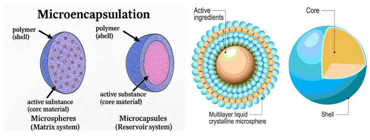
Microspheres are spherical particles with a diameter of 1–1000 μm. Microspheres are essential for improving the way conventional drugs are absorbed and diminishing their side effects. The main advantage of employing microspheres as a drug delivery system is the regulated release of the medicinal contents. By postponing the release of the medication from dosage forms, microencapsulation reduces adverse effects and enhances patient adherence. Using emulsion solvent diffusion evaporation, this method covers an aqueous insoluble core (drugs) with an aqueous insoluble coat (polymer) to produce a sustained release drug delivery system. Phase separation, spray-drying, and emulsification using single or double solvent evaporation systems are some of the methods used to produce microspheres. One method for producing microspheres is to dissolve the precursor components in volatile solvents and then disperse them in a different solvent that isn't miscible with the first one. A tiny particle known as microspheres that dissolve in water will be produced once the last solvent has completely evaporated. Medication with a short half-life that is easily carried from the gastrointestinal tract (GIT) is instantly removed from the bloodstream. [1] Controlled release (CR) has been developed to get around this problem. This process will keep the drug's intensity in the plasma constant for a longer period of time and release it gradually into the gastrointestinal tract. If a dose formulation reaches the necessary plasma therapeutic drug concentration and remains stable during treatment, it is considered appropriate. This can be accomplished by administering a conventional dosage form at a predetermined frequency and dosage. Nanoparticles have the advantage of not being microcarriers because they move into the interstitium inside the lymphatic system's 100 nm range. Undoubtedly, it is possible to carry potentially hazardous items. Dried microparticles can be thought of as solids rather than liquids once they are encapsulated. Since the intake dose is administered as a series of discrete, small, multiarticulate particles, each of which holds and releases a portion of the dosage, a single subunit failure has no effect on the overall dosage failure. To help release medication into the skin, microparticles are employed in skin treatments. By doing this, the drug remains confined at the application site and is kept out of the bloodstream. To reduce undesirable side effects and keep medication in the skin at the right concentration, they function as a reservoir that releases an active ingredient gradually over time. Consequently, cycles of over- and under-medication are less frequent. The treatment of infectious diseases is particularly affected by the decrease in antibiotic resistance. By putting the product in the right vehicle, these distribution techniques can help improve product safety. [2] Chemical microencapsulation emerged as a promising substitute drug delivery method in the 1940s and 1960s, while polymer/membrane technology rose to prominence in the 1980s. [3]

Characteristics of Microspheres: -

The size of the microsphere may be important for an experiment or less important than other factors. Traditionally, the format of the test or assay determines the particle size. Smaller spheres (~0.1-0.4μm) are utilized for lateral flow testing, but bigger spheres (~4-10μm) are needed for bead-based flow cytometric studies. [4] Polystyrene (PS), polymethyl methacrylate (PMMA), and silica are frequently used to make microspheres. Certain materials have distinct optical and physical properties that may be advantageous or disadvantageous for particular uses. Polymer beads have a significant ability to bind proteins and are hydrophobic. Storage buffers frequently contain surfactants, including SDS (Sodium Dodecyl Sulphate) or 0.01-0.1% Tween® 20, to make handling easier. During the synthesis process, functional monomers can co-polymerize with methyl methacrylate or styrene to produce beads with reactive groups. Suspensions can be stabilized and covalent binding processes facilitated by functional groups. Microspheres of silica are inherently negatively charged and hydrophilic. Surfactants and stabilizers are rarely needed for aqueous silica solutions. While plain silica microspheres can be treated with silanes to provide functional groups or alter surface properties, carboxyl- and amine-functionalized silica spheres are frequently employed in covalent coating procedures. [5,6]

Advantages of microsphere: [7,8]

  • Improved compliance with patients
  • Boost the bioavailability
  • Constant concentration of drug in plasma
  • Decrease in negative consequences & adverse effects
  • Boosts stability
  • Formulation administered parenterally
  • Targeted drug delivery

Disadvantages of microsphere: -

  • Cost of production: Compared to conventional dosage forms, the cost of manufacturing-controlled medication dosage forms is higher.
  • The ability to reproduce; Microspheres are difficult to replicate since their production necessitates specialized knowledge and technologies.
  • Possible toxicity: Due to their high drug concentration, microspheres run the risk of dose dumping, which could be harmful.
  • The toxicity of polymers; Depending on how the formulation is designed, polymeric additives like plasticizers, stabilizers, and antioxidants may also undergo oxidation, hydrolysis, or harmful reactions with biological agents.
  • Swallowing: Oral microspheres should be eaten, not chewed or broken, as they are meant to prolong medication release.
  • Maintaining conditions; Microsphere processing parameters, including pH, temperature, agitation, solvent evaporation, and heating, can impact the stability of encapsulated drugs [9,10].

Ingredients of Microspheres: -

Polymers: -

While producing microspheres, researchers most commonly use both biodegradable and non-biodegradable polymers. Two types of polymers are used to create microspheres: Synthetic and Natural. Before choosing the polymer for the microsphere formulation, we must take into account a number of aspects, such as availability, biocompatibility, biodegradability, and nontoxicity. It should be readily available, non-toxic, biocompatible, and biodegradable. These polymers that satisfy these selection criteria have a number of advantages over conventional drug delivery systems, including increased drug bioavailability due to the longer duration of the drug's residency in the body. In contrast to carbohydrates like agarose, carrageenan, chitosan, and starch, as well as proteins like albumin, collagen, and gelatine, natural polymers include poly (acryl) dextran, poly starch, and DEAE. Chemically modified carbohydrates include sodium alginate, cellulose ether, xanthan gum, Scheroglucan, Gum Arabica, tamarind seed polysaccharide, beeswax, and carnauba wax. Chitin and maize protein (Zien) make up the other class. Lactides, glycosides and their copolymers, polyanhydrides, and poly alkyl cyanoacrylates are examples of synthetic polymers that are biodegradable, while the other class includes polysebacic anhydrides, poly esters/poly lactides, poly ortho-esters, polycarbonates, polylactic glycolic acid (PLGA), polycaprolactones, poly-phosphazenes, ethyl cellulose, Eudragit L100, Eudragit S100, HPMC, Eudragit RS100, and RL100. [11-16]

Surfactant: -

The emulsification and extrusion procedures that produce microspheres depend heavily on surfactants. Surfactants are essential for the creation of stable emulsions because they lessen the interfacial tension between hydrophilic and hydrophobic molecules. The application of surfactant creates unique microspheres by preventing the emulsion droplets from sticking together. The right emulsifier is selected using the hydrophilic–lipophilic balance indicator (HLB). Hydrophilic surfactants with an HLB value of 3.5–6 are known as lipophilic surfactants. A smaller size dispersion is produced by decreasing the size of the microspheres' particles by increasing the amount of surfactant. Non-ionic surfactants include Brij5, Polysorbate 80, Tween 40, Tween 20, Span 85, Span 80, Span 20, Polyxamer188, and anionic surfactants include sodium lauryl sulphate and sodium dodecyl sulphate and sorbitan. [17-23]

Oil: -

The ratio of the water phase's viscosity to the oil phase's affects the microspheres' particle size, size distribution, and homogeneity. For example, because olive oil has a higher viscosity than liquid paraffin, it has been observed that microspheres created with olive oil have bigger particle sizes than those made with liquid paraffin. A range of oils are used in the emulsification/gelation process to create microspheres. Examples include groundnut oil, castor oil, soybean oil, olive oil, sunflower oil, rapeseed oil, rapeseed methyl esters, and liquid paraffin. [24]

Crosslinkers: -

The most common crosslinkers for creating microspheres are the ions Ca2+, Sr2+, and Ba2+. However, because Ca2+ ions are non-toxic and Sr2+ and Ba2+ ions are only mildly toxic, they are commonly used crosslinkers to produce microspheres. Agglomeration of microspheres happens at low concentrations of Ca2+ ions. The microsphere's entrapment efficiency slowly rises with an increase in Ca2+ ion concentration. However, crosslinker overload causes the entrapment efficiency to decrease if more crosslinker is added after the concentration of crosslinker reaches its ideal level. Examples include glutaraldehyde, sulphuric acid, and calcium carbonate. [25-28]

Solvent: -

The solvent evaporation method of creating microspheres is the main application for solvents. Ethanol, methylene chloride, methanol, acetonitrile, dichloromethane (DCM), and polyvinyl alcohol (PVA) are a few examples. [29-31]

Types Of Microspheres: -

Microspheres are classified into different types. They are of following: -

1. Bio-adhesive Microspheres

2. Magnetic Microspheres

3. Floating Microspheres

4. Radioactive Microspheres

5. Polymeric Microspheres:

(a) Biodegradable Polymeric Microspheres

(b) Synthetic Polymeric Microspheres

  1. Bio-adhesive Microspheres: -

The process by which a drug attaches to a membrane via the stickiness of water-soluble polymers is called adhesion. The transport of therapeutic agents to the site of action is extended by bio-adhesive microspheres with mucoadhesive properties, which allow the medication coated on the polymer's surface to stick to the intended organ. The term "bio-adhesion" refers to the process by which a medication delivery device adheres to a mucosal membrane, including the nasal, buccal, ophthalmic, and rectal membranes. These microspheres provide better therapeutic effects, make close contact with the absorption site, and have a longer half-life at the site of action. [32]

  1. Magnetic Microspheres: -

This sort of delivery system is essential since it localizes the drug to the site of ailments. A larger amount of a drug that is currently on the market can be replaced with a smaller amount of a drug with a magnetic target. Because magnetic microspheres contain chitosan, dextran, and other ingredients, magnetic carriers react magnetically to a magnetic field. Chemotherapeutic chemicals are administered to liver cancers using a range of therapy magnetic microspheres. Drugs like proteins and peptides can potentially be targeted by this method. Diagnostic microspheres are used to assess liver metastases and distinguish intestinal loops from other abdominal structures by producing supra-magnetic iron oxide nanoparticles. [33]

  1. Floating Microspheres: -

Floating types remain buoyant in the stomach without affecting the pace of gastric emptying because their bulk density is lower than that of gastric fluid. As the medication is delivered gradually and at the desired rate, the system is seen to be floating on stomach contents, increasing gastric residency, and increasing variability in plasma concentration. It also reduces the likelihood of dosage dumping. It requires fewer dosages because its therapeutic effect lasts longer. Ketoprofen, the drug, is given as floating microspheres. [34]

  1. Radioactive Microspheres: -

The initial capillary bed is tapped with 10–30 nm radio embolization treatment microspheres because they are larger than the capillaries' diameter. Because they are injected into the arteries that supply the tumour of interest, radioactive microspheres provide a high dosage of radiation to the targeted areas in each of these situations without endangering the healthy surrounding tissues. It differs from drug delivery systems in that radiation acts from within a typical distance of radioisotopes rather than being released from microspheres. Radioactive microspheres come in three varieties: α, β, and γ emitters. [35,36]

  1. Polymeric Microspheres: -

The different types of polymeric microspheres can be classified as follows and they are biodegradable polymeric microspheres and Synthetic polymeric microspheres [37].

  1. Biodegradable Polymeric Microspheres: -

Since they are naturally bio-adhesive, biodegradable, and biocompatible, natural polymers like starch are used. When biodegradable polymers come into contact with mucosal membranes, their high degree of swelling with aqueous media causes gel formation and prolongs the residence time. The rate and extent of the drug's release over time are controlled by the polymer concentration and the release pattern. The main drawback is that controlling medication release is difficult due to the complex drug loading efficiency of biodegradable microspheres in clinical settings. Nonetheless, they offer a variety of uses in treatment that is depending on microspheres. [38]

 (b) Synthetic Polymeric Microspheres: -

The ability of synthetic polymeric microspheres to move away from the injection site is their main disadvantage, since it raises the possibility of embolism and further organ injury. These microspheres have been demonstrated to be safe and biocompatible and are also utilised as bulking agents, fillers, embolic particles, drug delivery vehicles, etc. [39]

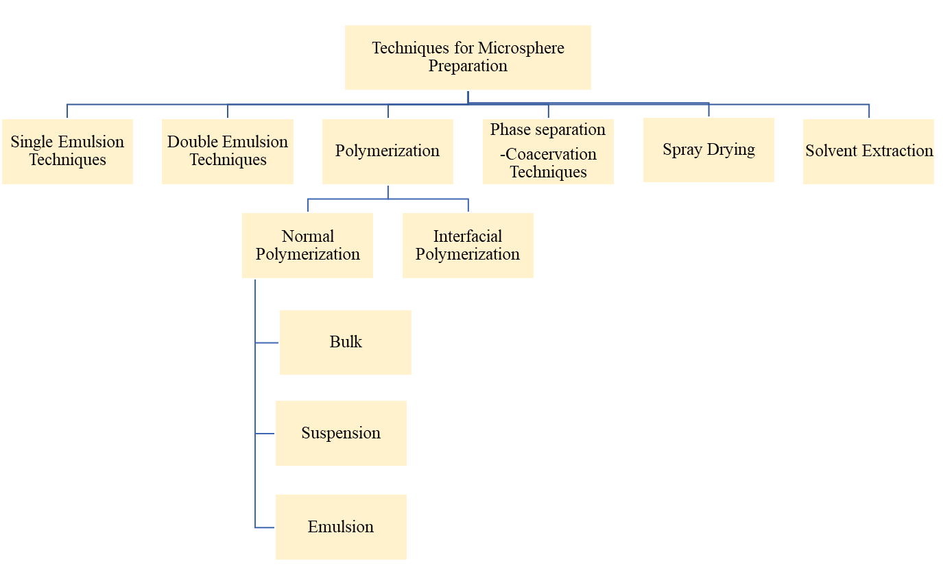
Methods Of Preparation: -

Various standard methods exist for microsphere preparation:

  1. Single Emulsion Techniques
  2. Double Emulsion Techniques
  3. Polymerization:
  1. Normal Polymerization
  • Bulk
  • Suspension
  • Emulsion
  1. Interfacial Polymerization
  1. Phase Separation-Coacervation Technique
  2. Spray Drying
  3. Solvent Extraction
  1. Single Emulsion Techniques: -

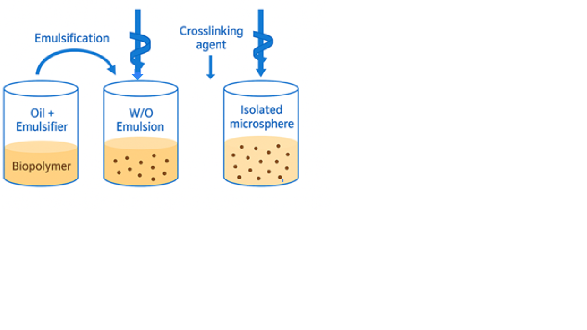
Proteins and carbohydrates are the main targets of this approach. Natural polymers are first dissolved in an aqueous medium before they are released in an oil phase. After that, the scattered globules are cross-linked using chemical cross-linkers such formaldehyde, glutaraldehyde, or acid chloride, or heat denaturation. Heat denaturation is not suitable for heat-sensitive compounds, and chemical cross-linking may expose the active ingredient to chemicals for prolonged periods of time. [40]

Double Emulsion Techniques: -

Using this technique, a double emulsion of the w/o/w or o/w/o types is made. The medication is present in the aqueous solution and dispersed throughout the organic phase. The primary emulsion forms when the organic phase containing the coated polymer encapsulates the medication in the dispersed aqueous phase. A secondary emulsion is then produced by adding this primary emulsion to a PVA aqueous solution after it has been homogenised or sonicated. A secondary emulsion is then produced by adding this first emulsion to a PVA aqueous solution after it has been homogenised or sonicated. After that, the microspheres are filtered and desiccated. [41]

  1. Polymerization: -

Microspheres are prepared using many polymerization processes, including:

Normal Polymerization, Interfacial Polymerization.

  1. Normal Polymerization: -

Bulk Polymerization:

A monomer or a mixture of monomer and initiator is frequently heated to begin the polymerisation and finish the process. The catalyst or initiator is added to the reaction mixture to aid in or accelerate the process. The resultant polymer can be moulded or broken up into microspheres. Two methods for drug loading include adsorption-based loading and drug addiction during the polymerisation process.

Suspension Polymerization: -

It is accomplished by heating the monomer or mixture of monomers that contain active ingredients (drugs) as droplets dispersed in a continuous aqueous phase. The droplets could possibly contain an initiator and additional chemicals.

Emulsion Polymerization: -

However, it differs from suspension polymerisation in that the initiator is present in the aqueous phase and subsequently diffuses to the surface of the micelle or emulsion globule [42].

  1. Interfacial Polymerization: -

 Two reactive monomers are used in the interfacial polymerisation process; one dissolves in the continuous phase and the other is distributed there. The alternate monomer is emulsified by the continuous phase, which is frequently watery. The monomers quickly scatter and polymerize upon the contact. Depending on how soluble the polymer is in the emulsion droplet, the carrier shape may alter.
Temperature, monomer concentration, vehicle makeup, and reactivity can all affect polymerization. Particle size can be controlled by adjusting the size of droplets or globules in the dispersed phase. Maintaining monomer concentration is necessary to control the polymerization process.

  1. Phase Separation-Coacervation Technique: -

This simple procedure separates a micromolecular solution into two immiscible liquid phases. The coacervation concept involves decreasing the solubility of the polymer in the organic phase, which results in the formation of polymer-rich phases known as coacervates. This method involves dispersing drug particles within a polymer solution, followed by the addition of an incompatible polymer. This addition causes the first polymer to phase separate, encasing the drug particles. [43]

  1. Spray Drying: -

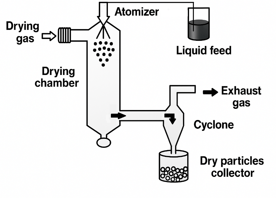
The drug is quickly homogenised and distributed in solid form inside the polymer solution after a polymer is dissolved in a volatile organic solvent, such as acetone or dichloromethane. The resulting dispersion is atomised by a hot air stream, forming minuscule droplets from which the solvent rapidly evaporates, leaving behind microspheres 1–100 μm in size. A cyclone separator is used to gather the generated microparticles from the hot air, and hoover drying is used to get rid of any remaining solvent. This technique involves dissolving the drug and polymers in a suitable solvent to produce a solution, which is then sprayed through a nozzle in a spray dryer under different experimental conditions. Research has shown that spray drying can create vitamin-D3 microspheres with different release patterns based on the kind of lactide-based polymer. [44]

  1. Solvent Extraction: -

This technique uses hydrophilic organic solvents, such as Isopropyl alcohol, to extract the organic solvent, which removes the organic phase. Water was then used to remove the organic phase.

Evaluation Of Microspheres: -

There are various ways to evaluate microspheres such as:

Particle Size and Shape: -

The two most popular techniques for seeing microspheres are scanning electron microscopy (SEM) and standard light microscopy (LM). Both approaches can be used to determine the shape and exterior structure of these microspheres. Light microscopy (LM) can be used to adjust coating parameters for double-walled microspheres. The microsphere forms can be observed before and after coating, and the change can be measured under a microscope. The SEM has a higher resolution than the LM. [45] Scanning electron microscopy (SEM), which may be used to analyse double-walled systems after particles have been cross-sectioned, can be used to analyse the microsphere's surface. Confocal fluorescence imaging is used to structurally characterise multiple-walled microspheres. [46] Other than instrumental methods, such as multi-size Coulter counters and laser light scattering, can be used to describe the microspheres' size, shape, and morphology.

Density Determination: -

The density of the microspheres can be determined using a device called a multi-volume pycnometer. The multi-volume pycnometer is then filled with a precisely weighed sample in a cup. Helium is pumped into the chamber at a constant pressure and allowed to expand. This expansion causes the pressure inside the chamber to decrease. At different initial pressures, two consecutive pressure drop measurements are noted. Two pressure measurements are used to determine the microsphere carrier's volume and, consequently, its density. [47]

Isoelectric Point: -

Using a method known as microelectrophoresis, the electrophoretic mobility of microspheres can be measured in order to estimate the isoelectric point. For different pH values between 3 and 10, the average velocity is calculated by timing the movement of particles across a distance of 1 mm. This information can also be used to calculate the particle's electrical mobility. The surface-contained charge, ionisable behaviour, or ion absorption characteristics of the microspheres may all have an impact on the electrophoretic mobility.

Angle of Contact: -

The wetting property of a microparticulate carrier can be evaluated by measuring the angle of contact. This determines the hydrophilia or hydrophobia of microspheres. This thermodynamic property is specific to solids and is influenced by the presence of the adsorbed component. The angle of contact is measured at the solid-air-water interaction. To measure the approaching and receding angle of contact, a droplet is placed in a circular cell above the objective of the inverted microscope. One minute after the microspheres are placed, the contact angle is measured at 20°C.

Electron Spectroscopy for Chemical Analysis: -

The surface chemistry of the microspheres can be determined using Electron Spectroscopy for Chemical Analysis (ESCA). The method known as Electron Spectroscopy for Chemical Analysis (ESCA) can be used to ascertain the surface's atomic composition. The spectra generated by ECSA can be used to identify the surface degradation of the biodegradable microspheres.

Fourier transform-infrared spectroscopy: -

The polymeric matrix degradation of the carrier system is evaluated using the FT-IR. The surface of the microspheres is examined by measuring Alternated Total Reflectance (ATR). The infrared beam that passes through the ATR cell is reflected multiple times through the sample to create IR spectra, primarily of surface material. The ATR-FTIR provides information regarding the surface composition of the microspheres, depending on the conditions and production methods.

Swelling Index: -

The swelling index is determined by measuring the amount that microspheres expand in a particular solvent. By letting 5 mg of dried microspheres swell overnight in a measuring cylinder with 5 ml of buffer solution, you can determine the equilibrium swelling degree [48].

The swelling index can be calculated using the given formula.

???????????????????????????????? ???????????????????? = ???????????????? × 10

                                               wo                               

 Entrapment Efficiency: -

One can determine the percentage of entrapment or the efficacy of the microspheres' capture by allowing the cleansed microspheres to lysate. Next, the lysate's active components are determined in compliance with the monograph's specifications. [49] Entrapment efficiency can be calculated using the formula below:

%???????? =     ???????????????????????? ???????????????? ????????????????????????????     × 100

               Theoretical drug content

 The Percentage Yield: -

The percentage yield is calculated by dividing the total weight of the medication and polymer needed to make each batch by the weight of the microspheres that were produced from that batch. The result is then multiplied by 100. [50]

% ???????????????????? = ???????????????????????????????????? ???????????????????? × 100

Theoretical Yield

Drug Loading Efficiency: - The percentage of nanoparticle weight that is attached to the encapsulated product is known as drug loading, or the quantity of drug loaded per unit nanoparticle weight. The entire weight of the nanoparticles divided by the total amount of drug contained yields the drug loading percentage. In drug delivery, yield, or the amount of drug provided per quantity, is stated as a percentage. [51]

%???????? =    ???????????????????????? ???????????????? ????????????????????????????    × 100

Total weight of microsphere

Applications of Microspheres: -

Various applications of Microspheres in therapy and drug delivery:

Microspheres have revolutionized various fields, particularly in drug delivery and therapeutic interventions, due to their unique properties and versatility.

  1. Revolutionizing Vaccination: By overcoming drawbacks and improving safety, effectiveness, and administration simplicity, microspheres present a possible substitute for conventional vaccinations. As parenteral carriers, biodegradable microspheres can enhance vaccine durability, modulate antigen release, and strengthen immune response.
  2. Targeted Drug Delivery: Microspheres can be engineered to target specific receptors, to deliver drugs to specific locations. This targeted approach minimizes side-effects and maximizes therapeutic effect.
  3. Monoclonal Antibody-Guided Targeting: Monoclonal antibodies (Mabs) can direct microspheres containing bioactive substances to specific sites due to their high selectivity. There are several ways to do this immunological targeting, which improves the accuracy of medication administration.
  4. In Tumour Treatment: Microspheres play a key role in the tumour-treatment method known as Chemoembolization. To achieve sustained drug levels and vascular occlusion, blood vessels are selectively blocked, and chemotherapy is directly delivered to tumour.
  5. Imaging with Radiolabelled Microspheres: Numerous cells, tissues, and organs can be visualised due to radiolabelled microspheres. The precise locations that can be scanned and provide useful diagnostic data depend on the size of the microspheres.
  6. Topical Delivery through Micro-sponges: Micro-sponges, which are porous microspheres, are added to topical formulations such as lotions and creams. Controlled release of ingredients like emollients and fragrances can be done.
  7. Improved Oral Drug Delivery: For the oral administration of medications with low bioavailability and poor water solubility, microspheres are useful. Their absorption and bioavailability can be enhanced by micronization through encapsulation in microspheres.
  8. Sublingual and Buccal Drug Delivery: Some peptide medications work best on the buccal mucosa. By avoiding the liver's first-pass metabolism, mucoadhesive microspheres can prolong the duration of a drug's residence in the buccal cavity.
  9. Gene Delivery with Non-Viral Vectors: Microspheres are being researched as non-viral gene therapy vectors because they have benefits over viral vectors, such as targeted administration, ease of production, and decreased immunogenicity.
  10. Colon-Specific Drug Delivery: It is possible to create microspheres that will release medications specific in the colon, preventing absorption in the stomach and small intestine. This is useful for medications that treat illnesses related to the colon.
  11. Breast Cancer Treatment: It is possible to directly inject cytotoxin-loaded microspheres into the arteries supplying breast tumours, symbolising the tumour vasculature and delivering the medication to the location.
  12. Brain Tumour Treatment: Agents for diagnosis and treatment are delivered to brain tumour cells via microspheres. For better results, controlled release microspheres have demonstrated potential in sustained drug delivery.
  13. Lung Cancer Treatment: Additionally, microspheres have been used to treat lung cancer. PEGylated microparticles have been shown in studies to have improved anti-cancer effectiveness and extended drug release.
  14. Ocular drug delivery: For the delivery of drugs into the eyes, microspheres are an excellent carrier. Compared to aqueous ocular preparations, the medication's bioavailability has been enhanced using microspheres for drug administration. Microspheres are employed for long-lasting medication release because of their regulated or sustained release mechanism, which lowers the frequency of doses.
  15. Vaginal drug delivery: Drug delivery microspheres are used to treat genital tract mycotic infections and other vaginal infections. Gelatine, chitosan, and PLGA polymers.

Future Prospects:

The topic of medication delivery using microspheres is developing quickly and presents a number of chances for formulation and therapeutic application innovation. Future research is anticipated to concentrate on stimuli-responsive microspheres, which offer better control over drug release kinetics and site-specific delivery by releasing pharmaceuticals in response to environmental cues like pH, temperature, or enzyme activity. [52,53]. It is expected that technological advancements like ligand-mediated targeting, surface modification, and nanotechnology would improve drug targeting effectiveness, especially in the treatment of chronic illnesses and cancer [54,55]. Moreover, long-acting injectable systems with lower dosing frequency will be made possible by the addition of biodegradable polymers with tenable breakdown patterns. [56]. In personalized medicine, where medication administration may be tailored to a patient's unique pharmacokinetic and pharmacodynamic profiles, microspheres are likewise becoming more and more popular [57]. By overcoming the drawbacks of traditional batch methods, the development of 3D printing and microfluidics for the manufacturing of microspheres offers potential scalability, precision, and reproducibility. [58] To convert lab-scale success into commercial viability, however, issues including batch variability, scale-up obstacles, high production costs, and regulatory compliance must be resolved.[59]. Overcoming these obstacles will need cooperative research combining engineers, pharmaceutical specialists, and regulatory bodies. Microspheres have the potential to be a key component of next-generation drug delivery systems with continued research and interdisciplinary efforts, greatly advancing safer, more efficient, and patient-friendly treatments.

CONCLUSION

Microspheres solve many of the drawbacks of traditional dosage forms and offer a very effective and versatile platform for targeted and sustained drug administration. In addition to reducing side effects and increasing patient compliance, their capacity to regulate the release rate, safeguard delicate medications, and target certain tissues improves therapeutic results. Developments in surface modification, production methods, and polymer science have significantly increased the range of possible uses for microspheres in different administration routes. Microsphere technologies are becoming more and more feasible for clinical usage as a result of continuous research, despite some formulation and scaling issues. Future developments in drug delivery and customized treatment could be further revolutionized by combining microspheres with intelligent delivery systems, such as ligand-targeted and stimuli-responsive designs.

REFERENCES

  1. Athira K, K Vineetha, Krishnananda Kamath K, A R Shabaraya, Microspheres as a Novel Drug Delivery System - A Review, International Journal of Pharmaceutical Sciences Review and Research, 2022;75(1):160-166. https://doi.org/10.47583/ijpsrr.2022.v75i01.027
  2. Dhadde Gurunath S, Mali Hanmant S., Raut Indrayani D., Nitalikar Manoj M., Bhutkar Mangesh A, A Review on Microspheres: Types, Method of Preparation, Characterization and Application, Asian Journal of Pharmacy and Technology,2021:11(2):1-7. https://doi.org/10.52711/2231-5713.2021.00025
  3. Gavhane P, Deshmukh M, Khopade AN, Kunjir VV, Shete RV. A review on microsphere. Journal of Drug Delivery and Therapeutics. 2021 Jan 15;11(1):188-9
  4. Pandey V.P., Manavalan R., Sundarrajan T., Ganesh K.S., ‘Formulation and release characteristics of sustained release diltiazem hydrochloride tablets”, Indian J. Pharm. Sci. 2003; 65: Page no.- 44-48.
  5.  S. Freiberg and X. X. Zhu, “International Journal of Pharmaceutics”, Volume 282, Issues 1-2, 10 September 2004, Page no.- 1-18.
  6. M. A. Bayomi, S. A. Al-Suwayeh, A. M. El Helw and A. F. Mesnad, Pharmaceutica Acta Helvetiae, Volume 73, Issue 4, December 1998, Page no.- 187-192.
  7. Prasad BS, Gupta VR, Devanna N, Jayasurya K. Microspheres as drug delivery system-a review. J Glob Trends Pharm Sci 2014; 5:1961-72.
  8. Virmani T, Gupta J. Pharmaceutical application of microspheres: An approach for the treatment of various diseases. Int J Pharm Sci Res 2017; 8:3252-60.
  9. Prasad BS, Gupta VR, Devanna N, Jayasurya K. Microspheres as drug delivery system-a review. J Glob Trends Pharm Sci 2014; 5:1961-72.
  10. Virmani T, Gupta J. Pharmaceutical application of microspheres: An approach for the treatment of various diseases. Int J Pharm Sci Res 2017; 8:3252-60.
  11. Ratnaparkhi M, Wattamwar M, Jadhav A, Chaudhari S. Mucoadhesive microsphere-review. Int J Drug Dev Res 2014; 6:975-1344.
  12. Degim IT, Çelebi N. Controlled delivery of peptides and proteins. Curr Pharm Des 2007; 13:99-117.
  13. Bansal H, Kaur S, Gupta A. Microsphere: Methods of prepration and applications; a comparative study. Int J Pharm Sci Rev Res 2011; 10:69-78.
  14. Wu L, Wang M, Singh V, Li H, Guo Z, Gui S, et al. Three-dimensional distribution of surfactant in microspheres revealed by synchrotron radiation X-ray microcomputed tomography. Asian J Pharm Sci 2017; 12:326-34.
  15. Avachat A, Bornare P, Dash R. Sustained release microspheres of ropinirole hydrochloride: Effect of process parameters. Acta Pharm 2011; 61:363-76.
  16. Ranjha NM, Khan H, Naseem S. Encapsulation and characterization of controlled release flurbiprofen loaded microspheres using beeswax as an encapsulating agent. J Mater Sci Mater Med 2010; 21:1621-30.
  17. Pachuau L, Mazumder B. A study on the effects of different surfactants on ethyl cellulose microspheres. Int J Pharm Tech Res 2009; 1:966-71.
  18.  Kim JC, Song ME, Lee EJ, Park SK, Rang MJ, Ahn HJ. Preparation of microspheres by an emulsification-complexation method. J Colloid Interface Sci 2002; 248:1-4.
  19. De Rosa G, Iommelli R, La Rotonda MI, Miro A, Quaglia F. Influence of the co-encapsulation of different non-ionic surfactants on the properties of PLGA insulin-loaded microspheres. J Control Release 2000; 69:283-95.
  20. Gaur PK, Mishra S, Bajpai M. Formulation and evaluation of controlledrelease of telmisartan microspheres: In vitro/in vivo study. J Food Drug Anal 2014; 22:542-8.
  21. Dinarvand R, Moghadam SH, Sheikhi A, Atyabi F. Effect of surfactant HLB and different formulation variables on the properties of poly-D, L-lactide microspheres of naltrexone prepared by double emulsion technique. J Microencapsul 2005; 22:139-51.
  22. Shiga K, Muramatsu N, Kondo T. Preparation of poly (D, L?lactide) and copoly (lactide?glycolide) microspheres of uniform size. J Pharm Pharmacol 1996; 48:891-5.
  23.  Wan LS, Heng PW, Chan LW. Surfactant effects on alginate microspheres. Int J Pharm 1994; 103:267-75.
  24. Khare P, Jain SK. Influence of rheology of dispersion media in the preparation of polymeric microspheres through emulsification method. AAPS Pharm Sci Tech 2009; 10:1295-300.
  25. Khare P, Jain SK. Influence of rheology of dispersion media in the preparation of polymeric microspheres through emulsification method. AAPS Pharm Sci Tech 2009; 10:1295-300.
  26. Khare P, Jain SK. Influence of rheology of dispersion media in the preparation of polymeric microspheres through emulsification method. AAPS Pharm Sci Tech 2009; 10:1295-300.
  27. Behera AL, Patil SV, Sahoo SK. Formulation and characteristics of 5 fluorouracil microspheres by solvent evaporation method. Int J Pharm Pharm Sci 2011; 3:32-5.
  28.  Kendre P, Chaudhari P. Formulation and evaluation of telmisartan microspheres by solvent evaporation technique. Indo Am J Pharm Res 2012; 2:651-7.
  29. Uyen NT, Hamid ZA, Tram NX, Ahmad N. Fabrication of alginate microspheres for drug delivery: A review. Int J Biol Macromol 2020; 153:1035-46.
  30. Jain, N.K., 2004. Controlled and Novel Drug Delivery. New Delhi: CBS Publishers.
  31. Bodmeier, R. and Chen, H., 1989. Preparation and characterization of microspheres containing the anti-inflammatory agents, indomethacin, ibuprofen, and ketoprofen. Journal of Controlled Release, 10(2), pp.167-175.
  32. T. Hekmatara, G. Regdon, P. Sipos, I. Er?s and K. Pintye- Hódi, “Journal of Thermal Analysis and Calorimetry”, Volume 86, No.- 2, November 2006, Page no.- 287-290.
  33. M.N. Reddy and A.A. Shirwalkar, “Indian Journal of Pharmaceutical Sciences”, April 2000, Volume 62, Issue 4, Page no.- 308-310.
  34. Reza Arshadya, “Journal of Controlled Release”, Volume 14, Issue 2, October 1990, Page no.- 111-131.
  35. Jin-Chul Kim, Dae Ho Kim, Min Chul Kwon, Hae Gon Chung, Seung-Jun Lee, Jong-Duk Kim, Hyeon Yong Lee, “Journal of Dispersion Science and Technology”, Volume 27, Issue 2, April 2006, Page no.- 165-169.
  36. Amita Bagal, Ravi Dahiya, Vance Tsai, Peter A. Adamson, “Archives of Facial Plastic Surgery”, Jul/Aug 2007, Vol 9, No. 4, Page no.- 275-280.
  37. Amita Bagal, Ravi Dahiya, Vance Tsai, Peter A. Adamson, “Archives of Facial Plastic Surgery”, Jul/Aug 2007, Vol 9, No. 4, Page no.- 275-280
  38. Kumaresh S. Soppimath, Anandrao R. Kulkarni, Walter E. Rudzinski, Tejraj M. Aminabhavi, Drug Metabolism Reviews, Volume 33, Issue 2, April 2001, Page no.- 149-160.
  39. T. Hekmatara, G. Regdon, P. Sipos, I. Er?s and K. Pintye- Hódi, “Journal of Thermal Analysis and Calorimetry”, Volume 86, No.- 2, November 2006, Page no.- 287-290.
  40. Gavhane P, Deshmukh M, Khopade AN, Kunjir VV, Shete RV. A review on microsphere. Journal of Drug Delivery and Therapeutics. 2021 Jan 15;11(1):188-9
  41. Yanga YY, Chiab HH, Chunga TS. Effect of preparation temperature on the characteristics and release profiles of PLGA microspheres containing protein fabricated by double-emulsion solvent extraction/ evaporation method. J Control Release 2000; 69:81-96.
  42. Wong CY, Al-Salami H, Crispin R Dass, Microparticles, microcapsules and microspheres: A review of recent developments and prospects for oral delivery of insulin, international journal of pharmaceutics, 2018; 537(1-2): 223-244. https://doi.org/10.1016/j.ijpharm.2017.12.036  PMid:29288095
  43. Gavhane P, Deshmukh M, Khopade AN, Kunjir VV, Shete RV. A review on microsphere. Journal of Drug Delivery and Therapeutics. 2021 Jan 15;11(1):188-9
  44. Sharma N, Purwar N, Gupta PC. Microspheres as drug carriers for controlled drug delivery: a review. International Journal of pharmaceutical sciences and research. 2015 Nov 1;6(11):4579.
  45. Sinha, V.R., Agrawal, M.K. and Kumria, R., 2005. Influence of formulation and excipient variables on the pellet properties prepared by extrusion spheronization. Current Drug Delivery, 2(1), pp.1-8
  46. Kawashima, Y., Niwa, T., Takeuchi, H., Hino, T., Itoh, Y. and Furuyama, S., 1991. Characterization of polymorphs of tranilast anhydrate and tranilast monohydrate when crystallized by two solvent change spherical crystallization techniques. Journal of Pharmaceutical Sciences, 80(5), pp.472-476.
  47. Tanaka, W., Akito, E., Yoshida, K., Terada, T. and Ninomiya, H., 1977. Pharmaceutical preparations for oral cavity administration. U.S.50,51. Ingle, T.G., Pande, S.D., Sawarkar, R. and Padole, D., 2023. The current trends in microspheres: A review. Journal of Drug Delivery and Therapeutics, 13(1), pp.183-194.
  48. Ingle, T.G., Pande, S.D., Sawarkar, R. and Padole, D., 2023. The current trends in microspheres: A review. Journal of Drug Delivery and Therapeutics, 13(1), pp.183-194.
  49. Ingle, T.G., Pande, S.D., Sawarkar, R. and Padole, D., 2023. The current trends in microspheres: A review. Journal of Drug Delivery and Therapeutics, 13(1), pp.183-194
  50. Ingle, T.G., Pande, S.D., Sawarkar, R. and Padole, D., 2023. The current trends in microspheres: A review. Journal of Drug Delivery and Therapeutics, 13(1), pp.183-194
  51. Ingle, T.G., Pande, S.D., Sawarkar, R. and Padole, D., 2023. The current trends in microspheres: A review. Journal of Drug Delivery and Therapeutics, 13(1), pp.183-194.
  52. Zhang L, Gu FX, Chan JM, Wang AZ, Langer RS, Farokhzad OC. Nanoparticles in medicine: therapeutic applications and developments. Clin Pharmacol Ther. 2008.
  53. Makadia HK, Siegel SJ. Poly lactic-co-glycolic acid (PLGA) as biodegradable controlled drug delivery carrier. Polymers. 2011.
  54. Danhier F, Ansorena E, Silva JM, Coco R, Le Breton A, Préat V. PLGA-based nanoparticles: an overview of biomedical applications. J Control Release. 2012.
  55. Soppimath KS, Aminabhavi TM, Kulkarni AR, Rudzinski WE. Biodegradable polymeric nanoparticles as drug delivery devices. J Control Release. 2001.
  56. Jain RA. The manufacturing techniques of various drug loaded biodegradable poly(lactide-co-glycolide) (PLGA) devices. Biomaterials. 2000.
  57. U.S. FDA. The Role of Drug Delivery in Precision Medicine. Guidance Document. 2018.
  58. Niu L, et al. Advances in microfluidic technology for microsphere production. Lab Chip. 2021.
  59. Dureja H, Kaushik D. Regulatory and commercial challenges in novel drug delivery systems. Drug Deliv Transl Res. 2015.

Reference

  1. Athira K, K Vineetha, Krishnananda Kamath K, A R Shabaraya, Microspheres as a Novel Drug Delivery System - A Review, International Journal of Pharmaceutical Sciences Review and Research, 2022;75(1):160-166. https://doi.org/10.47583/ijpsrr.2022.v75i01.027
  2. Dhadde Gurunath S, Mali Hanmant S., Raut Indrayani D., Nitalikar Manoj M., Bhutkar Mangesh A, A Review on Microspheres: Types, Method of Preparation, Characterization and Application, Asian Journal of Pharmacy and Technology,2021:11(2):1-7. https://doi.org/10.52711/2231-5713.2021.00025
  3. Gavhane P, Deshmukh M, Khopade AN, Kunjir VV, Shete RV. A review on microsphere. Journal of Drug Delivery and Therapeutics. 2021 Jan 15;11(1):188-9
  4. Pandey V.P., Manavalan R., Sundarrajan T., Ganesh K.S., ‘Formulation and release characteristics of sustained release diltiazem hydrochloride tablets”, Indian J. Pharm. Sci. 2003; 65: Page no.- 44-48.
  5.  S. Freiberg and X. X. Zhu, “International Journal of Pharmaceutics”, Volume 282, Issues 1-2, 10 September 2004, Page no.- 1-18.
  6. M. A. Bayomi, S. A. Al-Suwayeh, A. M. El Helw and A. F. Mesnad, Pharmaceutica Acta Helvetiae, Volume 73, Issue 4, December 1998, Page no.- 187-192.
  7. Prasad BS, Gupta VR, Devanna N, Jayasurya K. Microspheres as drug delivery system-a review. J Glob Trends Pharm Sci 2014; 5:1961-72.
  8. Virmani T, Gupta J. Pharmaceutical application of microspheres: An approach for the treatment of various diseases. Int J Pharm Sci Res 2017; 8:3252-60.
  9. Prasad BS, Gupta VR, Devanna N, Jayasurya K. Microspheres as drug delivery system-a review. J Glob Trends Pharm Sci 2014; 5:1961-72.
  10. Virmani T, Gupta J. Pharmaceutical application of microspheres: An approach for the treatment of various diseases. Int J Pharm Sci Res 2017; 8:3252-60.
  11. Ratnaparkhi M, Wattamwar M, Jadhav A, Chaudhari S. Mucoadhesive microsphere-review. Int J Drug Dev Res 2014; 6:975-1344.
  12. Degim IT, Çelebi N. Controlled delivery of peptides and proteins. Curr Pharm Des 2007; 13:99-117.
  13. Bansal H, Kaur S, Gupta A. Microsphere: Methods of prepration and applications; a comparative study. Int J Pharm Sci Rev Res 2011; 10:69-78.
  14. Wu L, Wang M, Singh V, Li H, Guo Z, Gui S, et al. Three-dimensional distribution of surfactant in microspheres revealed by synchrotron radiation X-ray microcomputed tomography. Asian J Pharm Sci 2017; 12:326-34.
  15. Avachat A, Bornare P, Dash R. Sustained release microspheres of ropinirole hydrochloride: Effect of process parameters. Acta Pharm 2011; 61:363-76.
  16. Ranjha NM, Khan H, Naseem S. Encapsulation and characterization of controlled release flurbiprofen loaded microspheres using beeswax as an encapsulating agent. J Mater Sci Mater Med 2010; 21:1621-30.
  17. Pachuau L, Mazumder B. A study on the effects of different surfactants on ethyl cellulose microspheres. Int J Pharm Tech Res 2009; 1:966-71.
  18.  Kim JC, Song ME, Lee EJ, Park SK, Rang MJ, Ahn HJ. Preparation of microspheres by an emulsification-complexation method. J Colloid Interface Sci 2002; 248:1-4.
  19. De Rosa G, Iommelli R, La Rotonda MI, Miro A, Quaglia F. Influence of the co-encapsulation of different non-ionic surfactants on the properties of PLGA insulin-loaded microspheres. J Control Release 2000; 69:283-95.
  20. Gaur PK, Mishra S, Bajpai M. Formulation and evaluation of controlledrelease of telmisartan microspheres: In vitro/in vivo study. J Food Drug Anal 2014; 22:542-8.
  21. Dinarvand R, Moghadam SH, Sheikhi A, Atyabi F. Effect of surfactant HLB and different formulation variables on the properties of poly-D, L-lactide microspheres of naltrexone prepared by double emulsion technique. J Microencapsul 2005; 22:139-51.
  22. Shiga K, Muramatsu N, Kondo T. Preparation of poly (D, L?lactide) and copoly (lactide?glycolide) microspheres of uniform size. J Pharm Pharmacol 1996; 48:891-5.
  23.  Wan LS, Heng PW, Chan LW. Surfactant effects on alginate microspheres. Int J Pharm 1994; 103:267-75.
  24. Khare P, Jain SK. Influence of rheology of dispersion media in the preparation of polymeric microspheres through emulsification method. AAPS Pharm Sci Tech 2009; 10:1295-300.
  25. Khare P, Jain SK. Influence of rheology of dispersion media in the preparation of polymeric microspheres through emulsification method. AAPS Pharm Sci Tech 2009; 10:1295-300.
  26. Khare P, Jain SK. Influence of rheology of dispersion media in the preparation of polymeric microspheres through emulsification method. AAPS Pharm Sci Tech 2009; 10:1295-300.
  27. Behera AL, Patil SV, Sahoo SK. Formulation and characteristics of 5 fluorouracil microspheres by solvent evaporation method. Int J Pharm Pharm Sci 2011; 3:32-5.
  28.  Kendre P, Chaudhari P. Formulation and evaluation of telmisartan microspheres by solvent evaporation technique. Indo Am J Pharm Res 2012; 2:651-7.
  29. Uyen NT, Hamid ZA, Tram NX, Ahmad N. Fabrication of alginate microspheres for drug delivery: A review. Int J Biol Macromol 2020; 153:1035-46.
  30. Jain, N.K., 2004. Controlled and Novel Drug Delivery. New Delhi: CBS Publishers.
  31. Bodmeier, R. and Chen, H., 1989. Preparation and characterization of microspheres containing the anti-inflammatory agents, indomethacin, ibuprofen, and ketoprofen. Journal of Controlled Release, 10(2), pp.167-175.
  32. T. Hekmatara, G. Regdon, P. Sipos, I. Er?s and K. Pintye- Hódi, “Journal of Thermal Analysis and Calorimetry”, Volume 86, No.- 2, November 2006, Page no.- 287-290.
  33. M.N. Reddy and A.A. Shirwalkar, “Indian Journal of Pharmaceutical Sciences”, April 2000, Volume 62, Issue 4, Page no.- 308-310.
  34. Reza Arshadya, “Journal of Controlled Release”, Volume 14, Issue 2, October 1990, Page no.- 111-131.
  35. Jin-Chul Kim, Dae Ho Kim, Min Chul Kwon, Hae Gon Chung, Seung-Jun Lee, Jong-Duk Kim, Hyeon Yong Lee, “Journal of Dispersion Science and Technology”, Volume 27, Issue 2, April 2006, Page no.- 165-169.
  36. Amita Bagal, Ravi Dahiya, Vance Tsai, Peter A. Adamson, “Archives of Facial Plastic Surgery”, Jul/Aug 2007, Vol 9, No. 4, Page no.- 275-280.
  37. Amita Bagal, Ravi Dahiya, Vance Tsai, Peter A. Adamson, “Archives of Facial Plastic Surgery”, Jul/Aug 2007, Vol 9, No. 4, Page no.- 275-280
  38. Kumaresh S. Soppimath, Anandrao R. Kulkarni, Walter E. Rudzinski, Tejraj M. Aminabhavi, Drug Metabolism Reviews, Volume 33, Issue 2, April 2001, Page no.- 149-160.
  39. T. Hekmatara, G. Regdon, P. Sipos, I. Er?s and K. Pintye- Hódi, “Journal of Thermal Analysis and Calorimetry”, Volume 86, No.- 2, November 2006, Page no.- 287-290.
  40. Gavhane P, Deshmukh M, Khopade AN, Kunjir VV, Shete RV. A review on microsphere. Journal of Drug Delivery and Therapeutics. 2021 Jan 15;11(1):188-9
  41. Yanga YY, Chiab HH, Chunga TS. Effect of preparation temperature on the characteristics and release profiles of PLGA microspheres containing protein fabricated by double-emulsion solvent extraction/ evaporation method. J Control Release 2000; 69:81-96.
  42. Wong CY, Al-Salami H, Crispin R Dass, Microparticles, microcapsules and microspheres: A review of recent developments and prospects for oral delivery of insulin, international journal of pharmaceutics, 2018; 537(1-2): 223-244. https://doi.org/10.1016/j.ijpharm.2017.12.036  PMid:29288095
  43. Gavhane P, Deshmukh M, Khopade AN, Kunjir VV, Shete RV. A review on microsphere. Journal of Drug Delivery and Therapeutics. 2021 Jan 15;11(1):188-9
  44. Sharma N, Purwar N, Gupta PC. Microspheres as drug carriers for controlled drug delivery: a review. International Journal of pharmaceutical sciences and research. 2015 Nov 1;6(11):4579.
  45. Sinha, V.R., Agrawal, M.K. and Kumria, R., 2005. Influence of formulation and excipient variables on the pellet properties prepared by extrusion spheronization. Current Drug Delivery, 2(1), pp.1-8
  46. Kawashima, Y., Niwa, T., Takeuchi, H., Hino, T., Itoh, Y. and Furuyama, S., 1991. Characterization of polymorphs of tranilast anhydrate and tranilast monohydrate when crystallized by two solvent change spherical crystallization techniques. Journal of Pharmaceutical Sciences, 80(5), pp.472-476.
  47. Tanaka, W., Akito, E., Yoshida, K., Terada, T. and Ninomiya, H., 1977. Pharmaceutical preparations for oral cavity administration. U.S.50,51. Ingle, T.G., Pande, S.D., Sawarkar, R. and Padole, D., 2023. The current trends in microspheres: A review. Journal of Drug Delivery and Therapeutics, 13(1), pp.183-194.
  48. Ingle, T.G., Pande, S.D., Sawarkar, R. and Padole, D., 2023. The current trends in microspheres: A review. Journal of Drug Delivery and Therapeutics, 13(1), pp.183-194.
  49. Ingle, T.G., Pande, S.D., Sawarkar, R. and Padole, D., 2023. The current trends in microspheres: A review. Journal of Drug Delivery and Therapeutics, 13(1), pp.183-194
  50. Ingle, T.G., Pande, S.D., Sawarkar, R. and Padole, D., 2023. The current trends in microspheres: A review. Journal of Drug Delivery and Therapeutics, 13(1), pp.183-194
  51. Ingle, T.G., Pande, S.D., Sawarkar, R. and Padole, D., 2023. The current trends in microspheres: A review. Journal of Drug Delivery and Therapeutics, 13(1), pp.183-194.
  52. Zhang L, Gu FX, Chan JM, Wang AZ, Langer RS, Farokhzad OC. Nanoparticles in medicine: therapeutic applications and developments. Clin Pharmacol Ther. 2008.
  53. Makadia HK, Siegel SJ. Poly lactic-co-glycolic acid (PLGA) as biodegradable controlled drug delivery carrier. Polymers. 2011.
  54. Danhier F, Ansorena E, Silva JM, Coco R, Le Breton A, Préat V. PLGA-based nanoparticles: an overview of biomedical applications. J Control Release. 2012.
  55. Soppimath KS, Aminabhavi TM, Kulkarni AR, Rudzinski WE. Biodegradable polymeric nanoparticles as drug delivery devices. J Control Release. 2001.
  56. Jain RA. The manufacturing techniques of various drug loaded biodegradable poly(lactide-co-glycolide) (PLGA) devices. Biomaterials. 2000.
  57. U.S. FDA. The Role of Drug Delivery in Precision Medicine. Guidance Document. 2018.
  58. Niu L, et al. Advances in microfluidic technology for microsphere production. Lab Chip. 2021.
  59. Dureja H, Kaushik D. Regulatory and commercial challenges in novel drug delivery systems. Drug Deliv Transl Res. 2015.

Photo
Tapas Kumar Mohapatra
Corresponding author

Gayatri College of Pharmacy, Sambalpur. Odisha, Pin-768200

Photo
Sayed Razaur Raheman
Co-author

Gayatri College of Pharmacy, Sambalpur. Odisha, Pin-768200

Photo
Abhijit Sahu
Co-author

Gayatri College of Pharmacy, Sambalpur. Odisha, Pin-768200

Photo
Shruti Naik
Co-author

Gayatri College of Pharmacy, Sambalpur. Odisha, Pin-768200

Photo
Rajesh Pothal
Co-author

Gayatri College of Pharmacy, Sambalpur. Odisha, Pin-768200

Tapas Kumar Mohapatra*, Sayed Razaur Raheman, Abhijit Sahu, Shruti Naik, Rajesh Pothal, Microspheres as Innovative Carriers for Controlled and Targeted Drug Delivery, Int. J. of Pharm. Sci., 2025, Vol 3, Issue 6, 77-92. https://doi.org/10.5281/zenodo.15569854

More related articles
Microspheres: Key Players in Targeted Drug Deliver...
Krupali Chaudhari , Mansi Dhankani, Amitkumar Dhankani , Sunil Pa...
Formulation and Characterization of Sustained-Rele...
Shaheen Shama, Eisha Ganju, Rajni Dubey, Bhaskar Kumar Gupta, ...
Advances in Buccal Film Technology: A Modern Drug Delivery Approach ...
Chandrika Khanolkar, Tushar GRukari, Vijay Jagtap, ...
Design, Development and Optimization of Floating Gastro-Retentive Drug Delivery ...
Satyam Pendor, Dr. A. V. Chandewar, Dr. M. A. Channawar, Gaurav Magar, ...
Formulation And Evaluation Of Floating Microspheres Of Gliclazide ...
Dr Divya Pingili, Shivani Boda, Archana Awasthi, Sowjanya, Pranay Teja, I. Bala Tripura Sundari, ...
Related Articles
Microspheres: A Promising Technology For Controlled Drug Delivery...
FARSANA P P, Dr.Preetha S Panicker, NASRIN NASIR, KEERTHI. R, ...
Microspheres as Novel Drug Delivery Advantages and Application: A Review ...
Eisha Ganju, Shaheen Shama, Rajni Dubey, Bhaskar Kumar Gupta, ...
Design, Development and Optimization of Floating Gastro-Retentive Drug Delivery ...
Satyam Pendor, Dr. A. V. Chandewar, Dr. M. A. Channawar, Gaurav Magar, ...
Development And Evaluation of Sustained Release Formulation by Spray Drying Tech...
Abhay Dhakare, Dr. Shilpa Gawande, Dr. A. V. Chandewar, Anisha Kohale, ...
Microspheres: Key Players in Targeted Drug Delivery and Controlled Release ...
Krupali Chaudhari , Mansi Dhankani, Amitkumar Dhankani , Sunil Pawar, ...