View Article

  • Innovations in Transdermal Drug Delivery: Challenges, Approaches and Future Perspectives

  • 1Department of Pharmaceutics, Konkan Gyanpeeth Rahul Dharkar College of Pharmacy and Research Institute, Karjat (India).
    2Department of Pharmaceutics, Dr. L. H. Hiranandani College of Pharmacy, Ulhasnagar (India)
     

Abstract

Transdermal Drug Delivery Systems (TDDS) represent a promising approach for non-invasive drug administration, providing controlled and sustained release of therapeutic agents through the skin. This method offers several advantages over conventional delivery routes, such as avoiding first-pass metabolism, reducing systemic side effects, and improving patient compliance. However, the development of effective TDDS is challenging due to the skin's barrier function, primarily the stratum corneum, which restricts drug permeation. Recent innovations in formulation strategies, including matrix, reservoir, and drug-in-adhesive systems, have enhanced the efficiency of transdermal patches. Advanced permeation techniques such as microneedles, iontophoresis, ultrasound, and chemical enhancers have further expanded the range of drugs suitable for transdermal delivery, including peptides and macromolecules. Evaluation methods, including in vitro release studies, permeation tests, and in vivo pharmacokinetic analyses, are critical for optimizing drug formulations and ensuring safety and efficacy. The incorporation of novel technologies such as vesicular systems (liposomes, niosomes) and nanocarriers, alongside the development of smart, responsive transdermal systems, highlights the future potential of TDDS. This review explores recent advancements, formulation strategies, and challenges in transdermal drug delivery, providing insights into the innovative approaches that are shaping the next generation of transdermal therapeutics. Emphasis is placed on overcoming the limitations of current systems and enhancing drug permeation and patient adherence, aiming to expand the clinical applications of TDDS across various therapeutic areas.

Keywords

Transdermal, Strategies, Drug Delivery Systems, Clinical Applications

Introduction

Transdermal Drug Delivery Systems (TDDS) have emerged as a crucial innovation in the pharmaceutical industry, offering a non-invasive method for delivering therapeutic agents through the skin directly into systemic circulation. The concept of transdermal delivery dates back to ancient times when herbal patches were used for therapeutic purposes. However, it was not until the 1970s, with the approval of the first transdermal patch (scopolamine for motion sickness), that this delivery method gained significant attention [1]. Since then, transdermal systems have evolved, with numerous drugs formulated as patches, providing an alternative to oral, injectable, and topical formulations. The skin, being the largest organ of the body, presents an attractive route for drug delivery due to its accessibility and vast surface area. Despite its potential, the skin also serves as a formidable barrier, primarily due to its outermost layer, the stratum corneum. This layer, consisting of dead keratinized cells and a lipid-rich matrix, acts as a protective barrier against external substances, preventing water loss and hindering the permeation of most drug molecules [2]. As a result, only drugs with specific physicochemical properties, such as low molecular weight (<500 Da), moderate lipophilicity, and an appropriate partition coefficient (log P between 1 and 3), can effectively penetrate the skin in significant quantities. These limitations have driven extensive research to develop strategies that enhance skin permeation and expand the range of drugs suitable for transdermal delivery [3,4].

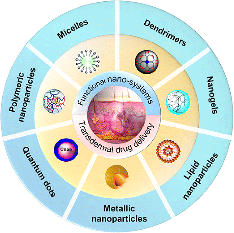
Figure 1: Transdermal Drug Delivery Systems

TDDS offer several significant advantages over traditional drug delivery methods. They bypass the gastrointestinal tract, avoiding issues such as drug degradation by gastric enzymes and first-pass hepatic metabolism, which can significantly reduce drug bioavailability. Additionally, transdermal systems provide controlled and sustained drug release, leading to stable plasma concentrations over extended periods. This reduces the need for frequent dosing and minimizes the risk of peak-and-trough fluctuations in drug levels, enhancing therapeutic efficacy and reducing the likelihood of side effects. The non-invasive nature of transdermal patches also improves patient compliance, particularly in the treatment of chronic conditions such as pain management, hormone replacement therapy, cardiovascular diseases, and smoking cessation [5]. Despite these advantages, the development of effective transdermal systems is challenging due to the skin's barrier properties and the need to maintain drug stability, efficacy, and patient safety [6]. Traditional formulations, such as matrix and reservoir systems, have been employed successfully in various marketed products. However, to overcome the limitations posed by the stratum corneum, several novel approaches have been developed. These include chemical permeation enhancers, which temporarily disrupt the lipid structure of the stratum corneum, and physical enhancement techniques such as iontophoresis, ultrasound (sonophoresis), and microneedle arrays, which create microchannels or use energy to facilitate drug permeation. Additionally, the use of vesicular carriers like liposomes, niosomes and transfersomes has been explored to encapsulate drugs and enhance their penetration through the skin [7]. The evaluation of TDDS involves a comprehensive assessment of drug release kinetics, skin permeation, adhesive properties, and safety profiles. In vitro and in vivo studies play a crucial role in optimizing formulations and predicting their performance in clinical settings. Techniques such as Franz diffusion cells are commonly used to study drug release and permeation, while pharmacokinetic and pharmacodynamic analyses are conducted to assess the therapeutic efficacy and safety of transdermal systems in vivo [8,9]. Recent advancements in materials science and nanotechnology have opened new avenues for enhancing the performance of transdermal drug delivery systems. The development of nanocarriers, such as nanoparticles, nanoemulsions, and solid lipid nanoparticles, has shown promise in improving drug solubility, stability, and skin penetration. Moreover, the integration of responsive materials and smart technologies in TDDS is paving the way for next-generation systems capable of delivering drugs on demand, based on physiological triggers such as pH changes or glucose levels [10]. This review aims to provide a comprehensive overview of the formulation strategies, permeation enhancement techniques, and evaluation methods for transdermal drug delivery systems. It discusses the challenges associated with transdermal delivery and explores innovative approaches that have been developed to overcome these barriers. Additionally, the review highlights recent trends and future perspectives in the field, focusing on novel materials, smart technologies, and the potential of TDDS in expanding the scope of drug delivery for various therapeutic applications. As the demand for non-invasive and patient-friendly drug delivery methods continues to grow, transdermal systems are expected to play an increasingly significant role in the landscape of modern therapeutics.

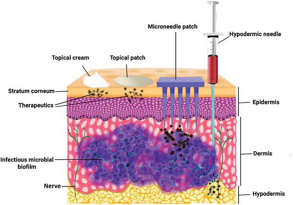
Figure 2: Comparison of The Skin Penetration Through TDDS

Advantages of Transdermal Drug Delivery

1. Non-Invasive Administration

  • Avoids Gastrointestinal Issues: TDDS bypasses the gastrointestinal (GI) tract, reducing the risk of GI irritation, degradation by digestive enzymes, and first-pass metabolism in the liver.
  • Improved Patient Compliance: Non-invasive patches are generally more convenient and less painful compared to injections, increasing patient adherence, especially in long-term therapies.

2. Controlled and Sustained Drug Release

  • Steady Plasma Drug Levels: TDDS allows for a controlled, continuous release of the drug over an extended period, helping to maintain a steady therapeutic plasma concentration, reducing the risk of peaks and troughs associated with oral or injectable formulations.
  • Reduced Dosing Frequency: The prolonged release from transdermal systems decreases the need for frequent dosing, improving patient convenience.

3. Bypassing First-Pass Metabolism

  • Enhanced Bioavailability: Drugs administered through the transdermal route avoid first-pass metabolism by the liver, potentially increasing bioavailability and reducing the required dosage.
  • Reduced Metabolite Formation: By avoiding hepatic metabolism, the formation of potentially harmful metabolites is minimized, reducing the risk of side effects.

4. Improved Safety Profile

  • Minimized Side Effects: By providing a controlled release directly into systemic circulation, TDDS reduces fluctuations in drug concentration, thereby minimizing side effects related to peak plasma levels.
  • Reversible Administration: Transdermal patches can be easily removed if adverse effects occur, providing a safer option for drug delivery compared to irreversible routes like injections.

5. Enhanced Patient Comfort and Compliance

  • Convenience: Patches are easy to apply and remove, leading to better compliance, especially in elderly patients or those with chronic conditions.
  • Non-Intrusive: The non-invasive nature of TDDS makes it a preferred option for individuals with needle phobia or those requiring long-term drug administration.

6. Flexibility in Formulation

  • Wide Range of Drug Candidates: Transdermal systems can be designed for various drug molecules, including those with short half-lives, narrow therapeutic indices, or requiring sustained release.
  • Customizable Dosage Forms: The dosage can be adjusted by varying the size of the patch or the concentration of the active ingredient, offering flexibility in tailoring treatment regimens.

7. Potential for Localized Treatment

  • Targeted Delivery: In certain cases, TDDS can be used for localized drug delivery (e.g., lidocaine patches for local pain relief), reducing systemic exposure and associated side effects.
  • Reduced Risk of Systemic Toxicity: For conditions requiring localized therapy, transdermal application limits systemic absorption, lowering the risk of toxicity.

8. Lower Risk of Drug Abuse and Misuse

  • Difficult to Tamper: Transdermal patches are less prone to abuse compared to oral or injectable forms, as it is challenging to extract the active drug without compromising its efficacy.
  • Reduced Risk of Overdosing: The controlled release mechanism minimizes the chances of accidental overdose, making it a safer option for potent drugs like opioids.

9. Ease of Termination

  • Immediate Cessation of Drug Delivery: In case of adverse reactions or toxicity, removing the transdermal patch stops drug absorption, providing a quick way to terminate the drug's effects.

10. Environmental and Economic Benefits

  • Reduced Need for Sterile Administration: Unlike injectables, TDDS does not require sterile conditions, reducing manufacturing costs.
  • Lower Healthcare Costs: The reduced need for healthcare professional intervention (e.g., injections) and fewer hospital visits contribute to overall cost savings [11-13].

Challenges in Transdermal Drug Delivery

TDDS face several challenges that limit their widespread application. The primary obstacle is the skin's stratum corneum, which serves as an effective barrier, restricting drug penetration, especially for large, hydrophilic molecules. This limits the range of suitable drugs to those with low molecular weight, high lipophilicity, and adequate potency. Additionally, skin permeability can vary significantly among individuals due to factors like age, skin condition, and environmental influences, leading to inconsistent drug absorption. Issues such as skin irritation, allergic reactions, and contact dermatitis from adhesives or permeation enhancers further complicate long-term use. The limited surface area of patches restricts the dosing capacity, making TDDS less effective for high-dose drugs [14]. Technological and manufacturing complexities, coupled with high production costs, add to the challenges of developing efficient systems. Risks such as dose dumping, poor adhesion, and uncontrolled release can lead to variable therapeutic outcomes. Moreover, regulatory hurdles and the need for patient adherence to application instructions can impact the reliability of TDDS. Finally, enhancing skin permeability through chemical or physical methods poses additional risks of irritation or damage, necessitating a balance between efficacy and safety. Despite these challenges, ongoing research aims to optimize TDDS, expanding their applicability in drug delivery [15].

Formulation Approaches in Transdermal Systems

1. Components of TDDS

  • Backing Layer: Provides structural support, protects the patch, and is made of impermeable materials like polyester or aluminum foil.
  • Drug Reservoir or Matrix: Contains the active drug and controls its release.
    • Matrix Systems: Drug dispersed in a polymer matrix for diffusion-controlled release.
    • Reservoir Systems: Liquid or gel drug compartment with a rate-controlling membrane for consistent release.
  • Adhesive Layer: Ensures the patch adheres to the skin; in drug-in-adhesive systems, the drug is integrated into the adhesive.
  • Release Liner: Protects the adhesive and drug layer during storage and is removed before application [16].

2. Design Approaches in TDDS

  • Matrix Systems:
    • Drug is embedded in a polymer matrix.
    • Simple, stable, and provides controlled release.
  • Reservoir Systems:
    • Drug is stored in a reservoir with a rate-controlling membrane.
    • Ensures consistent delivery but requires careful handling to prevent leakage.
  • Drug-in-Adhesive Systems:
    • Drug is incorporated into the adhesive layer.
    • Thin, flexible, and user-friendly.
  • Multilayer Systems:
    • Composed of multiple drug-containing layers for complex release profiles or combination therapies [16,17].

3. Role of Excipients

  • Polymers: Provide structure and regulate drug release (e.g., HPMC, PVA).
  • Adhesives: Facilitate adherence to the skin; common types include acrylics and silicones.
  • Penetration Enhancers: Improve drug transport through the stratum corneum (e.g., ethanol, oleic acid).
  • Plasticizers: Enhance patch flexibility (e.g., propylene glycol).
  • Stabilizers: Prevent drug degradation (e.g., antioxidants like BHT, BHA) [17].

4. Drug Release Mechanisms

  • Diffusion-Controlled Release: Drug diffuses through the polymer matrix or rate-controlling membrane.
  • Adhesion-Controlled Release: Drug diffuses through the adhesive layer.
  • Osmosis-Controlled Release: Osmotic pressure regulates drug delivery.
  • Bio-Responsive Release: Drug release is triggered by external stimuli like pH or temperature.

5. Factors Influencing TDDS Formulation

  • Drug Properties: Ideal for drugs with molecular weight <500 Da, moderate lipophilicity (log P 1–3), and adequate solubility.
  • Skin Properties: Hydration, thickness, and site of application affect absorption.
  • Patient Compliance: Comfort, non-irritation, and ease of use improve adherence.

6. Advanced Approaches

  • Microneedle-Assisted Systems: Create micropores in the skin to bypass the stratum corneum, suitable for macromolecules.
  • Nanotechnology-Based Systems: Use nanoparticles or nanoemulsions to enhance penetration and control release.
  • Smart Patches: Integrate biosensors for drug monitoring and personalized medicine [16-18].

Permeation Enhancement Techniques for TDDS

1. Chemical Enhancement Techniques

Chemical permeation enhancers work by temporarily disrupting the integrity of the stratum corneum or altering its physicochemical properties to improve drug diffusion.

1.1. Solvents

Solvents are widely used for their ability to interact with skin lipids, increasing their fluidity and enhancing drug solubility.

  • Examples: Ethanol, isopropanol, propylene glycol, and dimethyl sulfoxide (DMSO).
  • Mechanism: These solvents extract lipids or disrupt the lipid bilayer structure of the stratum corneum, creating pathways for drug permeation [19].

1.2. Surfactants

Surfactants reduce the surface tension and increase the wettability of the skin, facilitating drug delivery.

  • Examples: Sodium lauryl sulfate, polysorbates (e.g., Tween), and cetrimide.
  • Mechanism: They interact with keratin and disrupt lipid organization [19,20].

1.3. Fatty Acids and Esters

Fatty acids enhance permeability by altering the lipid arrangement of the stratum corneum.

  • Examples: Oleic acid, lauric acid, and isopropyl myristate.
  • Mechanism: These compounds disrupt the lipid bilayer, increasing its fluidity [20].

1.4. Ionic Liquids

Ionic liquids enhance drug solubility and skin permeability.

  • Examples: Combinations of organic salts like choline and geranic acid.
  • Mechanism: They dissolve lipophilic drugs and reduce skin resistance.

1.5. Terpenes

Terpenes like menthol and limonene are effective penetration enhancers.

  • Mechanism: They interact with lipid bilayers, disrupting the barrier without significant damage to skin integrity [20].

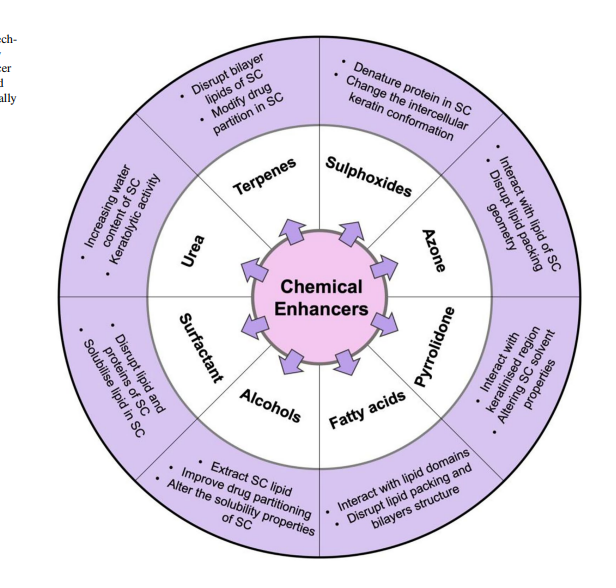
Figure 3: Chemical Enhancement Techniques for TDDS

2. Physical Enhancement Techniques

Physical methods bypass or disrupt the skin's natural barrier using external forces or devices.

2.1. Microneedles

Microneedles create tiny, temporary pores in the stratum corneum, allowing drugs to pass through.

  • Types: Solid, coated, dissolvable, or hollow microneedles.
  • Advantages: Pain-free, minimally invasive, and suitable for hydrophilic and macromolecular drugs.

2.2. Iontophoresis

This technique uses a low electrical current to drive charged drug molecules through the skin.

  • Mechanism: The electrical field reduces skin resistance and facilitates the movement of ionic drugs.
  • Applications: Commonly used for peptides, proteins, and charged small molecules.

2.3. Sonophoresis (Ultrasound)

Sonophoresis uses ultrasonic waves to enhance skin permeability.

  • Mechanism: High-frequency ultrasound disrupts lipid bilayers and enhances fluid movement.
  • Applications: Delivery of macromolecules like insulin.

2.4. Electroporation

Electroporation uses short, high-voltage electrical pulses to create transient micropores in the skin.

  • Mechanism: Electrical pulses disrupt lipid bilayers without causing long-term damage.
  • Applications: Delivery of DNA, vaccines, and peptides.

2.5. Thermal Techniques

Heat-based methods enhance drug delivery by increasing skin temperature and fluidizing lipid bilayers.

  • Techniques: Radiofrequency heating, infrared, or direct thermal patches.
  • Mechanism: Heat reduces viscosity and increases drug diffusivity [21,22].

3. Vesicular Systems

Vesicular systems are specialized delivery carriers that encapsulate drugs, protecting them and improving skin penetration.

3.1. Liposomes

  • Structure: Phospholipid vesicles that mimic biological membranes.
  • Mechanism: Fusion with the lipid layers of the stratum corneum to enhance penetration.
  • Applications: Delivery of both hydrophilic and lipophilic drugs.

3.2. Niosomes

  • Structure: Non-ionic surfactant vesicles similar to liposomes but more stable.
  • Mechanism: Penetrate the lipid bilayer and facilitate sustained release.
  • Applications: Anti-inflammatory drugs and skin diseases.

3.3. Transfersomes

  • Structure: Ultra-deformable vesicles made of phospholipids and edge activators.
  • Mechanism: They deform and squeeze through narrow skin pores, enhancing drug penetration.
  • Applications: Large molecules like proteins and vaccines [23-25].

4. Novel Technologies

Recent advancements have introduced novel methods to overcome the limitations of traditional techniques.

4.1. Nanocarriers

Nanotechnology offers nanoparticles, nanogels, and nanoemulsions for improved drug delivery.

  • Examples: Solid lipid nanoparticles (SLNs), polymeric nanoparticles.
  • Mechanism: Nanocarriers enhance drug stability and target deeper skin layers.

4.2. Chemical-Nano Synergies

Combining chemical enhancers with nanocarriers has shown synergistic effects on drug permeation.

  • Example: Use of DMSO-loaded nanoparticles for improved lipophilic drug delivery.

4.3. Peptides and Polymers

Peptides and polymer-based systems interact with skin proteins or lipids to enhance permeability.

  • Example: Cell-penetrating peptides (CPPs) that transport hydrophilic molecules.

4.4. Smart Systems

Responsive systems like pH-sensitive and temperature-sensitive formulations release drugs in response to specific stimuli.

5. Enzymatic and Biochemical Modulation

This approach targets the skin's biochemical pathways or enzymatic processes to enhance permeation.

  • Proteolytic Enzymes: Break down proteins in the stratum corneum to reduce resistance.
  • Peptide Modulators: Modify skin's natural lipid-protein interactions to facilitate drug diffusion.

6. Combination Approaches

Often, a combination of methods is used to achieve synergistic effects, improving both the extent and rate of drug permeation.

  • Example: Microneedles with iontophoresis or chemical enhancers with vesicular carriers [21-25].

Table 1: Permeation Enhancement Techniques for TDDS [25-28]

Technique

Description

Examples

Mechanism

Applications

Chemical Techniques

Use of chemical agents to modify skin barrier or enhance drug solubility

Solvents

Solubilize drugs and disrupt lipid bilayers.

Ethanol, DMSO, Propylene glycol

Extracts lipids, increases fluidity of the stratum corneum.

Lipophilic drugs, hydrophilic drugs

Surfactants

Lower surface tension and increasewettability.

Sodium lauryl sulfate, Tween

Disrupts keratin and lipid arrangement in the skin barrier.

Delivery of hydrophilic molecules

Fatty Acids and Esters

Alter lipid organization in the stratum corneum.

Oleic acid, Isopropyl myristate

Disrupts lipid bilayer, enhances fluidity.

Lipophilic drugs

Ionic Liquids

Enhance drug solubility and permeability.

Choline-geranate

Dissolve drugs and reduce skin resistance.

Peptides, small hydrophilic molecules

Terpenes

Naturally occurring enhancers that interact with lipids.

Menthol, Limonene

Disrupts lipid bilayers without causing significant skin damage.

Anti-inflammatory drugs, analgesics

Physical Techniques

Devices or methods to bypass or modify the skin barrier

Microneedles

Create microchannels in the skin without pain.

Solid, Coated, Dissolvable

Physically disrupts the stratum corneum to allow direct drug transport.

Macromolecules, vaccines, insulin

Iontophoresis

Uses electrical current to drive drug ions into the skin.

Transdermal iontophoresis patches

Reduces skin resistance and facilitates ionic drug movement.

Peptides, proteins, charged drugs

Sonophoresis (Ultrasound)

Uses ultrasonic waves to increase permeability.

Low-frequency ultrasound devices

Disrupts lipid layers and increases skin hydration.

Insulin, macromolecules

Electroporation

Applies electrical pulses to create micropores in the skin.

High-voltage pulse generators

Temporarily disrupts lipid bilayers to create pathways.

Vaccines, DNA delivery

Thermal Techniques

Apply heat to enhance drug diffusion.

Heat patches, infrared devices

Fluidizes lipids and increases diffusivity of the stratum corneum.

Analgesics, anti-inflammatory drugs

Vesicular Systems

Drug carriers encapsulate and transport drugs through the skin.


Liposomes

Phospholipid vesicles that mimic cell membranes.

Phosphatidylcholine-based liposomes

Fuse with lipid bilayers of the skin, facilitating drug transport.

Hydrophilic and lipophilic drugs

Niosomes

Non-ionic surfactant vesicles similar to liposomes.

Span, Tween-based vesicles

Penetrate skin barriers and provide sustained drug release.

Anti-inflammatory agents, antibiotics

Transfersomes

Highly deformable vesicles that squeeze through narrow pores.

Phospholipids with edge activators

Deform under mechanical stress, allowing deeper penetration.

Vaccines, proteins, large molecules

Novel Technologies

Advanced systems for improved efficiency and delivery

Nanocarriers

Use nanotechnology for drug encapsulation and delivery.

SLNs, Nanogels, Nanoemulsions

Stabilize drugs, enhance permeation, and provide sustained release.

Anti-cancer drugs, peptides

Chemical-Nano Synergies

Combine chemical enhancers with nanocarriers for synergistic effects.

DMSO-loaded nanoparticles

Enhance drug solubility and bypass skin barrier limitations.

Lipophilic drugs, peptides

Peptides and Polymers

Use peptides or polymers to interact with skin proteins or lipids.

Cell-penetrating peptides (CPPs)

Facilitate the transport of hydrophilic drugs across the stratum corneum.

Hydrophilic drugs, biologics

Enzymatic and Biochemical

Modify enzymatic pathways to enhance permeation.

Proteolytic enzymes, Peptide modulators

Break down proteins or modify skin biochemistry for easier transport.

High-molecular-weight drugs

Combination Techniques

Combine multiple methods to maximize drug delivery.

Microneedles with iontophoresis

Synergize the benefits of multiple techniques for enhanced efficiency and control.

Vaccines, macromolecules, biologics

Evaluation of Transdermal Drug Delivery Systems

Physicochemical Evaluation

Thickness of the Patch

The thickness of the drug loaded patch is measured in different points by using a digital micrometer and determines the average thickness and standard deviation for the same to ensure the thickness of the prepared patch [29].

Uniformity of weight

Weight variation is studied by individually weighing 10 randomly selected patches and calculating the average weight. The individual weight should not deviate significantly from the average weight.

Drug content determination

An accurately weighed portion of film (about 100 mg) is dissolved in 100 ml of suitable solvent in which drug is soluble and then the solution is shaken continuously for 24 h in shaker incubator. Then the whole solution is sonicated. After sonication and subsequent filtration, drug in solution is estimated spectrophotometrically by appropriate dilution [29,30].

Moisture content

The prepared films are weighed individually and kept in a desiccator containing calcium chloride at room temperature for 24 h. The films are weighed again after a specified interval until they show a constant weight. The percent moisture content is calculated using following formula.

% Moisture Content=Initial Wt.- Final Wt.Final Wt. X 100

Uptake Moisture

Weighed films are kept in a desiccator at room temperature for 24 h. These are then taken out and exposed to 84% relative humidity using saturated solution of Potassium chloride in a desiccator until a constant weight is achieved. % Moisture uptake is calculated as given below.

% Moisture Uptake= Final Wt.-  Initial Wt. Initial Wt. X 100

Flatness

A transdermal patch should possess a smooth surface and should not constrict with time. This can be demonstrated with flatness study. For flatness determination, one strip is cut from the centre and two from each side of patches. The length of each strip is measured and variation in length is measured by determining percent constriction. Zero percent constriction is equivalent to 100 percent flatness [30,31].

% Constriction= L1-L2 L2 X 100

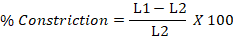
L1 = Initial length of each strip

L2 = Final length of each strip.

Folding Endurance

Evaluation of folding endurance involves determining the folding capacity of the films subjected to frequent extreme conditions of folding. Folding endurance is determined by repeatedly folding the film at the same place until it breaks. The number of times the films could be folded at the same place without breaking is folding endurance value [32].

Tensile Strength

To determine tensile strength, polymeric films are sandwiched separately by corked linear iron plates. One end of the films is kept fixed with the help of an iron screen and other end is connected to a freely movable thread over a pulley. The weights are added gradually to the pan attached with the hanging end of the thread. A pointer on the thread is used to measure the elongation of the film. The weight just sufficient to break the film is noted. The tensile strength can be calculated using the following equation [33].

Tensile Stress (s)=Applied forceCross sectional area=m X gb X t

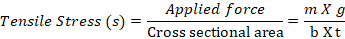
Where, S = tensile stress in 980 dynes/cm2, m = mass in grams, g = acceleration due to gravity (980 dynes/cm 2) b = breadth of strip in centimeters, t = thickness of strip in centimeters.

Water vapor transmission studies (WVT)

For the determination of WVT, Rao et al., (1997) weighed one gram of calcium chloride and placed it in previously dried empty vials having equal diameter. The polymer films were pasted over the brim with the help of adhesive like silicon adhesive grease and the adhesive was allowed to set for 5 minutes. Then, the vials were accurately weighed and placed in humidity chamber maintained at 68 % RH. The vials were again weighed at the end of every 1st day, 2nd day, 3rd day up to 7 consecutive days and an increase in weight was considered as a quantitative measure of moisture transmitted through the patch [34].

In other reported method, desiccators were used to place vials, in which 200 mL of saturated sodium bromide and saturated potassium chloride solution were placed. The desiccators were tightly closed and humidity inside the desiccator was measured by using hygrometer. The weighed vials were then placed in desiccator and procedure was repeated [34,35].

Water vapor transmission rate= Final Wt.-  Initial Wt. Time X Area X 100

Adhesive studies

The therapeutic performance of TDDS can be affected by the quality of contact between the patch and the skin. The adhesion of a TDDS to the skin is obtained by using PSAs, which are defined as adhesives capable of bonding to surfaces with the application of light pressure [36]. The adhesive properties of a TDDS can be characterized by considering the following factors:

  • Peel Adhesion properties
  • Tack properties
  • Thumb tack test
  • Rolling ball test
  • Quick stick (Peel tack) test
  • Probe tack test
  • Shear strength properties or creep resistance.

Swellability

The patches of 3.14 cm² was weighed and put in a petridish containing 10 ml of double distilled water and were allowed to imbibe. Increase in weight of the patch was determined at preset time intervals, until a constant weight was observed. The degree of swelling (S) was calculated using the formul

a: % Swelling= Wt-Wo Wo X 100

Where S is percent swelling, Wt is the weight of patch at time t and W0 is the weight of patch at time zero.

In Vitro Release Studies

There are various methods available for determination of drug release rate of TDDS.

  • The Paddle over Disc
  • The Cylinder modified USP Basket
  • The reciprocating discs
  • Diffusion Cells e.g., Franz Diffusion Cell and its modification Keshary- Chien Cell [37].

In Vitro Permeation Studies

Usually, permeation studies are performed by placing the fabricated transdermal patch with rat skin or synthetic membrane in between receptor and donor compartment in a vertical diffusion cell such as franz diffusion cell or keshary-chien diffusion cell. The transdermal system is applied to the hydrophilic side of the membrane and then mounted in the diffusion cell with lipophillic side in contact with receptor fluid. The receiver compartment is maintained at specific temperature (usually 32±5°C for skin) and is continuously stirred at a constant rate. Samle analyzed by spectrophotometric method [38].

Recent Advances in Transdermal Drug Delivery

TDDS have significantly improved their efficiency and expanded their applications [39]. Innovations in materials, such as biodegradable polymers, stimuli-responsive systems, and lipid-based carriers like transfersomes and solid lipid nanoparticles, have enhanced drug stability and penetration. Emerging technologies, including microneedle systems, iontophoresis, sonophoresis, and electroporation, have addressed the challenges posed by the skin's stratum corneum barrier, enabling the delivery of macromolecules and poorly permeable drugs [40]. Nanotechnology has further revolutionized TDDS with liposomes, polymeric nanoparticles, nanoemulsions, and carbon-based nanomaterials offering improved solubility, targeted delivery, and sustained release. Integration with wearable devices has led to smart patches capable of real-time monitoring and drug administration, especially for chronic diseases like diabetes and hypertension. Combination approaches, such as chemical enhancers with physical techniques, have shown synergistic effects, while dual-action systems improve therapeutic outcomes [41]. These advancements have extended TDDS applications to pain management, hormone replacement therapy, and vaccine delivery, paving the way for personalized medicine, sustainable formulations, and efficient delivery of biologicals like peptides and nucleic acids [42,43].

Applications of Transdermal Drug Delivery Systems (TDDS)

Transdermal drug delivery systems have found widespread applications across various therapeutic areas due to their non-invasive nature, ability to bypass first-pass metabolism, and capacity for sustained and controlled drug release. Below are the key applications of TDDS:

1. Pain Management

  • Analgesics: Transdermal patches delivering drugs like fentanyl and buprenorphine are widely used for chronic pain and post-operative pain management.
  • Non-Steroidal Anti-Inflammatory Drugs (NSAIDs): Diclofenac and ketoprofen patches offer localized relief for musculoskeletal disorders.

2. Hormone Replacement Therapy (HRT)

  • Estrogen and Progesterone: TDDS is extensively used for managing menopausal symptoms and preventing osteoporosis.
  • Testosterone: Transdermal patches or gels provide a non-invasive option for treating hypogonadism in men.

3. cardiovascular diseases

  • Nitroglycerin Patches: Used for the prevention and management of angina pectoris.
  • Clonidine Patches: Effective in managing hypertension through controlled drug delivery.

4. Neurological and Psychiatric Disorders

  • Parkinson’s Disease: Rotigotine patches improve symptoms by delivering dopamine agonists.
  • ADHD: Methylphenidate patches provide controlled drug release, improving focus and reducing hyperactivity.
  • Depression: Selegiline patches for monoamine oxidase inhibitor (MAOI) therapy.

5. Diabetes Management

  • Insulin Delivery: Emerging transdermal approaches using microneedles and iontophoresis for non-invasive insulin administration.
  • Glucose Monitoring: Integrated wearable systems combining transdermal patches with sensors.

6. Smoking Cessation

  • Nicotine Patches: Provide a controlled release of nicotine to reduce withdrawal symptoms and aid smoking cessation.

7. Vaccination

  • Microneedle-Based Vaccines: Painless delivery of vaccines (e.g., COVID-19, influenza) through the skin, enhancing patient compliance and reducing cold-chain dependency.

8. Contraception

  • Hormonal Patches: Deliver estrogen and progestin to prevent pregnancy, offering an alternative to oral contraceptives.

9. Dermatological Treatments

  • Topical Agents: Delivery of drugs like corticosteroids and antifungal agents for localized skin disorders such as eczema, psoriasis, and fungal infections.
  • Cosmetic Applications: Transdermal systems for anti-aging treatments, skin hydration, and delivery of vitamins.

10. Oncology

  • Chemotherapeutic Agents: Experimental TDDS for localized and systemic delivery of anticancer drugs, minimizing systemic toxicity.

11. Neurodegenerative diseases

  • Alzheimer’s Disease: Investigational TDDS for delivering cholinesterase inhibitors or neuroprotective agents to improve cognitive function.

12. Motion Sickness and Nausea

  • Scopolamine Patches: Provide long-lasting relief from motion sickness by delivering anticholinergic drugs through the skin.

13. Wound Healing

  • Antimicrobial Agents: Patches releasing antibiotics for infected wounds.
  • Growth Factors: Experimental systems delivering factors like VEGF for accelerated wound healing.

14. Pediatric and Geriatric Applications

  • Ease of Administration: TDDS is particularly beneficial for children and the elderly, avoiding challenges associated with swallowing pills or enduring injections [44-49].

Future Perspectives

The future of transdermal drug delivery systems (TDDS) is poised for transformative advancements driven by breakthroughs in materials science, nanotechnology, and digital health integration. Emerging technologies, such as microneedles, wearable systems, and hybrid nanocarriers, will broaden the scope of deliverable drugs, including peptides, proteins, and nucleic acids. Smart TDDS integrated with biosensors and artificial intelligence offer the potential for personalized medicine by providing real-time monitoring and dynamic control over drug delivery profiles. Sustainable formulations, including biodegradable and eco-friendly materials, are expected to address environmental concerns associated with conventional systems. Additionally, innovations in physical enhancement techniques, such as electroporation and sonophoresis, may further improve the bioavailability of challenging molecules. As TDDS progress, regulatory frameworks must evolve to accommodate these novel technologies, ensuring safety and efficacy while fostering innovation.

CONCLUSION

Innovations in transdermal drug delivery have transformed the field, addressing key challenges such as poor skin permeability and limited drug applicability. Advanced materials, novel drug delivery techniques, and integration with wearable devices have significantly enhanced the versatility, efficiency, and patient compliance of TDDS. Despite the progress, hurdles such as delivering high molecular weight drugs and achieving cost-effective scalability persist. The convergence of nanotechnology, bioengineering, and digital health is expected to overcome these barriers, paving the way for next-generation TDDS capable of revolutionizing therapeutic practices. By continuously addressing current limitations and embracing emerging opportunities, transdermal systems hold immense promise for improving global healthcare outcomes.

REFERENCES

  1. Prausnitz MR, Langer R. Transdermal drug delivery. Nat Biotechnol. 2008;26(11):1261–8.
  2. Donnelly RF, Singh TR, Woolfson AD. Microneedle-based drug delivery systems: Microfabrication, drug delivery, and safety. Drug Deliv Transl Res. 2012;2(5):311–21.
  3. Benson HAE, Namjoshi S. Proteins and peptides: Strategies for delivery to and across the skin. J Pharm Sci. 2008;97(9):3591–610.
  4. Ita K. Transdermal drug delivery: Progress and challenges. J Drug Deliv Sci Technol. 2014;24(3):245–56.
  5. Barry BW. Novel mechanisms and devices to enable successful transdermal drug delivery. Eur J Pharm Sci. 2001;14(2):101–14.
  6. Brown MB, Martin GP, Jones SA, Akomeah FK. Dermal and transdermal drug delivery systems: Current and future prospects. Drug Deliv. 2006;13(3):175–87.
  7. Wang M, Hu L, Xu C, Zhang Y, Zeng W, Wei X, et al. Advances in transdermal drug delivery systems: Microneedles and biomaterials. MedComm. 2022;3(3).
  8. Alkilani AZ, McCrudden MT, Donnelly RF. Transdermal drug delivery: Innovative pharmaceutical developments based on disruption of the barrier properties of the stratum corneum. Pharmaceutics. 2015;7(4):438–70.
  9. Verma DD, Verma S, Blume G, Fahr A. Liposomes increase skin penetration of hydrophilic substances into human skin. Eur J Pharm Biopharm. 2003;55(3):271–7.
  10. Liu Y, Zhao J, Zhang X. Advances in stimuli-responsive drug delivery systems for transdermal delivery. Front Bioeng Biotechnol. 2021; 9:650192.
  11. Prausnitz MR, Mitragotri S, Langer R. Current status and future potential of transdermal drug delivery. Nat Rev Drug Discov. 2004;3(2):115–24.
  12. Dhawan S, Aggarwal G, Harikumar SL. Enhanced transdermal permeability of insulin by transfersomes. Indian J Pharm Sci. 2009;71(6):735–9.
  13. Kim YC, Park JH, Prausnitz MR. Microneedles for drug and vaccine delivery. Adv Drug Deliv Rev. 2012;64(14):1547–68.
  14. Puri A, Loomis K, Smith B, Lee JH, Yavlovich A, Heldman E, et al. Lipid-based nanoparticles as pharmaceutical drug carriers: From concepts to clinic. Crit Rev Ther Drug Carrier Syst. 2009;26(6):523–80.
  15. Morrow DI, McCarron PA, Woolfson AD, Donnelly RF. Innovative strategies for enhancing topical and transdermal drug delivery. Open Drug Deliv J. 2007; 1:36–59.
  16. Strambini LM, Longo A, Candini A, Ortolani A, Posati T, De Grazia A, et al. Highly integrated flexible sensing patch for on-demand delivery of transdermal drugs. Adv Funct Mater. 2020;30(10):1906309.
  17. Lane ME. Skin penetration enhancers. Int J Pharm. 2013;447(1-2):12–21.
  18. Prausnitz MR. Microneedles for transdermal drug delivery. Adv Drug Deliv Rev. 2004;56(5):581–7.
  19. Kalluri H, Banga AK. Transdermal delivery of proteins. AAPS PharmSciTech. 2011;12(1):431–41.
  20. Donnelly RF, Singh TRR, Alkilani AZ, McCrudden MT, O’Neill S, O’Mahony C, et al. Hydrogel-forming microneedle arrays exhibit antimicrobial properties. Int J Pharm. 2013;451(1-2):76–91.
  21. Benson HA, Watkinson AC. Topical and Transdermal Drug Delivery: Principles and Practice. Hoboken: Wiley; 2012.
  22. Bos JD, Meinardi MM. The 500 Dalton rule for the skin penetration of chemical compounds and drugs. Exp Dermatol. 2000;9(3):165–9.
  23. Joung YH. Development of vaccine-delivery systems using microneedles. Biomater Res. 2013;17(1):10.
  24. Ghosh TK, Pfister WR, Yum SI, editors. Transdermal and Topical Drug Delivery Systems. Buffalo Grove: Interpharm Press; 1997.
  25. Mitragotri S. Synergistic effect of enhancers for transdermal drug delivery. Pharm Res. 2000;17(11):1354–9.
  26. Karande P, Jain A, Mitragotri S. Insights into synergistic interactions in chemical penetration enhancers for transdermal drug delivery. J Control Release. 2006;115(2):272–80.
  27. Cevc G, Blume G. Lipid vesicles penetrate into intact skin owing to the transdermal osmotic gradients and hydration effects. Biochim Biophys Acta Biomembr. 1992;1104(1):226–32.
  28. Rzhevskiy AS, Singh TR, Donnelly RF. Microneedles as the technique of drug delivery enhancement in diverse organs. J Control Release. 2018; 270:184–202.
  29. Zhang Y, Brown MB, Jones SA. Influence of liposomes on topical drug delivery to the skin. Int J Pharm. 2010;390(1):53–62.
  30. Birchall JC, Clemo R, Anstey A, John DN. Microneedles in clinical practice: recent advances and opportunities. Pharm Res. 2011;28(8):1634–42.
  31. Sabri AH, Kim Y, Marlow M, Scurr DJ, Segal J, Marlow N, et al. Enhancing skin permeability using novel surfactants and advanced formulation strategies. J Control Release. 2020; 322:95–104.
  32. Godin B, Touitou E. Ethosomes: Novel vesicular carriers for enhanced delivery: Characterization and skin penetration properties. Drug Dev Res. 2000;50(3-4):406–15.
  33. Moghassemi S, Hadjizadeh A. Nano-niosomes as nanoscale drug delivery systems: An illustrated review. J Control Release. 2014; 185:22–36.
  34. Wu XM, Todo H, Sugibayashi K. Advances in transdermal systemic drug delivery. Drug Discov Today. 2012;17(15-16):848–53.
  35. Keleb E, Sharma RK, Mosa EB, Aljahwi AA. Transdermal drug delivery system-design and evaluation. Int J Adv Pharm Sci. 2010;1(2):201–11.
  36. Vemula SK, Lagishetty V, Lingala S. Design and development of transdermal drug delivery system. J Pharm Res. 2010;3(5):901–8.
  37. Dujaili JA, Hameed BH, Sakeer L. Transdermal patches: Current developments and future prospects. Pharma Innov. 2018;7(10):21–8.
  38. Larrañeta E, McCrudden MT, Courtenay AJ, Donnelly RF. Microneedles: A new frontier in nanomedicine delivery. Pharm Res. 2016;33(5):1055–73.
  39. Fresta M, Puglisi G. Application of liposomes as potential cutaneous drug delivery systems. Adv Drug Deliv Rev. 1996;18(3):343–53.
  40. Seto JE, Polat BE, Lopez RF, Blankschtein D, Langer R. Effects of ultrasound and chemical enhancers on transdermal drug delivery: A mechanistic study. J Control Release. 2010;145(1):26–32.
  41. Ramezanli T, Seidi F, Jenjob R, Santos HA. Enhancing the efficiency of transdermal drug delivery with nanomaterials. Nanomedicine. 2018;13(16):2033–51.
  42. Ma H, Wu B, Bai Y, Liu Z, Shu C, Zhong Y. Recent advances in nanotechnology for transdermal drug delivery. Nanotechnology. 2020;31(12):125102
  43. Kathe AA, Kathpalia H. Film forming systems for topical and transdermal drug delivery. Asian J Pharm Sci. 2017;12(6):487–97.
  44. Vhora I, Patil S, Chaudhary S, Patel A, Bhatt P. Nanocarriers for transdermal delivery: A comprehensive review. Curr Pharm Des. 2015;21(22):3217–35. 
  45. Raj GM, Raveendran R. Introduction to basics of pharmacology and toxicology. 1st vol. Singapore: Springer Nature Singapore Pte Ltd. 2019.
  46. Marschütz MK, Bernkop-Schnürch A. Oral peptide drug delivery: polymer-inhibitor conjugates protecting insulin from enzymatic degradation in vitro. Biomaterials. 2000; 21:1499–507.
  47. Dahan A, Miller JM, Amidon GL. Prediction of solubility and permeability class membership: provisional BCS classifcation of the world’s top oral drugs. AAPS J. 2009; 11:740–6.
  48. Patel A, Cholkar K, Mitra AK. Recent developments in protein and peptide parenteral delivery approaches. Ther Deliv. 2014; 5:337–65.
  49. Wen NX, Venkatraman S. Protein delivery options: how well have we succeeded? Ther Deliv. 2015; 6:537–9.

Reference

  1. Prausnitz MR, Langer R. Transdermal drug delivery. Nat Biotechnol. 2008;26(11):1261–8.
  2. Donnelly RF, Singh TR, Woolfson AD. Microneedle-based drug delivery systems: Microfabrication, drug delivery, and safety. Drug Deliv Transl Res. 2012;2(5):311–21.
  3. Benson HAE, Namjoshi S. Proteins and peptides: Strategies for delivery to and across the skin. J Pharm Sci. 2008;97(9):3591–610.
  4. Ita K. Transdermal drug delivery: Progress and challenges. J Drug Deliv Sci Technol. 2014;24(3):245–56.
  5. Barry BW. Novel mechanisms and devices to enable successful transdermal drug delivery. Eur J Pharm Sci. 2001;14(2):101–14.
  6. Brown MB, Martin GP, Jones SA, Akomeah FK. Dermal and transdermal drug delivery systems: Current and future prospects. Drug Deliv. 2006;13(3):175–87.
  7. Wang M, Hu L, Xu C, Zhang Y, Zeng W, Wei X, et al. Advances in transdermal drug delivery systems: Microneedles and biomaterials. MedComm. 2022;3(3).
  8. Alkilani AZ, McCrudden MT, Donnelly RF. Transdermal drug delivery: Innovative pharmaceutical developments based on disruption of the barrier properties of the stratum corneum. Pharmaceutics. 2015;7(4):438–70.
  9. Verma DD, Verma S, Blume G, Fahr A. Liposomes increase skin penetration of hydrophilic substances into human skin. Eur J Pharm Biopharm. 2003;55(3):271–7.
  10. Liu Y, Zhao J, Zhang X. Advances in stimuli-responsive drug delivery systems for transdermal delivery. Front Bioeng Biotechnol. 2021; 9:650192.
  11. Prausnitz MR, Mitragotri S, Langer R. Current status and future potential of transdermal drug delivery. Nat Rev Drug Discov. 2004;3(2):115–24.
  12. Dhawan S, Aggarwal G, Harikumar SL. Enhanced transdermal permeability of insulin by transfersomes. Indian J Pharm Sci. 2009;71(6):735–9.
  13. Kim YC, Park JH, Prausnitz MR. Microneedles for drug and vaccine delivery. Adv Drug Deliv Rev. 2012;64(14):1547–68.
  14. Puri A, Loomis K, Smith B, Lee JH, Yavlovich A, Heldman E, et al. Lipid-based nanoparticles as pharmaceutical drug carriers: From concepts to clinic. Crit Rev Ther Drug Carrier Syst. 2009;26(6):523–80.
  15. Morrow DI, McCarron PA, Woolfson AD, Donnelly RF. Innovative strategies for enhancing topical and transdermal drug delivery. Open Drug Deliv J. 2007; 1:36–59.
  16. Strambini LM, Longo A, Candini A, Ortolani A, Posati T, De Grazia A, et al. Highly integrated flexible sensing patch for on-demand delivery of transdermal drugs. Adv Funct Mater. 2020;30(10):1906309.
  17. Lane ME. Skin penetration enhancers. Int J Pharm. 2013;447(1-2):12–21.
  18. Prausnitz MR. Microneedles for transdermal drug delivery. Adv Drug Deliv Rev. 2004;56(5):581–7.
  19. Kalluri H, Banga AK. Transdermal delivery of proteins. AAPS PharmSciTech. 2011;12(1):431–41.
  20. Donnelly RF, Singh TRR, Alkilani AZ, McCrudden MT, O’Neill S, O’Mahony C, et al. Hydrogel-forming microneedle arrays exhibit antimicrobial properties. Int J Pharm. 2013;451(1-2):76–91.
  21. Benson HA, Watkinson AC. Topical and Transdermal Drug Delivery: Principles and Practice. Hoboken: Wiley; 2012.
  22. Bos JD, Meinardi MM. The 500 Dalton rule for the skin penetration of chemical compounds and drugs. Exp Dermatol. 2000;9(3):165–9.
  23. Joung YH. Development of vaccine-delivery systems using microneedles. Biomater Res. 2013;17(1):10.
  24. Ghosh TK, Pfister WR, Yum SI, editors. Transdermal and Topical Drug Delivery Systems. Buffalo Grove: Interpharm Press; 1997.
  25. Mitragotri S. Synergistic effect of enhancers for transdermal drug delivery. Pharm Res. 2000;17(11):1354–9.
  26. Karande P, Jain A, Mitragotri S. Insights into synergistic interactions in chemical penetration enhancers for transdermal drug delivery. J Control Release. 2006;115(2):272–80.
  27. Cevc G, Blume G. Lipid vesicles penetrate into intact skin owing to the transdermal osmotic gradients and hydration effects. Biochim Biophys Acta Biomembr. 1992;1104(1):226–32.
  28. Rzhevskiy AS, Singh TR, Donnelly RF. Microneedles as the technique of drug delivery enhancement in diverse organs. J Control Release. 2018; 270:184–202.
  29. Zhang Y, Brown MB, Jones SA. Influence of liposomes on topical drug delivery to the skin. Int J Pharm. 2010;390(1):53–62.
  30. Birchall JC, Clemo R, Anstey A, John DN. Microneedles in clinical practice: recent advances and opportunities. Pharm Res. 2011;28(8):1634–42.
  31. Sabri AH, Kim Y, Marlow M, Scurr DJ, Segal J, Marlow N, et al. Enhancing skin permeability using novel surfactants and advanced formulation strategies. J Control Release. 2020; 322:95–104.
  32. Godin B, Touitou E. Ethosomes: Novel vesicular carriers for enhanced delivery: Characterization and skin penetration properties. Drug Dev Res. 2000;50(3-4):406–15.
  33. Moghassemi S, Hadjizadeh A. Nano-niosomes as nanoscale drug delivery systems: An illustrated review. J Control Release. 2014; 185:22–36.
  34. Wu XM, Todo H, Sugibayashi K. Advances in transdermal systemic drug delivery. Drug Discov Today. 2012;17(15-16):848–53.
  35. Keleb E, Sharma RK, Mosa EB, Aljahwi AA. Transdermal drug delivery system-design and evaluation. Int J Adv Pharm Sci. 2010;1(2):201–11.
  36. Vemula SK, Lagishetty V, Lingala S. Design and development of transdermal drug delivery system. J Pharm Res. 2010;3(5):901–8.
  37. Dujaili JA, Hameed BH, Sakeer L. Transdermal patches: Current developments and future prospects. Pharma Innov. 2018;7(10):21–8.
  38. Larrañeta E, McCrudden MT, Courtenay AJ, Donnelly RF. Microneedles: A new frontier in nanomedicine delivery. Pharm Res. 2016;33(5):1055–73.
  39. Fresta M, Puglisi G. Application of liposomes as potential cutaneous drug delivery systems. Adv Drug Deliv Rev. 1996;18(3):343–53.
  40. Seto JE, Polat BE, Lopez RF, Blankschtein D, Langer R. Effects of ultrasound and chemical enhancers on transdermal drug delivery: A mechanistic study. J Control Release. 2010;145(1):26–32.
  41. Ramezanli T, Seidi F, Jenjob R, Santos HA. Enhancing the efficiency of transdermal drug delivery with nanomaterials. Nanomedicine. 2018;13(16):2033–51.
  42. Ma H, Wu B, Bai Y, Liu Z, Shu C, Zhong Y. Recent advances in nanotechnology for transdermal drug delivery. Nanotechnology. 2020;31(12):125102
  43. Kathe AA, Kathpalia H. Film forming systems for topical and transdermal drug delivery. Asian J Pharm Sci. 2017;12(6):487–97.
  44. Vhora I, Patil S, Chaudhary S, Patel A, Bhatt P. Nanocarriers for transdermal delivery: A comprehensive review. Curr Pharm Des. 2015;21(22):3217–35. 
  45. Raj GM, Raveendran R. Introduction to basics of pharmacology and toxicology. 1st vol. Singapore: Springer Nature Singapore Pte Ltd. 2019.
  46. Marschütz MK, Bernkop-Schnürch A. Oral peptide drug delivery: polymer-inhibitor conjugates protecting insulin from enzymatic degradation in vitro. Biomaterials. 2000; 21:1499–507.
  47. Dahan A, Miller JM, Amidon GL. Prediction of solubility and permeability class membership: provisional BCS classifcation of the world’s top oral drugs. AAPS J. 2009; 11:740–6.
  48. Patel A, Cholkar K, Mitra AK. Recent developments in protein and peptide parenteral delivery approaches. Ther Deliv. 2014; 5:337–65.
  49. Wen NX, Venkatraman S. Protein delivery options: how well have we succeeded? Ther Deliv. 2015; 6:537–9.

Photo
Sakshi Bhoir
Corresponding author

Department of Pharmaceutics, Konkan Gyanpeeth Rahul Dharkar College of Pharmacy and Research Institute, Karjat (India).

Photo
Dr. Sushma Singh
Co-author

Department of Pharmaceutics, Dr. L. H. Hiranandani College of Pharmacy, Ulhasnagar (India)

Photo
Swapnil Phalak
Co-author

Department of Pharmaceutics, Konkan Gyanpeeth Rahul Dharkar College of Pharmacy and Research Institute, Karjat (India).

Sakshi Bhoir*, Dr. Sushma Singh, Swapnil Phalak, Innovations in Transdermal Drug Delivery: Challenges, Approaches and Future Perspectives. Sci., 2025, Vol 3, Issue 4, 57-75. https://doi.org/10.5281/zenodo.15118663

More related articles
Microneedle For the Transdermal Delivery of Hypert...
Monika Bhosale, Dr. V. Y. Lokhande , Dr. Karande, ...
An Overview of Covid-19 Transmission, Current Trea...
Ritu Soni, Vaishnavi Soni, Utkarsh Shrivastava, Talib Khan, Trilo...
Bilayer Floating Tablet : A Novel Floating Tablet...
Mayur Aware, Sheetal Gondkar, Rishikesh Bachhav, ...
Analyzing Antihypertensive Drugs Floating Times for Efficiency Evaluation: A Rev...
Dr. Rama Rao Vadapalli, Darisi Saketh, Gangolu Yohan, Gurram Lokeswari, Bodapati Meghana, Jayanth Ka...
An Overview on Preparation Methods and Evaluation of Transdermal Patches ...
D. Rama Brahma Reddy , D. Tejaswi, K. Hima Sankar, K. Venkatesh, G. Trinath Babu, V. Suresh, B. Upen...
Related Articles
Navigating the Launching of New Pharmaceutical Products: Promotional Challenges ...
Prajwal Bhadke, Ishwar Kale, Vaishnavi Dere, Aakansha Usare , Shubham Kamdi, ...
Formulation of Dosage Forms with Rabeprazole: Challenges and Future Perspectives...
Aman Kumar Singh, Alka Verma, Ram Sevak Verma, Ramnivas, Suraj Mishra, Pranjul Verma, ...
Comprehensive Review on Ethosomes: A Novel Vesicular Approach for Topical Drug D...
Shilpa Tigote, Rashmi Mahabal, Namita Bhosale, Vijay Jagtap, ...
Microneedle For the Transdermal Delivery of Hypertensive Drugs...
Monika Bhosale, Dr. V. Y. Lokhande , Dr. Karande, ...
More related articles
Microneedle For the Transdermal Delivery of Hypertensive Drugs...
Monika Bhosale, Dr. V. Y. Lokhande , Dr. Karande, ...
An Overview of Covid-19 Transmission, Current Treatment, And Future Therapeutic ...
Ritu Soni, Vaishnavi Soni, Utkarsh Shrivastava, Talib Khan, Triloki Kurmi, ...
Microneedle For the Transdermal Delivery of Hypertensive Drugs...
Monika Bhosale, Dr. V. Y. Lokhande , Dr. Karande, ...
An Overview of Covid-19 Transmission, Current Treatment, And Future Therapeutic ...
Ritu Soni, Vaishnavi Soni, Utkarsh Shrivastava, Talib Khan, Triloki Kurmi, ...