1Professor & Research Director, Department of Pharmaceutical Quality Assurance, Channabasweshwar College (Degree) Latur - 413512, Maharashtra, India.
2,3M. Pharmacy Second Year Student, Channabasweshwar College (Degree) Latur - 413512, Maharashtra, India
4Professor, Department of Pharmaceutical Chemistry, Channabasweshwar College (Degree) Latur - 413512, Maharashtra, India.
5Assistant Professor, Department of Pharmaceutical Quality Assurance, Channabasweshwar College (Degree) Latur - 413512, Maharashtra, India.
6Principal, Channabasweshwar pharmacy college (DEGREE), Latur – 413512, Maharashtra, India.
7Professor Hyun Tae Janga Lab 413, Department of Aeronautical Chemical En
The electrospun PVP and PEG fibers loaded with gallic acid were prepared effectively. The fiber with a smooth surface that contained 0.2 grams of gallic acid showed an increased medication content for the purpose of treating wounds. Strong antioxidant, antifungal, and somewhat anticancer in wounds, gallic acid is also useful for various ailments. In comparison to the equivalent films, the thermal, mechanical, and drug release characteristics of the gallic acid fibers were examined. With this understanding, efforts were made to optimize the manufacturing of electrospun R-RDP with gallic acid, a more effective antioxidant medication. In this work, we report the creation of an efficient nanofibre (NF) formulation that releases the best antioxidant medication, gallic acid, for use in wound dressing or wound healing. It does this by embedding the polymers PVP and PEG, which are inexpensive and widely available.
The 48 nanomaterials known as polymer nanofibers (NFs) have been thoroughly investigated for a variety of biomedical applications. Notably, NFs have been used as scaffolds for tissue engineering, wound healing, and drug delivery. Electrospinning (ES) was a popular method for creating NFs because it was easy to use and very flexible. A phenolic substance called gallic acid (3,4,5-trihydroxybenzoic acid) is mostly present in red wine, berries, citrus fruits, and tea leaves. Gallic acid has been shown to possess biological activities such as anti-inflammatory, anti-tumor, antimicrobial, antityrosinase, and antioxidant capabilities in addition to its potent antioxidant capacity. Gallic acid's extreme bitterness and astringency, however, restrict its application to functional foods. Furthermore, gallic acid degrades due to its sensitivity to light, pH, temperature, and oxygen. Encapsulation is a potentially effective way to distribute gallic acid since it can hide its unpleasant taste and stop it from oxidizing. Electrospinning is a straightforward and incredibly adaptable technique that is frequently utilized in drug delivery, tissue engineering scaffolds and filtration, immobilization of enzymes, and biosensors. To create sub-micron fibers, an electric field is used in the electrospinning process to pull polymer solution from a syringe needle towards a grounded collector. The Taylor cone is a conical shape created at the tip of the syringe by the electrostatic repulsion of mutual charges and the Columbic force of the external electric field, which act in opposition to the surface tension of the polymer solution. At the tip of the Taylor cone, a charged jet of the polymer solution is ejected as electrostatic force overcomes surface tension caused by the electric field. The solvent in the solution evaporates as the jet passes through the collector, depositing the fibers there.

Figure No. 1 Typical Lab-Scale Electrospinning Set-Up. This Set-Up Uses A Needle And A Flat Horizontal Metallic Collector
The manufactured fiber mats exhibit substantial For many reasons, electrospinning has shown promise as an antioxidant encapsulation technique. It can prevent the active component from degrading by producing nanofibers at normal temperature. Additionally, nanoscale capsule production is feasible, and settings for electrospinning can be adjusted to regulate the release of active ingredients. Furthermore, the release of active chemicals is slowed down by a small surface area. Fibers give a very wide surface area for controlled release as they go smaller and smaller, from micron to nanosize. Thus, the application of nanofiber. Research on electrospun mats with encapsulated bioactive chemicals as active packaging material is expanding.
2. Experimental Section:
2.1 Materials
Polyvinylpyrrolidone K90(M.W 360000), Polyethelyene glyacol (M.W 4000) purchased from Research lab fine chem. Industries Mumbai India. Gallic acid (GA) were purchased from Sigma Aldrich, USA. They were employed as a crosslinking agent and a bioactive compound, respectively.
2.2 Preparation of GA loaded polymeric solution (PVPK90/PEG/GA)
Table No. 1: Preparation conditions for Pvpk90/PEG polymer solutions
|
Sr. No. |
Sample No. |
Formulation of polymer ratio |
Ratio(ml) |
|
|
PVPK90(ml) |
PEG (ml) |
|||
|
1 |
R1 |
10 |
0 |
10 |
|
2 |
R2 |
9 |
1 |
9:1 |
|
3 |
R3 |
8 |
2 |
8:2 |
|
4 |
R4 |
7 |
3 |
7:3 |
|
5 |
R5 |
6 |
4 |
6:4 |
|
6 |
R6 |
5 |
5 |
5:5 |
2.4 Fabrication of nanofibers via electrospinning process
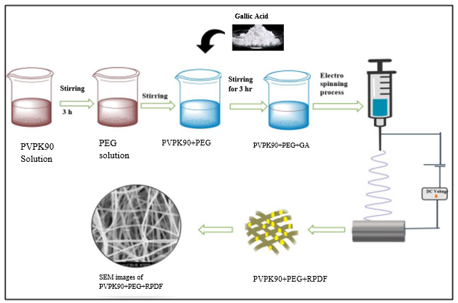
Figure No.2 Schematic Representation for Preparation Of GA-Loaded Electrospun Nanofibers Mat.
2.4 Characterization of nanofibre:
Fourier-transform infrared spectroscopy (FT-IR)7
FT-IR of blank nanofibers PVPK90/PEG and drug-loaded PVPK90/PEG/GA was performed for analysing the compatibility between polymer-excipient and drug-polymer-excipient. Each sample was scanned over a wavenumber region of 4000-400 cm-1 and typical bands were noted shows in figure no. 4-8.
Morphology
Scanning electron microscopy (SEM) 8
SEM was utilised to assess the morphology of the prepared nanofibers such as fiber diameter, fiber consistency, and diameter distribution. To boost electronic conductivity, nanofibers mat was put on a sample holder and coated with gold for 120 seconds. The samples were then focused and images were captured at different magnifications using SEM microscope (S3400N).
X-ray diffraction (XRD) spectroscopy 9
X-ray diffraction is an important tool to investigate the possible variations in the crystalline nature of a drug during electrospinning. Pure drug and all nanofibrous formulations and optimized drug loaded formulation i.e. 15% PVPK90/PEG/GA, were analyzed by X-ray diffractometer system X-600 (PANalytical, Empyrean Netherlands) with Cu Kα radiation, wavelength 1.540 Å, and scanned speed was 5° per min. from 10° to 60° of 2θ.
Differential Scanning Calorimetry (DSC)
DSC can determine the glass transition temperature (Tg), melting temperature (Tm), and crystallization temperature (Tc) of nanofibers. These thermal properties are crucial in understanding the stability and processing conditions of nanofibers.
Entrapment Efficiency 10
The drug entrapment efficiency of nanofibers was measured by drying drug-enriched nanofibers in a hot air oven for 5 min at 41 °C, then the nanofibers scaffold of known area 1 cm2 was removed and dissolved it in water. The amount of drug loaded into these fibres was estimated by UV analysis. The Entrapment Efficiency was calculated by eq. 1.
Entrapment efficiency %=Mass of drug releasedMass of total drug added ×100

In Vitro Drug Release Study11
An in vitro study was conducted using a magnetic stirrer to investigate the release of a drug from a fiber. The experiment began by preparing a stock solution, where 100 mg of the drug-loaded fiber was added to 100 ml of phosphate buffer solution (pH 7.2), and stirred at 100 rpm for 18 hours. At hourly intervals, 1 ml of the solution was withdrawn from the stock, and an equal volume of fresh buffer solution was added to maintain the volume. Each withdrawn sample was then analyzed for absorbance at 270nm using a spectrophotometer as shown in figure no. 14
3. RESULTS AND DISCUSSION:
RESULTS:
3.1 Scanning Electron Microscopy (SEM)
The surface morphology was determined using scanning electron microscope (JEOL JSM-6400). The samples were cut into circular shape with an average diameter of 1.5 cm. Specimens were coated with a thin gold layer using a sputter. The fiber diameters were measured using a SemAphore 4.0 software at 20000 magnification. In fig (a) the average fibre diameter of R1PVPK90 mat were found to be 487.7±125.39, (b) avg. mat fibre of R2 is found to be 513±223.43, (c) 440±223.43, (g)main drug embedded fibre mat found to be smooth continous fibre structured 627.9±149.78 nm respectively.
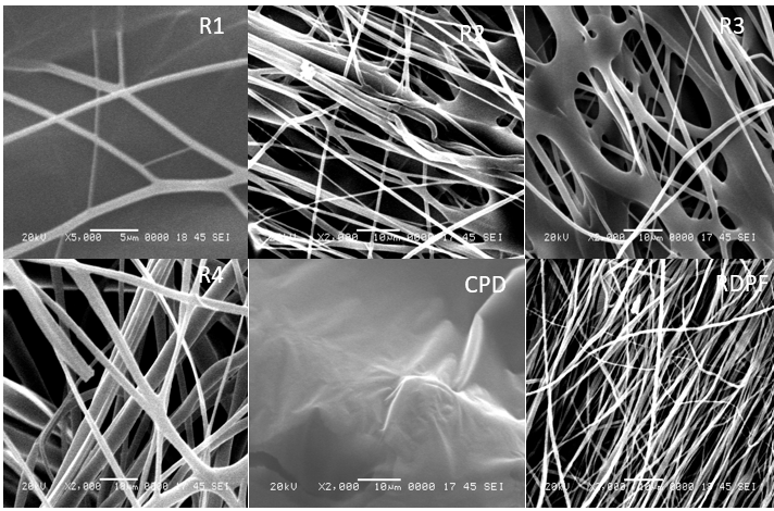
Figure No: 3. SEM Images of drug, polymer with their ratios and final optimized NF
Morphological properties of the GA/PVP electrospun_NF were investigated by SEM. With the magnification of 20000, the diameter of composite fibers was estimated to be 1e5 micron. Highly uniform and smooth composite fibers were prepared without the occurrence of bead defects. For the GA/PVP electrospun fibers, some bulges could be detected. It was believed to be the phenolic group, showing the shape of spindle and sphere. On the other hand, with the increment of gallic acid, the diameter of the electrospun fibers increased. While the supply voltage and electric field were constant, the process became more difficult. This discussion was strongly associated with the previous article reported by Yang et al.
3.2 FTIR Drug – Polymer Compatibility Studies:
Figure No. 4: FTIR spectra of Gallic acid
Table No. 2: Interpretation of FTIR spectra of GA
|
Sr. no. |
Absorption bands at cm? 1 |
vibrations |
|
1 |
1243.06 |
OH Carboxy/(-OH) |
|
2 |
2400 |
OH |
|
3 |
1600 |
C-C Stretching |
|
4 |
1702 |
Carboxyalic acid (strong & broad band) |
|
5 |
1411.09 |
C-C bending |
(B) PVPK90
Figure No. 5: FTIR spectra of PVPK90
Table No.3: Interpretation of FTIR spectra of PVPK90
|
Sr. no. |
Absorption bands at cm? 1 |
vibrations |
|
1 |
1217.02 |
Propolis/C-O |
|
2 |
1285.40 |
Propolis |
|
3 |
1371.02 |
C-O |
|
4 |
1421.11 |
C-H beading of CH2 |
|
5 |
1654.74 |
Propolis |
|
6 |
1738.74 |
C=O |
(C)PEG
Figure No:6. FTIR spectra of PEG
Table No. 4: Interpretation of FTIR spectra of PEG
|
Sr. no. |
Absorption bands at cm? 1 |
vibrations |
|
1 |
2882.33 |
CH2 |
|
2 |
1340.79 |
C-O |
|
3 |
1279.05 |
C-O Stretching |
|
4 |
1240.56 |
C-O Stretching |
|
5 |
1059.62 |
C-O-C |
|
6 |
1099.85 |
C-O-C |
|
7 |
958.04 |
Si-OH |
|
8 |
841.28 |
CH2 |
|
9 |
528.31 |
C-Cl |
(D) CPD
Figure No. 7: FTIR spectra of CPD
Table No. 5: Interpretation of FTIR spectra of CPD
|
Sr. no. |
Absorption bands at cm? 1 |
vibrations |
|
1 |
2874.95 |
C-H stretching |
|
2 |
1692.11 |
prpolis |
|
3 |
1609.26 |
prpolis |
|
4 |
1535.50 |
C=O |
|
5 |
1451.75 |
C-H beading |
|
6 |
1196.28 |
C-O-C |
|
7 |
1090.51 |
S=O |
|
8 |
1035.57 |
CH |
|
9 |
947.34 |
C-C |
(E) RDPF
Figure No.8: FTIR spectra of RDPF
Table No.6: Interpretation of FTIR spectra of RDPF
|
Sr. no. |
Absorption bands at cm? 1 |
vibrations |
|
1 |
1646.88 |
Propolis |
|
2 |
1289.75 |
C-O stretching |
|
3 |
543.84 |
-CCL |
|
4 |
471.33 |
C-CL |
Compatibility of polymer with GA was studied by furrier transform infrared spectroscopy (PerkinElmer). The drug and carrier separately and combination with each other were mixed ATr for determination of spectrum. The range selected was from 4000 – 400 cm-1. The FT-IR spectrum of formulation nanofiber show that there is no significant shift in the peaks or significant difference in spectra and characteristic peaks of formulation are same the pure drug. There is no any interaction between drug and polymer. The FT-IR spectrum of formulation shows same peak values when compared with the characteristic’s peak values of pure drug.
3.3 X-Ray Diffraction (XRD)
XRD spectra of the prepared scaffold was further used to characterize nanofibrous mat. There are no usual spikes for obtained GA in the region of 5o to 60o range. Moreover, the XRD results of the PVP revealed sharp peaks of the PVP crystalline structure at 2θ of 19.40o and 20.02o due to its high degree of crystallinity. Furthermore, crystalline peaks of 2θ at 44.20o were due to the presence GA in XRD pattern. The findings for the PVPK90/GA scaffold indicated that crystalline nature of PVP was changed during the electrospinning. Characteristic signal of PEG was weakened, indicating significant intermolecular and intra molecular hydrogen bonding between DEX and PVP chains. In the same way, there was no distinct peak observed in the XRD pattern of PVP/PEG/GA. It illustrated that GA was entirely coated after addition to the PVP/PEG solution. The results showed that the electrospinning technique did not favor nanofibers crystallinity, additionally slowed the production of crystalline microstructures that resulted in a more amorphous nature of the prepared nanofibers scaffolds. The electrospun PVP/PEG/GA fibers were amorphous, as seen by lack of a distinctive diffraction peak, and had the same peaks, demonstrating that GA loading had no effect on the PV/DEX nanofibers nature. It was possible due to the low weight ratio of GA to PVP/PEG. It revealed that PVP/PEG and GA were compatible also.
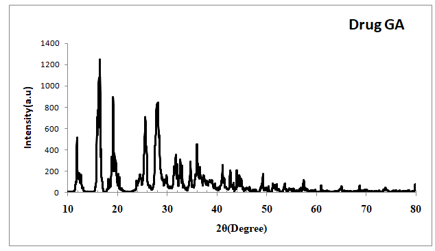
Figure No:9. Overlay of Powder XRD pattern of GA
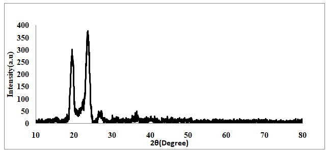
Figure No: 10. Overlay of Powder XRD pattern of CPD
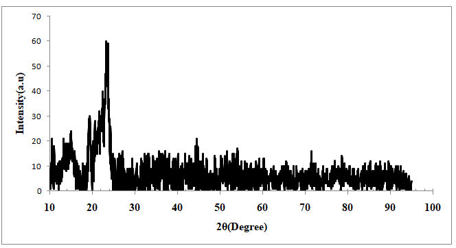
Figure No:11. Overlay of Powder XRD pattern of RDPF
3.4 Differential Scanning Calorimetry (DSC)
DSC studies were performed to characterize the solid state of drugs and polymers. Further, compatibility between drug and excipients can be evaluated by observing the thermal behavior of compounds such as appearance or disappearance of an endothermic or exothermic peak. If all the peaks remain the same, compatibility can be expected. DSC thermogram of GA, PVPK90, CPD and optimized nanofiber membrane are depicted in Figure No.13. DSC thermogram of GA showed a sharp endothermic peak at 95± 2 ºC which is attributed to its melting point. The sharp melting peaks exhibited by GA confirmed their existence as a crystalline form. Thermogram of PVPK90 exhibited a characteristic peak at 40ºC due to its semi-crystalline nature. The PEG also exhibited characteristic peaks of all components indicating physical compatibility between excipients and drug. Whereas, optimized RPDF membrane showed flat curve without sharp endothermic or exothermic peaks of drug. This indicates that transformation of phase i.e. crystalline state to amorphous state has taken place, during the entrapment process. This might be due to shear stress provided by the stirrer and electrospinning during the fabrication process of nanofiber which may prevent the recrystallization of GA, leaving GA in molecular dispersion form inside the RPDF membrane are shown in Fig 12 & 13.

Figure No:12. DSC thermograms of GA, PVPK90
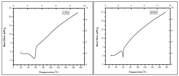
Figure No: 13. CPD and optimized formulation (RPDF)
3.5 In Vitro Study
An in vitro study was conducted using a magnetic stirrer to investigate the release of a drug from a fiber. The experiment began by preparing a stock solution, where 100 mg of the drug-loaded fiber was added to 100 ml of phosphate buffer solution (pH 7.2), and stirred at 100 rpm for 18 hours. At hourly intervals, 1 ml of the solution was withdrawn from the stock, and an equal volume of fresh buffer solution was added to maintain the volume. Each withdrawn sample was then analyzed for absorbance at 270nm using a spectrophotometer. By analyzing the absorbance data, it was determined that approximately 95.00% of the drug had been released into the buffer solution by the end of the 18-hour period. This study provides insights into the release kinetics of the drug from the fiber in vitro, which is crucial for understanding its potential applications in drug delivery systems.
Table No.7: % Of Drug Release on Each Hour
|
Sr. No. |
Time/ HR |
% Drug Release |
|
|
1 |
1.40 |
|
|
2 |
3.40 |
|
|
3 |
6.80 |
|
|
4 |
10.00 |
|
|
5 |
14.00 |
|
|
6 |
18.40 |
|
|
7 |
20.60 |
|
|
8 |
25.40 |
|
|
9 |
32.20 |
|
|
10 |
41.40 |
|
|
11 |
50.40 |
|
|
12 |
55.40 |
|
|
13 |
63.20 |
|
|
14 |
73.20 |
|
|
15 |
79.60 |
|
|
16 |
84.80 |
|
|
17 |
89.00 |
|
|
18 |
95.00 |

Figure No: 14. The % of drug release at each hour
3.6 Drug Entrapment Efficiency (UV)
The drug entrapment efficiency of nanofibers was measured by drying drug-enriched nanofibers in a hot air oven for 5 min at 41 °C, then the nanofibers scaffold of known area 1 cm2 was removed and dissolved it in water. The amount of drug loaded into these fibres was estimated by UV analysis. The Entrapment Efficiency was calculated by eq. 1.
Entrapment efficiency %=Mass of drug releasedMass of total drug added ×100

(1)
DISCUSSION:
The nanofibre formulation carried by the electrospinning method as per proper manner and effectively nanofibers made by standard procedures set and work carried out. The effect of drug: polymer on the physical characteristics of the formulated nanofibres was examined for various drug: polymer ratios of nanofibers at stirring speed of 400-600rpm for 1hrs by electrospinning method. The mean particle size of nanofibers can be influenced by drug: polymer ratio. It was observed that as drug: polymer ratio increases, the particle size decreased. The effect of stirring rate on the physical characteristics of the formulated nanofibers was examined for 9: 1 polymer ratio nanofibers. The stirring rate was varied in the range of 400-600rpm. Polymer concentration has a crucial role in making characterized nanofibers given concentration of polymer gives uniform and structured nanofibers and with optimum release of drug with the help of accurate set ratios of polymer concentration, flow rate, distance between needle and collector, voltage these parameter setted. PVPK90 proportion in NF resulted in an increase in the viscosity of the solution higher viscosities resulted into formation of dense network of the polymer preventing the drug from leaving the matrix. In FTIR analysis obtained results shows that no interaction between drug, polymer and formulation occurred because no change in the characteristics peak was seen. Hence drug and selected polymer were compatible with each other.In SEM analysis result shows that fibre mat found to be smooth continous fibre structured 527.9±149.78 nm respectively. As the viscosity of solution increased, entanglement in polymer chain also increased. Fibers were prepared without the occurrence of bead defects. It was remarkable to note. In in-vitro drug release studies for all Gallic acid nanofibers, formulations along with pure Gallic acid were performed and are reported in result section fig. All gallic acid loaded nanofibers formulations have shown significant enhancement in dissolution rate compared to pure gallic acid in PBS dissolution media at pH 7.2. Using standard least squares regression analysis model, a predictive tool was used to predict % drug release. To determine the reliability of the predictive model, a summary of the fit is shown in Figure No. 24. The regression model for the reduction of % drug release data had an R2 of 0.94, which means that 94% of the predicted % drug release are within the confidence intervals of the actual % drug release recorded. Similarly, the predicted beading regression analysis, R2 = 0.94, also had a very positive correlation with the actual % drug release recorded, showing that 94% of the model predictions are within the confidence intervals of the actual % drug release data recorded. Both confidence curves cross the horizontal lines, which indicate that the predictions are statistically significant to the actual measurements. The main effects of polymer concentration, applied voltage, flow rate, have so far been discussed in the electrospinning of polymer concentration. The DoE completed for manufacturing optimization of polymer concentration aimed at screening for the most influential factors. Multifactorial interactions are useful to gain a deeper understanding of the electrospinning process. Thirty-Four experiments of drug-loaded polymer conc. were completed. The effect of each parameter and multifactorial interactions were investigated on % drug release.
REFERENCES


Dr. Omprakash Bhusnure*, Rajashri Biradar, Shivam Vyavahare, Mani Ganesh, P. Thaware, Vijayendra Swammy, Hyun Tae Jang, Gallic Acid Nanofibre Loaded in Polymer PVP and PEG by Electrospun, Int. J. of Pharm. Sci., 2025, Vol 3, Issue 6, 5169-5183. https://doi.org/10.5281/zenodo.15753563
 10.5281/zenodo.15753563
