Sagar Institute of Research & Technology Pharmacy, SAGE University, Bhopal.
The purpose of topical drug delivery system is to allow therapeutic quantity of drug to correct place in body and to achieve and sustain desired effect of drug for while, in present investigation, we have designed Transfersomes loaded with luliconazole to enhance skin permeation and controlled drug release at targeted site and incorporate them in topical gel of carbopol 934 with good skin retention time. physicochemical property of prepared gel determined as per standards protocol to overcome compliance after patient use. Even spectroscopical analysis reveals no chemical interactivity between luliconazole and excipients. microscopic examination (optical microscopy and scanning electron microscopy) of gel showed uniform distribution of transfersomes inside gel with good order of kinetics of drug release. Hence, it can be concluded that transfersomes gel provides controlled release of drug and these systems can be good source as drug carriers for lipophilic drugs, bioavailability enhancer for poorly water-soluble drugs by nanoparticles, drug delivery system.
Fungal infection is generally characterized by progressive onsets of species of fungi and causes severe health problems in immune-restricted individuals with high morbidity and mortality. It is greatly associated with patients having hematologic, allergenic, prolonged leucopenia and antilogous grafts disorders. Fungal infections generally curve whole body’s system and lead serious lethality to body’s cellular system. The subcutaneous mycosis and is caused by chronic fungal infection which targets dermis and subcutaneous tissue and it is then termed as subcutaneous mycosis.10 Sporotrichosis is one of most important types of tropical infection caused by progressive onsets of fungus Sporothrix schenckii. For inhibition of subsequent progression of any fungal infection, drug should be much effective without having liabilities to produce any seriousharm. Obvious and palliative choice for patients is only way to cure progressive prevalence of fungal infection. Although, large number of pharmaceuticals are available in market which are conventionally utilized as tropical medicaments for treatment of cutaneous and subcutaneous fungal infections. Pharmaceuticals are available in form of creams, lotions, gels, etc. Due to bioavailability barriers or lack of availability of drug to therapeutic site is major concern of patient compliance. Therefore, in light of therapeutic concern of topical anti-fungal drug, drug absorption rate of drug should be controlled by type of formulation to achieve sufficient therapeutic value and can provide an extended pharmacological effect. Luliconazole is contemporary and wide-spectrum anti-fungal agent that is approved by FDA (USA). Due to bioavailability barrier of luliconazole, it does not encompass topical delivery system. In fungal infection, cutaneous and subcutaneous encompassment is required to customize drug permeation ability which can situate high drug concentrations at site of therapeutic action. However, many topical pharmaceuticals of luliconazole are present in markets that have minor skin permeability with shorter skin retention and it leads major patient compliance In current time, nano formulation have gained exponential growth in field of pharmaceuticals due to high complexity in drug load capacity, limited excipients quantity, steadiness in drug stability, lesser harmfulness, and easy scale-up and processing
2. METHOD AND MATERIAL:
Reagents and chemicals
Luliconazole received as gift sample from SMS Pharmaceuticals, INDIA, Carbopol 934, Steric acid, Ethanol, n-octanol, Methanol, Poloxomer 188, Sodium Hydroxide, Potassium Dihydrogen orthophosphate, and Disodium hydrogen orthophosphate Received from SIRT-Pharmacy, SAGE University Bhopal.
Preformulation studies :
Determination of the absorption maximum of luliconazole in ethanol The absorption maximum of luliconazole determined as per standard protocol with some modification. In brief, stock solution of luliconazole developed at concentration of 1 mg/ml in methanol. Further, it followed by serial dilution to get concentration of luliconazole as 2, 4, 6, 8, 10 μg/ml, and then it proceeded to UV spectrophotometric analysis at λmax of 299 nm. Qualification is taken in triplicate and obtained data were analyzed statistically.
Determination of Aqueous Solubility: The determination of aqueous solubility of luliconazole estimated through Saturation shake - flask method. An optimum amount of luliconazole dissolved in distilled water and acetate buffer pH 5.5 then followed by vortex and centrifugation at 50 rpm at 37 °C for 48 hrs, resulting solution filtered and analyzed spectrophotometrically at 299 nm. Qualification is taken in triplicate.
Determination of lipophilicity: Lipophilicity of luliconazole determined through traditional shake flask method as described in protocol with some modification. In brief, an optimum uniform amount of luliconazole poured inside three different volumetric flasks and then measured quantity of lipids such as stearic acid, prectrol, dynasan 114 placed to each flask simultaneously. Resulting heterogeneous mixture proceeded to vortex and then centrifugation at 50 rpm at 37 °C for 48 hrs. Supernatant are separate and filtered using syringe filter of 0.22 μm. Filtrate then analyzed spectrophotometrically at 299 nm. The further partition coefficient of luliconazole determined using n-octanol and water partition system. Measured amount of luliconazole placed inside conical flask containing measured volumes of an n-octanol and aqueous buffer solution. flask shaken with uniform time interval for 48h to attain equilibrium and then resulting mixture placed to separating flask with final shaking and kept remains undisturbed to be separated inside two layers. Targeted measurement proceeded to be analyzed spectrophotometrically at 299 nm. Resulting values of both phases were determined in form of log10P of ratio calculated. All qualification is taken in triplicate. 16
Fourier Transform Infrared (FTIR) Spectroscopy:
The spectral analysis is for luliconazole and stearic acid performed by Win-IR, Bio-Rad FTS spectrophotometer. Individual sample assorted with potassium bromide and later proceeds for spectroscopical observation under range of 4000 to 400 cm−1. 29
Preparations of luliconazole loaded Transfersomes :
The Transfersomes prepared using referenced protocol,Soya-phosphatidylcholine was taken in a round bottom flask. Span80 (sp) or Tween 80 (Tw) was put in the same round bottom flask. Methanol was then added to the same flask. The drug was also loaded in the same RBF. These were then dissolved by shaking. Thin film was then formed by keeping it in the rotatory vacuum evaporator at 600C. To prepare small vesicles, resulting LMVs were sonicated at room temperature or 500C for 30min. the sonicated vesicles were homogenized by manual extrusion 10 times through a sandwich of 200 and 100nm polycarbonate membrane. Then finally we got the Transferosome were transferred to 2% w/v Carbopol gel.
Evaluation of Transfersomes:
Evaluation of entrapment efficiency:
The EE of Transfersomes loaded with luliconazole estimated through described method with some modification. In brief, prepared Transfersomes dried at room temperature then 5 mg of dried Transfersomes were dissolved in 10 ml HPLC grade ethanol and further proceeds by filtration through syringe filter of 0.22 μm capacity. Concentration of luliconazole determined spectrophotometrically at 299 nm. 52 The qualification taken in triplicate and based on percentage entrapment, best one selected for further evaluation. Entrapment efficiency has been determined according to following equation:
EE % = W (Added drug) −W(free drug) / W(Added drug) x 100
Where, W(added drug) is quantity of drug added during preparation of SLN, W(free drug) is quantity of free drug measured in supernatant after centrifugation.
Physicochemical property:
Physicochemical Properties of Transfersomes dispersions were characterized as color, odor, pH, and solubility of Transfersomes F6 in aqueous medium.
Particle size and zeta potential:
The average particle size and zeta potential were determined as per described protocol with some modification. Analysis performed at room temperature by zeta potential/ particle size analyzer. Transfersomes F6 diluted with phosphate-buffered saline and pH of solution stabilized at 7.4 and then sample proceeded for analysis.75
Optical microscopy
Optical microscopic analysis of optimized formulation Transfersomes F6 analyzed with help of digital light optical microscope equipped with fluorescent lamp (Labomed LX-400) at 100x magnification. It aimed to determine whether luliconazole Transfersomes is effectively localized with homogenous and uniform texture within SLN dispersion.
FTIR spectra of Transfersomes F6
The spectral is analysis for Transfersomes F6 performed by Win-IR, Bio-Rad FTS spectrophotometer. Individual sample assorted with potassium bromide and later proceeds for spectroscopical observation under range of 4000 to 400 cm−1.
Preparation Of Gel: The gel developed as per referenced protocol with slight modification. Briefly, Carbopol 934P placed in defined quantity of distilled water while constant stirring at 600 rpm and followed by adding of methylparaben sodium (0.02% w/v) and propylparaben sodium (0.1% w/v) and remained undisturbed with continuous stirring for 30 min. Prepared gel base set aside for 24 hrs. Next, Transfersomes F6 disseminated with measured quantity of propylene glycol (5% w/w) and 1% ethanol (20% w/w) and far ahead it added to carbopol gel bases with continuous shaking at 1000 rpm and followed by churning for 30min. Tri-ethanol amine (TEA) subjected to final stage to maintain pH (5.5 - 6.5) for drug stabilization and stirred thoroughly to obtain clear gel.52. The same procedure applied to get four formulations having varying amount of Carbopol and aim is associated to prepare different forms of gel is to obtain best homogeneous and uniform texture with stable physicochemical reliability in respect of % release of leading moiety. Different formulations of Transfersomes gel are enlisted as in Table;
Table 1: Preparation of different formulations of Transfersomes containing gel.
|
Formulation code |
Carbopol 934 % (w/v) |
|
G1 |
0.5 |
|
G2 |
1 |
|
G3 |
1.5 |
|
G4 |
2 |
3. Characterization of gel :
Determination of pH:
The pH of gel evaluated as per standard protocol with help of digital pH meter. Glass electrode of pH meter immersed in optimized Transfersomes gel formulation and revolved to determine pH of gel.
Determination of viscosity:
The viscosity of gel evaluated as per standard protocol with some modification as follows. In brief, obtained Transfersomes gel evaluated based on physical appearance and then viscosity of Transfersomes gel evaluated through Brookfield Viscometer.
Determination of the entrapment efficiency:
The % EE of different prepared batches of gel estimated by quantitating free mass drug in diffused phase of gel solution after centrifugation. In brief, 1g of gel diffused with ethanol and vortexes for 5 minutes to ensure proper extraction of drugs in ethanol. Then, obtained mixture proceeded for centrifugation at 15000 rpm for 60 minutes at 4 °C temperature. Supernatant collected from centrifuged mixture and allowed to analyze for quantitative analysis spectrophotometrically at 299 nm. 55 The EE percentage calculated from equation as follows;
EE % = W(Added drug) −W(free drug) / W(Added drug) x 100
Where, W(initial drug) is mass of drug added initially, W(free drug) is mass of free drug detected in supernatant after centrifugation.
Spread ability: The spread ability of gel determined as per described protocol with some modification. In brief, 500 mg of optimized formulation put on acrylic plate at middle center and second plate concentrically situated above it. Width of circle in which gel spread measured as primary width. An approx. 500 g weight applied on above plate for few mins. spread ability of gel estimated as per rises in diameter due to dissemination of gels and obtained diameter of disseminated gel noted.
In-vitro drug release and kinetics study:
The drug release and kinetics profiling of optimized formulation (Transfersomes G3) gel were evaluated by in-vitro drug release profiling methods using dialysis bag technique.18 1g of gel sample accurately weighed and placed to cellulose dialysis membrane. Membrane tied with thread and placed to flask containing 50 ml ethanol and phosphate buffer solution. Container placed to magnetic stirrer at 37 °C with constant stirring at 50 rpm. Thereafter, 1 ml of sample withdrawn at regular intervals of 0.25, 0.5, 1, 2, 3, 4, 6, 8, 12, and 24, and withdrawn amount replenished with dissolution media at same time withdrawn. Released mass of Transfersomes entrapped luliconazole quantitated spectrophotometrically at 299 nm in respect of blank. Each measurement is taken in triplicate. In-vitro drug release profiles of prepared luliconazole loaded Transfersomes gel formulation evaluated statistically by various kinetic models named as zero- order, first-order, and Higuchi and Korsemeyer–Peppas model. Kinetics models were determined statistically to enlighten mechanism of drug release profiling. The high regression coefficient value considered to be much effective for initialization and acceptance of kinetics orders.
Fourier transforms infrared spectroscopy:
The spectral analysis is Transfersomes G3 performed by Win-IR, Bio-Rad FTS spectrophotometer. Individual sample assorted with potassium bromide and later proceeds for spectroscopical observation under range of 4000 to 400 cm−1.
Scanning Electron Microscopy:
The morphological analysis is Transfersomes G3 examined by SEM using standard protocol with some modification. Little sample of Transfersomes gel placed on glass stub and vacuum dried. After that, stub having sample sited to SEM chamber coated with gold-palladium and then sample observed microscopically at an accelerating voltage of 10 kV.52.
4. RESULT & DISCUSSION:
RESULTS AND DISCUSSION
Preformulation study of drug:
Determination of the absorption maximum of luliconazole in ethanol:
The potential drug absorption calculated as per standard protocol through absorption maximum of luliconazole at 299 nm λmax against concentration 2-10 μg/ml. regression equation and coefficient were found to be 0.0664x - 0.0478 and 0.998 respectively. Associated aim to determine luliconazole absorption maxima and method validation is for qualitative and quantitative analysis.
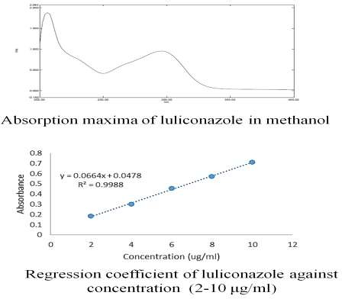
Figure1: Absorption maxima of luliconazole and regression coefficient against the different concentration of luliconazole (μg/ml)
Physicochemical studies of luliconazole were conducted to evaluate physicochemical properties of drug. Studies conducted to evaluate luliconazole hydrophilic and lipophilic compatibility. Result shows that luliconazole having poor solubility potential with water which found to be 0.00585 ± 0.293 mg/ml, and solubility of luliconazole in stearic acid, prectrol, dynasan 114 obtained 23.754±0.47, 18.314± 0.85, 22.875±0.32 mg/ml. Besides, non-aqueous solubility obtained 17.984 ± 0.52 mg/ml for luliconazole in n-octanol. log10P value of luliconazole in stearic acid, prectrol, dynasan 114, and n-octanol obtained as 3.98, 3.30, 3.87, and 3.65 simultaneously. The FTIR analysis performed of luliconazole and stearic acid for better compatibilityanalysis of leading moiety before and after formulation. FTIR spectra of luliconazole is shown in Figure 7; Table 6. principal IR absorption peaks of luliconazole at 2979.43 cm-1 (C-H stretch), 2198.82 cm-1 (C≡N stretch), 1554.35 cm-1 (C-H aromatics stretch), 1471.35 cm-1 (C=C-C aromatic ring stretch), 820.41 cm-1 (para C–H distribution) and 759.46cm-1 (C-Cl stretch) were all detected in spectra of luliconazole. These detected principal peaks confirmed purity and authenticity of luliconazole as similar to referenced report.
Table 2: FTIR Interpretation of Luliconazole
|
Characteristics Peaks |
Reported (cm-1) |
Observed(cm-1) |
|
C-H stretch |
2850 - 3000 |
2979.43 |
|
C≡N Stretch |
2100 - 2400 |
2198.82 |
|
C=C aromatic stretch |
1450 - 1650 |
1554.35 |
|
C=C-C Aromatic ring stretch |
1510 - 1450 |
1471.35 |
|
para C-H distribution |
860 - 800 |
820.41 |
|
C-Cl stretch |
600 - 800 |
759.46 |
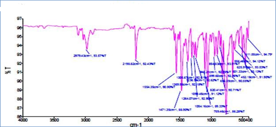
Figure 2: FTIR spectrum of luliconazole
The FTIR spectra are interpretation of stearic acid shown in Figure 9 and Table 7. principal IR absorption peaks of stearic acid at 2915.20cm-1 (C–H stretch alkanes), 2848.02 cm-1 (C-H stretch aldehyde), 1700.89 cm-1 (C=O stretch saturated), 1471.59cm-1(C-C stretch), 1295.47 cm-1 (C-O stretch, aromatic aster), 936.42 cm-1(O-H bend), 719.89cm-1(C=C bend) and 547.03 cm-1 (C-I stretch) were all detected in spectra of stearic acid. These detected principal peaks confirmed purity and authenticity of stearic acid as similar to reported data.
Table 3: FTIR interpretation Stearic acid
|
Characteristics Peaks |
Reported (cm-1) |
Observed(cm-1) |
|
C–H stretch alkanes |
2850 - 3000 |
2915.20 |
|
C-H stretch aldehyde |
2800 - 2860 |
2848.02 |
|
C=O stretch saturated |
1700 – 1730 |
1700.89 |
|
C-C stretch |
1400 - 1500 |
1471.59 |
|
C-O stretch, aromatic aster |
1250 - 1310 |
1295.47 |
|
O-H bend |
910 – 950 |
936.42 |
|
C=C bend |
665 - 730 |
719.89 |
|
C-I stretch |
500 - 600 |
547.03 |
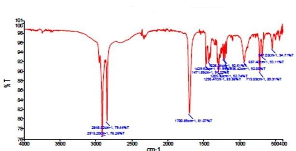
Figure 3 FTIR Spectrum Of Stearic Acid
Development of the method for luliconazole Transfersomes:
The method deals with various modified nano-precipitation methods for optimization of transfersomes in respect of EE of luliconazole at both treatment segments, i.e. nano-precipitation and cooling sonication probe. Temperature controlled by 4°C and 25°C in both segments. Instant adding of organic phase to aqueous phase conserved at 4°C which gives immediate precipitation due to hyphenation with anti-solvent. Temperature controlled at initial phase of nano-precipitation which helped to achieve homogeneity. High-pressure homogenization supports to get uniform homogeneity by decreasing larger crystals size and bead milling aggregation.87 Further, in optimization of transfersomes, method archived step by step with alternate changes in concentration of stearic acid and poloxomer 188 (w/v) ranging from 0.5-2 %. All prepared groups of transfersomes were coded successfully and proceed to quantitate percent entrapment of active moiety spectrophotometrically at 299 nm. Obtained data were evaluated statistically. transfersomes which deal with high entrapment of luliconazole chosen as optimized transfersomes and proceed for further evaluation.
Table 4: Preparation of different luliconazole Transfersomes.
|
|
|
Different concentration of leading reagents for the formation of transfersomes. |
|
|
Tran fersomes code |
Luliconazole% (w/v) |
Stearic acid % (w/v) |
Poloxomer 188 % (w/v) |
|
F1 |
1 |
0.5 |
1 |
|
F2 |
1 |
0.7 |
1 |
|
F3 |
1 |
1 |
1 |
|
F4 |
1 |
2 |
1 |
|
F5 |
1 |
1 |
0.5 |
|
F6 |
1 |
1 |
0.7 |
|
F7 |
1 |
1 |
1.5 |
|
F8 |
1 |
1 |
2 |
Evaluation of Transfersomes
Evaluation of entrapment efficiency of Initially, in pre-formulation studies, luliconazole characterized physiochemically and spectroscopically. After successful formation of different batches of nanoparticles, percentage EE of luliconazole determined. Percentage of EE evaluated spectrophotometrically at 299 nm. Thereafter results reveal that transfersomes F6 and transfersomes F1 have highest and lowest % EE of luliconazole loaded transfersomes by 92.13%±0.975 and 53.78%±1.052 w/w respectively. Similarly, study cited by Ige et al. reported maximum % EE by 90–95% w/w.39 Therefore, based on percent drug entrapment, transfersomes F6 selected as an optimized transfersomes and proceed for further evaluation includes physicochemical properties andgel formation. Percent drug entrapment of all transfersomes groups have been shown graphically in Figure 10 Initially, in pre-formulation studies, luliconazole characterized physicochemically and spectroscopically. After successful formation of different batches of nanoparticles, percentage EE of luliconazole determined. Percentage of EE evaluated spectrophotometrically at 299 nm. Thereafter results reveal that transfersomes F6 and transfersomes F1 have highest and lowest % EE of luliconazole loaded transfersomes by 92.13%±0.975 and 53.78%±1.052 w/w respectively. Similarly, study cited by Ige et al. reported maximum % EE by 90–95% w/w.39 Therefore, based on percent drug entrapment, ransfersomes F6 selected as an optimized transfersomes and proceed for further evaluation includes physicochemical properties andgel formation. Percent drug entrapment of all transfersomes groups have been shown graphically in Figure 10
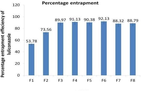
Figure 4: Percentage entrapment efficiency of luliconazole in Transfersomes
Physicochemical property: The Transfersomes F6 evaluated based on their physicochemical characteristics such as color, odor, pH stability, and aqueous solubility. physicochemical results reveal that transfersomes has white transparent color with homogeneous and uniform texture, aromatic odor, better stability at pH, and water solubility found 0.01819 ± 0.035 mg/ml, i.e. much enough than luliconazole solubility.
Zeta potential and particle size and size distribution identification:
The particle size analysis and zeta potential measurement of luliconazole transfersomes were identified successfully using nano ZS90 zetasizer system. Zeta potential is one of important parameters used to forecast physical stability of transfersomes. Stability of nanoparticle system depends on high zeta potential value which points toward better stability of transfersomes since it could deliver deterring force between nanoparticles.81As shown in Fig. 11, SLN shows quite high value of zeta potential by ~18.8 mV and states to high stability of transfersomes. In particle size analysis, transfersomes unveiled with mean particle diameter by ~344.3 nm, unimodal size distribution, polydispersity index (PDI) by 0.168, intercept value 0.98 and 92% peak intensity. PDI is parameter that represents dissemination factor with low aggregation of nanoparticles when PDI value would be < 0.5.88
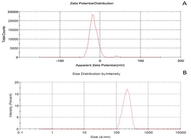
Figure 5: zeta potential, particle size and size distribution of luliconazole transfersomes F6
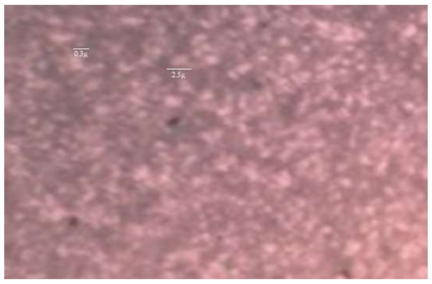
Optical microscopy:
Optical microscopy of optimized preparation i.e. transfersomes F6 defined with help of digital light optical microscope at 100x magnification and observation shows that luliconazole transfersomes is effectively localized with homogenous and uniform texture within transfersomes dispersion. It states that only particles with mean diameter higher than 2.5 μm which were visualized clearly against microscopy resolution power. Moreover, transfersomes preparation has even no self- assembled structures observed. Micellar structures were not observed during observation of optical microscopy. Optical microscopy images of luliconazole loaded transfersomes F6
Drug-excipient comparability study by FTIR:
FTIR analysis of transfersomes F6 performed to determine possible interaction between drug and drug additives. spectral data reveal principal absorption peaks of luliconazole at 2955.75 cm-1 for C-H stretching, 2523 & 2647 cm-1 for S-H stretching, 2201.52 cm-1 for C≡N stretching, 1556.90 cm-1 for C=N stretching, 1471.88 cm-1 for C=C aromatic ring stretching and 720.33and 1101.29 cm-1 for C-Cl stretching. Whereas, principal absorption peaks of stearic acid were found at 2914.97cm-1& 2848.05 cm-1 in high-frequency region attributed to -CH2- band asymmetric and symmetric stretching vibrations, whereas and 1698.03 cm-1 for -COOH stretching is attributed in low-frequency region. Spectral analysis of optimized transfersomes confirmed that there are no more changes in luliconazole after successful formation of transfersomes. Spectral data strongly supports referenced values as reported.
Table 5: FTIR interpretation of transfersomes F6
|
Characteristics Peaks |
Reported (cm-1) |
Observed(cm-1) |
|
C-H stretch |
2850 - 3000 |
2955.75 2914.97 2848.05 |
|
C≡N Stretch |
2100 - 2400 |
2201.52 |
|
C=C alkene stretch |
1650 - 2000 |
1698.03 |
|
C=C Aromatic stretch |
1450 - 1650 |
1463.82 |
|
C-Cl stretch |
550 - 850 |
609.29 |
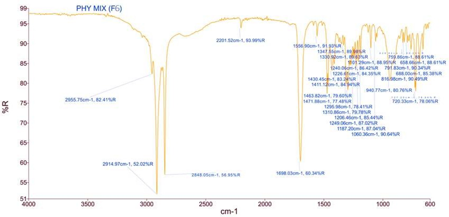
Figure 6: FTIR spectra of transfersomes F6
Optimization And Evaluation of Transfersomes Gel: The topical gel containing transfersomes loaded with luliconazole prepared successfully by stirring method using carbopol 934 as gelling agent. Method of preparation of different transfersomes gel found unpretentious and robust. Initially, all four different preparations of transfersomes gel coded as G1, G2, G3, and G4, were evaluated to quantitate percent entrapment of luliconazole spectrophotometrically at 299 nm. Resulting data shows that transfersomes G3 with 1.5% carbopol w/w showing highest percentage of drug entrapment with 91.39% ± 0.187. Thereafter optimized formulation is further evaluated to access physiochemical parameters includes visual appearance, pH, viscosity and spreadability. Resulting data reveals that viscosity of G3 gel as found to be 369cP, similar to gel viscosity as reported by Jana et al and pH found to be 6.12±0.255.41 further in spreadability evaluation, spreadability factor of prepared transfersomes gel found to be 4.5 and it states that prepared gel produces excellent spreadability as an ideal topical formulation. Spreadability is one of important physical properties of any topical formulation from patient’s compliance point of view.
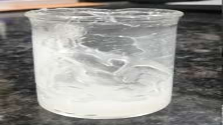
Figure 7: visual appearance of transfersomes G3 gel.
In vitro drug release and kinetics study:
Statistical models are commonly used to forecast release mechanism and compare release profile. in-vitro release profile of drug performed in prepared buffer system using dialysis bag technique for 24 hrs. desolvation percentages of luliconazole from transfersomes are increased in proportion of time as illustrated in Figure 15 and Table 10. Pieces of evidence of release profiles show that developed transfersomes is proficient to release drug in regulated manner. Slow release of leading moiety from most transfersomes form is based on homogeneous drug entrapment throughout systems.62 Ekambaram et al. state same concept and claimed that controlled drug desolvation profile can be attained when drug is uniformly distributed in lipid matrix. Poloxamer 407 having immense efficacy against drug release rate from transfersomes then Cremophor RH40 because of its higher HLB value than cremophor RH 40.20 Besides, Poloxamer 407 has high exterior spreadability so that it eases effects of interfacial tension between transfersomes and dissolution medium. It also reduces accumulation of drug particles and increases drug dissolution rate. Moreover, lipid mass in transfersomes can control size of nanoparticle and increase drug desolvation strength. Thickness of lipid surrounded nanoparticle increases length of drug disassociation resulting prolonged effect of drug release.
Table 6: Percentage drug release profile of G3 and control gel
|
Sr. no. |
Time (Hrs.) |
Percentage drug release of G3 |
Percentage drug release of control gel |
|
1 |
0 |
0 |
0 |
|
2 |
0.25 |
7.375±0.153 |
1.923± 0.011 |
|
3 |
0.5 |
14.002±0.185 |
2.052± 0.155 |
|
4 |
1 |
22.064±0.102 |
3.042± 0.158 |
|
5 |
2 |
32.289±0.173 |
3.182± 0.162 |
|
6 |
3 |
40.622±0.165 |
5.094± 0.122 |
|
7 |
4 |
47.048±0.151 |
7.815± 0.205 |
|
8 |
6 |
55.582±0.163 |
8.706± 0.215 |
|
9 |
8 |
62.309±0.134 |
9.387± 0.118 |
|
10 |
12 |
69.939±0.115 |
9.035± 0.205 |
|
11 |
24 |
79.578±0.213 |
9.773± 0.158 |
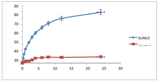
Figure 8: In-vitro drug release profile of transfersomes gel and control gel.
Furthermore, an in-vitro drug release profile for optimized formulation applied to various kinetic models (zero-order, first-order, Higuchi, and KrosmayerPeppas model). To state kinetics profiling of drug release, obtained data were analyzed statistically in respect of rate constant and highest correlation. Best-fitted line found in all models except little suitability in zero-order equation. Resulting data describe dissemination of drug in controlled or regular manner from homogenous matrix systems and it states why drug disseminates at slower rate. Observations concluded that transfersomes G3 is far efficient as potential topical formulation for sustained drug delivery. This finding is almost similar to virtuous covenant as per previous shreds of evidence. Graphical representation of kinetics order of transfersomes G3 gel shown.
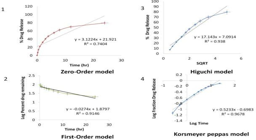
Figure 9: Kinetics Order of Transfersomes G3 Gel
FTIR spectral analysis of transfersomes G3 gel.
The FTIR spectral analysis of transfersomes gel G3 performed successfully to determine possible interaction between drug and drug additives and obtained spectral data matched with spectral data of luliconazole and stearic acid. Findings of spectral analysis show principal absorption peaks at 3331.36cm-1 for N–H stretching, 2961.88 cm-1 for C-H stretching, 2193.49 cm-1 for C≡N stretching, 609.26 & 1044.56 cm-1 for C-Cl stretching for luliconazole. Whereas, principal absorption peaks of stearic acid were ascribed to 2932.49cm-1& 2863.16 cm-1 in high-frequency region attributed to -CH2- band asymmetric and symmetric stretching vibrations, and 1639.31 cm-1 for -COOH stretching is attributed in low-frequency region. Spectral analysis of optimized formulation G3 reveals that no more possible interaction between drug and drug additives even after successive formation of topical gel. Hence it can be said that spectra show purity and authenticity of transfersomes G3 gel.
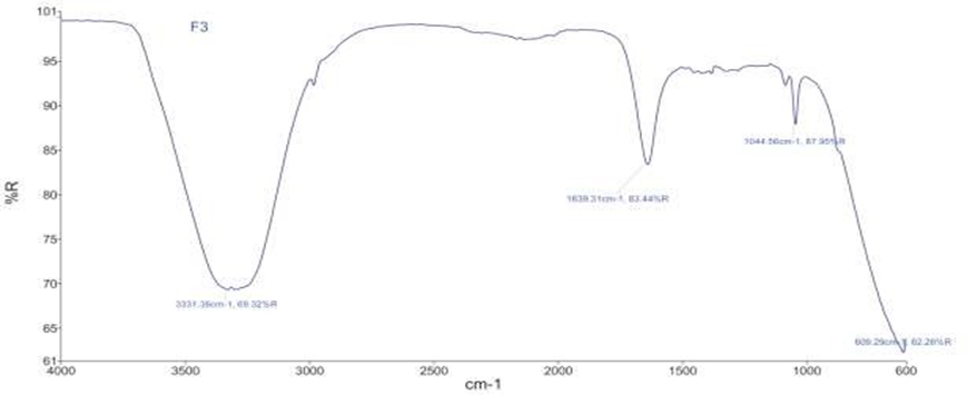
Figure 10: FTIR spectra of gel G3
Table 7: FTIR Interpretation of transfersomes G3 gel.
|
Characteristics Peaks |
Reported (cm-1) |
Observed(cm-1) |
|
N-H stretch |
3300 – 2400 |
3331.36 |
|
C=C stretch |
1638 – 1648 |
1639.31 |
|
CO - O – CO stretch |
1040 – 1050 |
1044. 56 |
|
C – Cl stretch |
550 – 850 |
609. 26 |
Scanning electron microscopy: The shape of enhanced formulation confirmed through SEM study and is shown in Figure18. Most of vesicles is well specified, spherical, and discreet having large internal aqueous space. Low density of nanoparticles is shown in SEM analysis which may lead due to factor of dilution of nanosuspension before preparing SEM photographs. SEM studies reveal that luliconazole loaded transfersomes in gel had spherical shape with smooth surface.

Figure 11: SEM analysis of transfersomes G3 gel
5.CONCLUSION:
The purpose of topical drug delivery system is to allow therapeutic quantity of drug to correct place in body and to achieve and sustain desired effect of drug for while, in present investigation, we have designed Transfersomes loaded with luliconazole to enhance skin permeation and controlled drug release at targeted site and incorporate them in topical gel of carbopol 934 with good skin retention time. physicochemical property of prepared gel determined as per standards protocol to overcome compliance after patient use. Even spectroscopical analysis reveals no chemical interactivity between luliconazole and excipients. microscopic examination (optical microscopy and scanning electron microscopy) of gel showed uniform distribution of transfersomes inside gel with good order of kinetics of drug release. Hence, it can be concluded that transfersomes gel provides controlled release of drug and these systems can be good source as drug carriers for lipophilic drugs, bioavailability enhancer for poorly water-soluble drugs by nanoparticles, drug delivery system.
REFERENCES


Aayushi Bhati*, Dr. Praveen Tahilani, Jitendra Banweer, Formulation and characterization of Transferosomes Gel for Effective Topical Fungal Treatment, Int. J. of Pharm. Sci., 2025, Vol 3, Issue 3, 3442-3457 https://doi.org/10.5281/zenodo.15113994
 10.5281/zenodo.15113994
