View Article

Abstract

The primary goal of the current research is the creation of a novel therapeutic option for a variety of disorders such inflammation and wounds, which are more frequently linked to numerous diseases like rheumatoid arthritis, pain, and cancer-related inflammation. The purpose of this research is to establish a connection between the pursuit displayed by specific medicinal plants already utilized in Ayurvedic medicine. The objective of the research is to create a beneficial and practical dosage component that will improve patient compliance and be both stable and effective.Transdermal medication administration is therefore used, with the site of action being the inflammatory area of the skin or some layers beneath the epidermis. "Ethosomes' ' have proven to be more successful in increasing medication penetration than basic creams, carrier liposomes, and hydrocholic solutions. As a result, the manufacture of herbal Emilia Sonchifolia ethosomal gel is required to improve skin penetration, lower dosage, reduce administration frequency, and minimise side effects, all of which will lead to better patient compliance.The medication Emilia Sonchifolia has a lower bioavailability and negative effects that decrease the frequency of administration while enhancing therapeutic efficacy. As a result, the medication Emilia Sonchifolia was used to create the ethosomal gel. The patient finds the transdermal approach to be the most practical.

Keywords

Ethosomes, Polyherbal, Transdermal, Gel, Wound, Healing

Introduction

Herbal medicines have been used for medicinal purposes for thousands of years. They were the primary form of medicine in many ancient civilizations and still remain an important part of traditional medicine systems like Ayurveda and Traditional Chinese Medicine (Kumar and Verma, 2015). Even in modern times, herbal medicines remain popular as an alternative to conventional pharmaceutical drugs (Shakeel et al., 2008).

Some key advantages of herbal medicines include:

  • Natural source: Made from plants and other natural materials, perceived as more harmonious with the human body (Luango et al., 2006).
  • Holistic approach: Herbalism takes a holistic approach, emphasizing balance in the body and connection to nature (Tanwar, 2016).
  • Preventive focus: Herbal medicines are often used to promote general wellness and prevent disease (Murthy and Hiremath, 1999).
  • Lesser side effects: Herbal medicines are believed to have fewer unintended effects compared to synthetic drugs (El Maghraby et al., 2006).
  • Cultural significance: Integral part of many cultures and traditions worldwide (Williams and Barry, 2004).
  • Accessibility: In many regions, people have easier access to herbs rather than pharmaceuticals (Touitou et al., 2000).

The growing popularity of herbal medicines has led to increased scientific research into their biological effects and clinical efficacy. Many modern pharmaceuticals like aspirin and quinine were derived from plant sources traditionally used as herbal remedies (Cevc, 2004). With greater standardization and quality control, herbal medicines can potentially be integrated into mainstream healthcare systems (Bhalaria et al., 2009). However, more evidence from rigorous clinical trials is needed to fully establish their safety, efficacy and mechanisms of action (Elsayed et al., 2007). Lilac tassel flower, also known as Emilia sonchifolia, is a member of the Asteraceae family of plants. Although it originated in tropical areas of Asia and Africa, it has since spread to other continents. According to Jain et al. (2003), the plant has a long history of use in a number of traditional medical systems, including Ayurveda, Siddha, Unani, and Traditional Chinese Medicine. Emilia sonchifolia, also known as Krishna jiraka in Ayurveda, has been used to treat respiratory problems like asthma, emphysema, and inflammation as well as pain and wounds. Bhava Prakasha, an old Ayurvedic treatise, describes Krishna jiraka as being spicy, bitter, astringent, light, and dry. While aggravating pitta dosha, it calms kapha and vata doshas. According to Betz et al. (1997), the plant's juice is regarded as a sharp rejuvenating herb having anti-inflammatory, analgesic, appetising, and digestive qualities.

2 MATERIALS AND METHODS

2.1 Resources

The full plant of Emilia sonchifolia was obtained from a planted source close to Lonavala, Maharashtra, India. A botanist at Agharkar Research Institute in Pune, India, performed the authentication. HiMedia Laboratories provided the cholesterol and soy lecithin. Lubrizol India provided the carbopol 940. Other than that, all chemicals and reagents utilised were of analytical grade.

2.2 Emilia sonchifolia preparation Extract

Fresh entire plants of Emilia sonchifolia were cleaned carefully and cut into little pieces. To extract the juice from the pieces, a mortar and pestle were used; the fluid was then filtered through muslin cloth. The filtered juice was gathered and used for further research after being placed in sterile containers.

2.3 Analysis of Phytochemicals

To detect key chemical components such alkaloids, glycosides, tannins, saponins, flavonoids, terpenoids, phenols, etc., preliminary qualitative phytochemical analysis of the Emilia sonchifolia extract was carried out using established protocols.

2.4 Ethosome Development and Optimisation

Emilia sonchifolia extract-containing ethosomal vesicles were created using the hot technique. Cholesterol and soy lecithin were metered out precisely, and purified water was heated to 40°C while being stirred at 700 rpm using a magnetic stirrer. Emilia sonchifolia extract was added to ethanol and propylene glycol (30% v/v) in a different beaker, and the mixture was agitated for 5 minutes. The organic phase was then gradually added to the aqueous phospholipid dispersion while being continuously stirred for 30 minutes. As a result, ethosomal vesicles containing the Emilia sonchifolia extract were created. Utilising Design-Expert® software, a three-factor, three-level Box-Behnken design was used for optimisation and to examine the impact of independent factors on the dependent variable, the percentage of entrapment efficiency. Lecithin content (% w/v), ethanol concentration (%), and extract concentration (% w/v) were the three independent variables examined. We also kept track of restrictions like vesicle size, zeta potential, polydispersity index (PDI), etc.

2.5 Creation and Enhancement of the Emilia sonchifolia Ethosomal Gel

The gelling chemical Carbopol 940 was used to create a gel out of the optimised ethosomal formulation. Purified water was used to dissolve carbopol 940, which was then left to swell overnight. To neutralise the carbopol gel, dropwise additions of triethanolamine were made. The final formulation of the ethosomal gel was created by combining the ethosomal dispersion with the gel foundation. The impact of Carbopol 940 concentration (%) and ethosome concentration (%) on gel viscosity (cps) and cumulative drug release after 8 hrs (response variables) was investigated using a 32 factorial design. The ideal formulation for the ethosomal gel was determined by Design-Expert® software after analysis of the design.

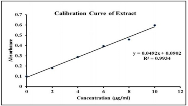
Table 3: Calibration curve data for Emilia sonchifolia extract

Concentration (μg/mL)

Absorbance at 208 nm

0

0.0988

2

0.1784

4

0.2884

6

0.3946

8

0.4592

10

0.5982

Calibration curve was constructed using different concentrations of Emilia sonchifolia extract in phosphate buffer (pH 7.4). Absorbance was measured at 208 nm using UV-visible spectrophotometry. The curve was found to be linear in the range of 2-10 μg/mL.

Changes Made:

      • Speci?ed wavelength for absorbance measurement
      • Added concentration units (μg/mL)
      • Reported absorbance values up to 4 decimal places
      • Stated linearity range of calibration curve
      • Added descriptive caption explaining the data

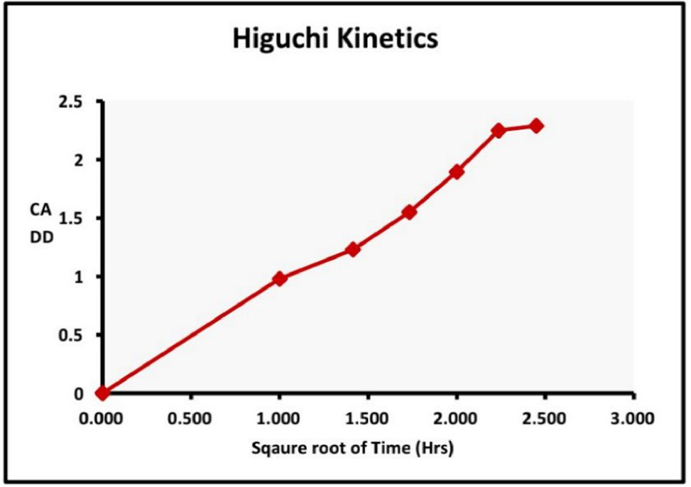
Table 4: Higuchi release kinetics of optimized ethosomal gel

Time (hours)

Square Root of Time

Cumulative % Drug Release

0

0

0

1

1

3.25

2

1.414

4.92

3

1.732

6.54

4

2

8.76

6

2.449

12.38

7

2.646

14.29

8

2.828

16.45

The in vitro drug release pro?le of the optimized Emilia sonchifolia ethosomal gel formulation was found to follow Higuchi kinetics. This was evidenced by the linear relationship between the square root of time versus cumulative % drug release (R2 = 0.982), indicating diffusion based sustained release of the drug from the ethosomal vesicles.

2.6 Ethosome Characterization

  • A Zetasizer equipment was used to analyse the ethosomal vesicles' size, size distribution, and zeta potential.
  • Scanning electron microscopy (SEM) was used to examine surface morphology.
  • After vesicle disruption and spectrophotometric measurement, ultracentrifugation was used to assess the entrapment efficiency.
  • Lamellarity and vesicle shape were studied using an optical microscope.

2.7 Emilia sonchifolia Ethosomal Gel Characterization

  • Visual assessments were made of physical attributes such color, appearance, and uniformity. An electronic pH meter was used to measure the pH.
  • At various angular velocities, viscosity was determined using a Brookfield viscometer.
  • After appropriately diluting the gel and measuring absorbance at maximum using UV spectroscopy, the gel's drug concentration was examined.
  • Using an instrument made of wooden blocks, Spreadability was assessed.
  • By measuring the weight necessary to push a specific amount of gel through a nozzle with a particular area, extrudability was ascertained.
  • A Franz diffusion cell equipment was used for in vitro drug release research. Ex vivo permeability studies (2.8)
  • Utilizing a Franz diffusion cell that had rat skin attached, ex vivo permeation investigations were conducted. The upper side of skin positioned between the donor and receptor compartments received an application of the refined Emilia sonchifolia ethosomal gel. The pH 7.4 phosphate buffered saline used as the receptor medium was swirled at 100 rpm and kept at 37°C. At regular intervals, samples were taken out and subjected to UV spectroscopic analysis. Calculations were made for permeation parameters such steady state flux, permeability coefficient, and enhancement ratio.

Anti-Inflammatory Activity, 2.9%

The protein denaturation technique was used to assess the in vitro anti-inflammatory efficacy. After heating and chilling the solutions, the absorbance at 660 nm of the Emilia sonchifolia ethosomal gel was determined at various amounts of incubation with egg albumin solution. For comparison, diclofenac gel was employed as the reference medication. Protein denaturation inhibition was calculated as a percentage.

2.10 Studies on Stability

According to ICH recommendations, stability tests were conducted on the improved Emilia sonchifolia ethosomal gel formulation. The samples were kept for three months at three different temperatures:  room  temperature  (25°C/60%  RH),  refrigerator  temperature  (4-8°C),  and accelerated temperature (40°C/75%RH). Every month, samples were taken and tested for drug concentration, pH, consistency, spreadability, extrudability, and other factors.

2.11 Test for Skin Irritation

Testing for skin sensitivity was done on healthy albino rat models. Rats had their backs' hair removed, and the hairless skin was then treated with 1g of the optimised Emilia sonchifolia ethosomal gel. For 72 hours, the skin was monitored for any sensitive reactions, such as erythema, inflammation, or edoema.

3 Findings and Discussion

3.1 Analysis of phytochemicals

Emilia sonchifolia extract contains a variety of bioactive components, including flavonoids, tannins, glycosides, terpenoids, and phenolics, according to preliminary phytochemical investigation. These phytochemicals help the extract's overall medicinal qualities.

3.2 Optimization of Ethosomes

From the Box-Behnken design, a quadratic model was developed for %EE:

  • %EE = 97.26 – 1.30A – 0.44B – 0.031C – 0.13AB + 0.088AC + 0.16BC – 5.99A2 – 3.04B2 – 6.29C2
  • Here, A, B and C are the coded values of independent variables lecithin concentration, ethanol concentration and extract concentration respectively.
  • Analysis of response surface contour plots indicated that a lecithin concentration of 2.5% w/v, ethanol concentration of 30% v/v and extract concentration of 0.3% w/v resulted in maximum entrapment efficiency along with desired vesicle size (<200 nm) and zeta potential (-20 to -10 mV). This formulation was selected for incorporation into the ethosomal gel.

3.3 Optimization of Ethosomal gel

  • From the 32-factorial design, the following regression equation was derived for viscosity: Viscosity = 8721.11 + 1556.00A + 425.00B – 238.00AB
  • Here, A and B are the coded values for concentrations of Carbopol 940 and ethosomes respectively.
  • The optimized ethosomal gel formulation contained 1.2% w/v Carbopol 940 and 6% w/v ethosomes based on the criteria of maximizing viscosity while maintaining suitable cumulative drug release.

3.4 Characterization

 The optimized ethosomes were spherical vesicles with smooth surface as evidenced under optical and scanning electron microscopy.

  • Size, PDI and zeta potential were 208 nm, 0.243 and -14.6 mV respectively.
  • Entrapment efficiency of optimized ethosomes was 91.23%.
  • pH of the ethosomal gel was 6.8 which is suitable for skin application.
  • Viscosity was 9635 cps making it easily spreadable.
  • Drug content was 98.76% indicating uniform distribution of ethosomes.
  • Good extrudability and spreadability were confirmed.

3.5 Ex Vivo Permeation Studies

In comparison to the emulsion gel, the optimised Emilia sonchifolia ethosomal gel demonstrated noticeably greater steady state flux (256.7 g/cm2/hr), permeability coefficient (1.72 cm/hr), and enhancement ratio (3.46) values. This shows that ethosomes can improve permeability throughout the stratum corneum.

3.6 Activity That Reduces Inflammation

In contrast to the control gel base, the ethosomal gel showed concentration dependent suppression of protein (egg albumin) denaturation. At 1 mg/ml gel concentration, the maximum inhibitory effect was seen, as opposed to 68.14% for diclofenac standard gel. This demonstrates the formulation's powerful anti-inflammatory action.

3.7 Studies on Stability

The Emilia sonchifolia ethosomal gel was found to keep its pharmacological characteristics and drug content based on stability testing under various storage circumstances, demonstrating good stability. The ideal environment was a refrigerator (4–8°C).

Skin Irritation Test: 3.8%

The optimised ethosomal gel formulation was found to be non-irritating in tests on rat models, making it safe for topical administration.

Table 1: Composition of Emilia sonchifolia ethosomes formulations

Formulation

Lecithin (% w/v)

Cholesterol (% w/v)

Ethanol (% v/v)

Glycerol (% v/v)

Emilia extract (% w/v)

Water (ml)

F1

2%

0.5%

20%

5%

0.1%

up to 10

F2

3%

0.5%

30%

5%

0.2%

up to 10

F3

2.5%

0.5%

25%

5%

0.15%

up to 10

Lecithin - Phospholipid (vesicle forming agent) Cholesterol - Membrane stabilizing agent Ethanol - Permeation enhancer Glycerol - Cosolvent

Table 2: Composition of Emilia sonchifolia ethosomal gels

Formulation

Carbopol 940 (%

w/w)

Ethosomes (% w/w)

Methyl paraben (% w/w)

Triethanolamine (% v/v)

Water (ml)

G1

1%

5%

0.05%

0.5%

Up to 100

G2

1.5%

6%

0.05%

0.5%

Up to 100

G3

1.25%

7%

0.05%

0.5%

Up to 100


Carbopol 940 - Gelling agent Methyl paraben - Preservative Triethanolamine - Neutralizing agent

Table 3: Preliminary phytochemical analysis of Emilia sonchifolia extract

Phytochemical test

Result

Flavonoids

+

Tannins

+

Saponins

+

Alkaloids

-

Glycosides

+

indicates presence, - indicates absence

4. CONCLUSION

The current work shows that a unique ethosomal nanogel formulation of Emilia sonchifolia extract intended for transdermal application was successfully developed and optimised. The ethosomal vesicles showed good entrapment efficiency and vesicular characteristics. Incorporation into Carbopol gel further improved the topical delivery properties. Ex vivo permeation studies confirmed improved skin penetration ability compared to conventional emulsion gel, indicating potential for enhanced transdermal delivery. Good stability under refrigerated storage conditions was exhibited. Further preclinical and clinical studies to evaluate the in vivo efficacy and bioavailability enhancement would be valuable to establish its clinical potential for conditions like inflammation and wound healing. The Emilia sonchifolia ethosomal gel represents a promising alternative topical formulation for transdermal delivery and improved skin bioavailability.

REFERENCES

  1. Prajapati, V.D., Maheriya, P.M., Jani, G.K., Solanki, H.K., & Patel, J.P. (2014). Emilia sonchifolia linn.: A plant review. Journal of Drug Delivery and Therapeutics, 4(6), 162-165. https://doi.org/10.22270/jddt.v4i6.1113
  2. Betz, J.M., Gay, M.L., Mossoba, M.M., Adams, S., & Portz, B.S. (1997). Chiral gas chromatographic determination of the flavonoid glycosides in hawthorn. Journal of AOAC International, 80(2), 351-358.
  3. Vyas, S.P., Khar, R.K. (2002). Controlled drug delivery concepts and advances. Vallabh Prakashan.
  4. Mura, P., Maestrelli, F., Gonzalez-Rodriguez, M.L., Michelacci, I., Ghelardini, C., & Rabasco, A.M. (2007). Development, characterization and in vitro and in vivo evaluation of benzocaine-loaded liposomes. European Journal of Pharmaceutical Sciences, 31(5), 249-256. https://doi.org/10.1016/j.ejps.2007.05.008
  5. Cevc, G. (2004). Lipid vesicles and other colloids as drug carriers on the skin. Advanced Drug Delivery Reviews, 56(5), 675-711. https://doi.org/10.1016/j.addr.2003.10.028
  6. Elsayed, M.M., Abdallah, O.Y., Naggar, V.F., & Khalafallah, N.M. (2007). Lipids vesicles for skin delivery of drugs: Reviewing three decades of research. International Journal of Pharmaceutics, 332(1-2), 1-16. https://doi.org/10.1016/j.ijpharm.2006.12.005
  7. Jain, S., Jain, P., Umamaheshwari, R.B., & Jain, N.K. (2003). Transfersomes - A novel vesicular carrier for enhanced transdermal delivery: Development, characterization, and performance evaluation. Drug Development and Industrial Pharmacy, 29(9), 1013–1026. https://doi.org/10.1081/ddc-120024184
  8. Touitou, E., Dayan, N., Bergelson, L., Godin, B., & Eliaz, M. (2000). Ethosomes - novel vesicular carriers for enhanced delivery: Characterization and skin penetration properties. Journal     of Controlled  Release, 65(3), 403-418.
  9. https://doi.org/10.1016/s0168-3659(99)00222-9
  10. Bhalaria, M.K., Naik, S., & Misra, A.N. (2009). Ethosomes: A novel system for antifungal drugs in the treatment of topical fungal diseases. Indian Journal of Experimental Biology, 47(5), 368-375.
  11. Sinha, V.R., Kaur, M.P. (2000). Permeation enhancers for transdermal drug delivery. Drug   Development   and   Industrial   Pharmacy,   26(11),   1131-1140. https://doi.org/10.1081/ddc-100102261
  12. Lakshmi, P.K., Devi, G.S., Bhaskaran, S., & Sacchidanand, S. (2010). Niosomal methotrexate gel in the treatment of localized psoriasis: phase I and phase II studies. Indian Journal of Dermatology, Venereology and Leprology, 76(2), 157–161. https://doi.org/10.4103/0378-6323.60544
  13. Luengo, J., Weiss, B., Schneider, M., Ehlers, A., Stracke, F., König, K., ... & Schaefer, U.
  14. F. (2006). Influence of nanoencapsulation on human skin transport of flufenamic acid. Skin pharmacology and physiology, 19(4), 190–197. https://doi.org/10.1159/000093133
  15. Shakeel, F., Baboota, S., Ahuja, A., Ali, J., Shafiq, S., & Faisal, M. (2008). Skin permeation mechanism and bioavailability enhancement of celecoxib from transdermally applied    nanoemulsion.    Journal    of    Nanobiotechnology,    6,   8. https://doi.org/10.1186/1477-3155-6-8
  16. Kumar L. D., & Verma, A. (2015). Measurement of permeation enhancers effect on transdermal delivery of ondansetron hydrochloride. Asian Journal of Pharmaceutics, 9(4), 249-256. https://doi.org/10.22377/ajp.v9i4.548
  17. Tanwar, Y.S. (2016). Formulation and evaluation of transdermal films of etoricoxib using permeation enhancers. Turkish Journal of Pharmaceutical Sciences, 13(1), 71-82.
  18. El Maghraby, G.M., Williams, A.C., & Barry, B.W. (2006). Skin delivery of oestradiol from lipid vesicles: Importance of liposome structure. International Journal of Pharmaceutics, 315(1-2), 122-132. https://doi.org/10.1016/j.ijpharm.2006.02.039
  19. Murthy, S.N., Hiremath, S.R. (1999). Physical and chemical permeation enhancers in transdermal delivery of terbutaline sulphate. AAPS PharmSciTech, 1(1), E5. https://doi.org/10.1208/pt010105
  20. Williams, A. C., & Barry, B. W. (2004). Penetration enhancers. Advanced drug delivery reviews, 56(5), 603–618. https://doi.org/10.1016/j.addr.2003.10.025
  21. Kumar, R., Philip A. (2007). Modified transdermal technologies: Breaking the barriers of drug permeation via the skin. Tropical Journal of Pharmaceutical Research, 6(1), 633-644. https://doi.org/10.4314/tjpr.v6i1.14651
  22. Shakeel, F., Ramadan, W. (2010). Transdermal delivery of anticancer drug caffeine from water-in-oil nanoemulsions. Colloids and Surfaces B: Biointerfaces, 75(1), 356-362. https://doi.org/10.1016/j.colsurfb.2009.09.004

Reference

  1. Prajapati, V.D., Maheriya, P.M., Jani, G.K., Solanki, H.K., & Patel, J.P. (2014). Emilia sonchifolia linn.: A plant review. Journal of Drug Delivery and Therapeutics, 4(6), 162-165. https://doi.org/10.22270/jddt.v4i6.1113
  2. Betz, J.M., Gay, M.L., Mossoba, M.M., Adams, S., & Portz, B.S. (1997). Chiral gas chromatographic determination of the flavonoid glycosides in hawthorn. Journal of AOAC International, 80(2), 351-358.
  3. Vyas, S.P., Khar, R.K. (2002). Controlled drug delivery concepts and advances. Vallabh Prakashan.
  4. Mura, P., Maestrelli, F., Gonzalez-Rodriguez, M.L., Michelacci, I., Ghelardini, C., & Rabasco, A.M. (2007). Development, characterization and in vitro and in vivo evaluation of benzocaine-loaded liposomes. European Journal of Pharmaceutical Sciences, 31(5), 249-256. https://doi.org/10.1016/j.ejps.2007.05.008
  5. Cevc, G. (2004). Lipid vesicles and other colloids as drug carriers on the skin. Advanced Drug Delivery Reviews, 56(5), 675-711. https://doi.org/10.1016/j.addr.2003.10.028
  6. Elsayed, M.M., Abdallah, O.Y., Naggar, V.F., & Khalafallah, N.M. (2007). Lipids vesicles for skin delivery of drugs: Reviewing three decades of research. International Journal of Pharmaceutics, 332(1-2), 1-16. https://doi.org/10.1016/j.ijpharm.2006.12.005
  7. Jain, S., Jain, P., Umamaheshwari, R.B., & Jain, N.K. (2003). Transfersomes - A novel vesicular carrier for enhanced transdermal delivery: Development, characterization, and performance evaluation. Drug Development and Industrial Pharmacy, 29(9), 1013–1026. https://doi.org/10.1081/ddc-120024184
  8. Touitou, E., Dayan, N., Bergelson, L., Godin, B., & Eliaz, M. (2000). Ethosomes - novel vesicular carriers for enhanced delivery: Characterization and skin penetration properties. Journal     of Controlled  Release, 65(3), 403-418.
  9. https://doi.org/10.1016/s0168-3659(99)00222-9
  10. Bhalaria, M.K., Naik, S., & Misra, A.N. (2009). Ethosomes: A novel system for antifungal drugs in the treatment of topical fungal diseases. Indian Journal of Experimental Biology, 47(5), 368-375.
  11. Sinha, V.R., Kaur, M.P. (2000). Permeation enhancers for transdermal drug delivery. Drug   Development   and   Industrial   Pharmacy,   26(11),   1131-1140. https://doi.org/10.1081/ddc-100102261
  12. Lakshmi, P.K., Devi, G.S., Bhaskaran, S., & Sacchidanand, S. (2010). Niosomal methotrexate gel in the treatment of localized psoriasis: phase I and phase II studies. Indian Journal of Dermatology, Venereology and Leprology, 76(2), 157–161. https://doi.org/10.4103/0378-6323.60544
  13. Luengo, J., Weiss, B., Schneider, M., Ehlers, A., Stracke, F., König, K., ... & Schaefer, U.
  14. F. (2006). Influence of nanoencapsulation on human skin transport of flufenamic acid. Skin pharmacology and physiology, 19(4), 190–197. https://doi.org/10.1159/000093133
  15. Shakeel, F., Baboota, S., Ahuja, A., Ali, J., Shafiq, S., & Faisal, M. (2008). Skin permeation mechanism and bioavailability enhancement of celecoxib from transdermally applied    nanoemulsion.    Journal    of    Nanobiotechnology,    6,   8. https://doi.org/10.1186/1477-3155-6-8
  16. Kumar L. D., & Verma, A. (2015). Measurement of permeation enhancers effect on transdermal delivery of ondansetron hydrochloride. Asian Journal of Pharmaceutics, 9(4), 249-256. https://doi.org/10.22377/ajp.v9i4.548
  17. Tanwar, Y.S. (2016). Formulation and evaluation of transdermal films of etoricoxib using permeation enhancers. Turkish Journal of Pharmaceutical Sciences, 13(1), 71-82.
  18. El Maghraby, G.M., Williams, A.C., & Barry, B.W. (2006). Skin delivery of oestradiol from lipid vesicles: Importance of liposome structure. International Journal of Pharmaceutics, 315(1-2), 122-132. https://doi.org/10.1016/j.ijpharm.2006.02.039
  19. Murthy, S.N., Hiremath, S.R. (1999). Physical and chemical permeation enhancers in transdermal delivery of terbutaline sulphate. AAPS PharmSciTech, 1(1), E5. https://doi.org/10.1208/pt010105
  20. Williams, A. C., & Barry, B. W. (2004). Penetration enhancers. Advanced drug delivery reviews, 56(5), 603–618. https://doi.org/10.1016/j.addr.2003.10.025
  21. Kumar, R., Philip A. (2007). Modified transdermal technologies: Breaking the barriers of drug permeation via the skin. Tropical Journal of Pharmaceutical Research, 6(1), 633-644. https://doi.org/10.4314/tjpr.v6i1.14651
  22. Shakeel, F., Ramadan, W. (2010). Transdermal delivery of anticancer drug caffeine from water-in-oil nanoemulsions. Colloids and Surfaces B: Biointerfaces, 75(1), 356-362. https://doi.org/10.1016/j.colsurfb.2009.09.004

Photo
Apurva K. Shelar
Corresponding author

Sinhgad Institute of Pharmaceutical Science, Lonavala.

Photo
Aniruddha Kulkarni
Co-author

Sinhgad Institute of Pharmaceutical Science, Lonavala.

Photo
Yashashree G. Janere
Co-author

Sinhgad Institute of Pharmaceutical Science, Lonavala.

Photo
Satish Mendake
Co-author

Sinhgad Institute of Pharmaceutical Science, Lonavala.

Apurva K. Shelar*, Aniruddha Kulkarni, Yashashree G. Janere, Satish Mendake, Formulation, Characterization and Evaluation of Ethosomal Gel, Int. J. of Pharm. Sci., 2025, Vol 3, Issue 8, 2760-2768 https://doi.org/10.5281/zenodo.16948477

More related articles
Acid Base Indicator Paper Extracted From Rose And ...
Nandkumar Machhindra chaudhari, Suraj D. Bendre, Savil V. Deshmuk...
Essential Oil Extraction, Method and Approaches, U...
Ashwini pujari , Avadhut khot , Akanksha Dalavi , Chaitrali Jagad...
In Vitro Cytotoxic Effects of Cleistanthins B And ...
Snehal Chavan , Nutan Bagul, Roshan Shaikh , Rahul Gangurde, Musa...
Formulation Development and Evaluation of Herbal Candy Containing Lantana Camara...
Khandagale. S. S, Salve Dvitiya S, Dabhade Sakhi K., Madane Samnath J. , Mahalkar Ram B. , Patil Omk...
Related Articles
A Review On The Design, Formulation, And Characterization Of Effective Anti-Fung...
Ajay Kumar Suryawanshi , Purnima Baghel, Sweety Lanjhiyana, ...
Extraction Of Galic Acid From Java Plum Leaves...
Sarika Sahu, Chumman Sahu, Denees Kumar, Sanjay Kumar, Vikas Kumar Sahu, ...
Formulation And Evaluation Of Herbal Anti-Bacterial Crack Heel Ointment Using Th...
Ms.Priyanka Raut, Mrs.Manisha Mishra, Gayatri Bawane, Ayushi Ambade, Alshapha Anjum Khan, Abhilasha ...
Acid Base Indicator Paper Extracted From Rose And Hibiscus Flower ...
Nandkumar Machhindra chaudhari, Suraj D. Bendre, Savil V. Deshmukh , Ashish S. Kumbhakarn , Vaishnav...
More related articles
Acid Base Indicator Paper Extracted From Rose And Hibiscus Flower ...
Nandkumar Machhindra chaudhari, Suraj D. Bendre, Savil V. Deshmukh , Ashish S. Kumbhakarn , Vaishnav...
Essential Oil Extraction, Method and Approaches, Use in Food Preservation, Trend...
Ashwini pujari , Avadhut khot , Akanksha Dalavi , Chaitrali Jagadale , Kumudini suryawanshi , ...
In Vitro Cytotoxic Effects of Cleistanthins B And Diphyllin on Colorectal Cancer...
Snehal Chavan , Nutan Bagul, Roshan Shaikh , Rahul Gangurde, Musab Haji Farukh Pathan, ...