View Article

Abstract

Early-onset Alzheimer’s disease (EOAD), defined by symptom onset before the age of 65, represents a rare but clinically significant subset of Alzheimer’s disease (AD). EOAD is marked by greater genetic influence, rapid progression, and frequent atypical variants including posterior cortical atrophy, logopenic primary progressive aphasia, frontal variant, and corticobasal presentations. These forms often present with non-amnestic symptoms such as visuospatial, language, or executive dysfunction, complicating diagnosis and management. Standard pharmacological treatments, including cholinesterase inhibitors and memantine, provide only symptomatic relief without altering disease course. In recent years, herbal and nutraceutical interventions have gained attention as adjunctive strategies due to their neuroprotective, antioxidant, anti-amyloid, and anti-inflammatory properties. Prominent candidates include Ginkgo biloba, curcumin, Bacopa monnieri, Withania somnifera (ashwagandha), and huperzine A, each with mechanistic plausibility and varying levels of preclinical and clinical evidence. While some clinical studies suggest modest cognitive benefits, trial heterogeneity, small sample sizes, and issues of standardization limit strong conclusions. This review outlines the epidemiology, clinical heterogeneity, and pathophysiology of EOAD, highlighting its atypical variants, and critically evaluates the current evidence for herbal managerial tools. Despite encouraging findings, rigorous clinical trials are urgently required to validate efficacy, establish safety, and define the role of herbal medicines as adjuncts in the comprehensive management of EOAD.

Keywords

Early-onset Alzheimer’s disease (EOAD); atypical Alzheimer’s variants; posterior cortical atrophy; logopenic primary progressive aphasia; frontal variant; corticobasal syndrome; APP; PSEN1; PSEN2; neuroimaging biomarkers; tauopathy; oxidative stress; herbal medicine; phytotherapy; Ginkgo biloba; curcumin; Bacopa monnieri; Withania somnifera; huperzine A; neuroprotection; adjunctive therapy

Introduction

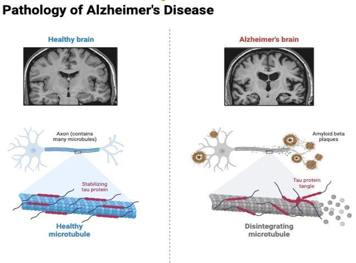
Alzheimer’s disease (AD) is the most common cause of dementia worldwide, affecting millions of people and posing a major global health burden. It is characterized by progressive cognitive decline, behavioral disturbances, and functional impairment, underpinned by neuropathological hallmarks such as extracellular amyloid-β plaques and intraneuronal tau neurofibrillary tangles. [1,5,]While most cases manifest after the age of 65 years (late-onset Alzheimer’s disease, LOAD), a significant minority—approximately 5–10% of cases—develop symptoms earlier, defined as early-onset Alzheimer’s disease (EOAD). EOAD is clinically important for several reasons. Firstly, its age of onset affects individuals in their most productive years, leading to profound personal, social, and economic consequences. Secondly, EOAD often presents with non-amnestic and atypical symptoms, including visuospatial deficits, language dysfunction, executive impairment, or behavioral changes, unlike the predominantly memory-led presentation seen in LOAD. [4]These atypical forms are categorized into distinct variants such as posterior cortical atrophy (PCA), logopenic primary progressive aphasia (lvPPA), frontal-behavioral variant, and corticobasal syndrome (CBS). This clinical heterogeneity complicates early recognition and often delays diagnosis.

From a pathophysiological perspective, EOAD differs from LOAD in its stronger genetic contribution. Mutations in the amyloid precursor protein (APP) and the presenilin genes (PSEN1 and PSEN2) account for a substantial proportion of familial EOAD cases, leading to abnormal amyloid processing and early, aggressive disease onset.[6]Beyond genetics, EOAD involves mitochondrial dysfunction, oxidative stress, neuroinflammation, synaptic failure, and cholinergic deficits, making it a complex, multi-factorial disorder. Advances in biomarkers— including cerebrospinal fluid (CSF) assays of amyloid and tau, plasma phosphorylated tau, and neurofilament light chain, as well as neuroimaging modalities (FDG-PET, amyloid PET, tau PET)—have improved diagnostic precision and facilitated earlier detection.Despite progress, treatment options remain limited. [13] Current pharmacological therapies such as cholinesterase inhibitors (donepezil, rivastigmine, galantamine) and NMDA receptor antagonist (memantine) provide symptomatic relief but do not halt disease progression.

Recently approved anti-amyloid monoclonal antibodies (aducanumab, lecanemab, donanemab) offer disease-modifying potential, but accessibility, cost, and safety concerns limit their widespread use. Consequently, there is growing interest in complementary and adjunctive therapies, particularly herbal medicines and nutraceuticals, that target oxidative stress, neuroinflammation, and synaptic dysfunction through multi-modal mechanisms.

Several herbal agents have been investigated for their neuroprotective roles. Ginkgo biloba extract has shown modest cognitive benefits and microcirculatory effects; curcumin, derived from turmeric, exerts anti-amyloid, anti-tau, and anti- inflammatory activity, particularly with novel bioavailability-enhanced formulations; Bacopa monnieri improves memory and synaptic plasticity; Withania somnifera (ashwagandha) demonstrates adaptogenic and neuroregenerative potential; and huperzine A, a natural cholinesterase inhibitor, offers cognitive improvements similar to synthetic drugs. While preclinical evidence is strong, clinical trials often yield inconsistent results due to small sample sizes, heterogeneous methodologies, and variability in herbal product standardization. Given the clinical heterogeneity of EOAD, limitations of existing therapies, and growing scientific interest in herbal interventions, a comprehensive review of EOAD and its herbal managerial tools is timely. This article aims to (i) summarize the epidemiology, clinical variants, and pathophysiology of EOAD; (ii) review current diagnostic and therapeutic approaches; and (iii) critically evaluate the evidence for herbal and nutraceutical strategies as adjunctive management tools in EOAD.

Early-Onset Alzheimer’s Disease (EOAD) :

Definition: EOAD is Alzheimer’s disease with clinical symptom onset before age 65. It represents a minority of AD cases but causes disproportionate social, economic and caregiving burdens because it affects people in their working years.

Epidemiology : EOAD accounts for roughly 5–10% of all AD cases, with prevalence estimates varying by age band and population; some studies report ~24 per 100,000 in mid-life age ranges and higher in certain cohorts. EOAD may be underrecognized because atypical, non-memory presentations lead to delayed diagnosis. [11,15]

Genetic architecture

  • Monogenic causes: A subset of EOAD is familial and autosomal dominant- most often caused by pathogenic variants in PSEN1 (most common), APP, and PSEN2. Detection rates vary by cohort (commonly ~10–16% in unselected EOAD; higher in familial cases), and PSEN1 mutations often cause the earliest ages of onset. [3]
  • Polygenic and sporadic EOAD: Many EOAD cases do not carry known pathogenic mutations. Emerging evidence suggests enrichment of common AD risk alleles (polygenic risk) and other risk factors contribute to sporadic EOAD. Thus EOAD is genetically heterogeneous — monogenic, oligogenic or polygenic. [2]
  • Clinical implication: Genetic testing (targeted gene panel / exome) is often recommended for patients with strong family history or very early onset; pre- and post-test genetic counseling is essential.[7]
  • Pathophysiology & neuropathology : Pathologic hallmarks are the same as late-onset AD: extracellular amyloid-β (Aβ) plaques and intracellular neurofibrillary tangles composed of hyperphosphorylated tau. In EOAD, amyloid dysmetabolism (especially with APP/PSEN mutations) can be more prominent and disease progression frequently faster. Regional patterns of tau and neurodegeneration may differ across EOAD phenotypes (e.g., more parietal/occipital involvement in PCA). [5,8,9]
  • Clinical presentation - full phenotypic spectrum : EOAD shows greater phenotypic heterogeneity than typical late-onset AD. Common clinical variants:
  1. Amnestic (typical) EOAD : Progressive episodic memory impairment (encoding/recall) similar to late-onset AD but occurring at younger ages. Often initially mistaken for stress, depression or sleep disorder. [23]
  2. Posterior Cortical Atrophy (PCA) : PCA is a visual-dominant syndrome: early visuospatial and visuoperceptual deficits (difficulty reading, visual mislocalization, simultanagnosia, dressing apraxia), with relative sparing of memory early on. PCA is commonly due to underlying AD pathology in EOAD cohorts. Recognition requires careful examination of higher-order visual complaints. [25]
  3. Logopenic variant Primary Progressive Aphasia (lvPPA) : Language- predominant presentation with word-finding pauses, impaired sentence repetition, phonologic errors. Neuroanatomy typically involves left temporoparietal cortex.
  4. Behavioral/dysexecutive variant : Early changes in planning, multitasking, decision-making, apathy or disinhibition; can be confused with frontotemporal dementia (FTD) and psychiatric disorders.
  5. Other atypical features : Early visual hallucinations, depression/anxiety as presenting features, or parkinsonian signs may appear in some EOAD cases and lead to misdiagnosis. [29]
  • Diagnostic workup (practical approach) : A timely, structured assessment helps differentiate EOAD from other causes and identify treatable contributors.
  1. Clinical assessment : Detailed history (onset, progression, family history), cognitive testing (MMSE, MoCA, domain-specific neuropsychological battery), functional assessment.
  2. Neuroimaging
  • Structural MRI: hippocampal/medial temporal atrophy in typical AD; atypical EOAD (PCA, lvPPA) shows focal parietal/occipital or left temporoparietal atrophy. Newer EOAD MRI signatures have been proposed to improve detection.
  • FDG-PET: regional hypometabolism matching clinical syndrome (parietotemporal in typical AD; occipital in PCA).
  • Amyloid PET / Tau PET: in-vivo visualization of pathological protein deposition — very helpful for diagnostic certainty.
  1. Fluid biomarkers CSF: low Aβ42 (or low Aβ42/Aβ40), elevated total tau and phospho-tau (p-tau) — classic AD profile.Plasma biomarkers: plasma p- tau181/p-tau217 and Aβ42/40 ratio are emerging as accessible, validated tools for screening and disease staging, increasingly useful in younger patients. Biomarker use should follow local availability and guidelines.
  2. Genetic testing : Offer genetic testing (APP, PSEN1, PSEN2 ± broader panels) when: strong autosomal dominant family history, onset in the 30s– 50s, or when patient/family requests. Always pair testing with genetic counseling..
  3. Differential diagnosis : Frontotemporal dementia (esp. behavioral variant), Lewy body disease, vascular cognitive impairment, primary psychiatric disorders, metabolic/toxic causes, and treatable structural lesions.
  • Natural history & prognosis : EOAD often progresses faster than late- onset AD, with earlier loss of independence and occupational decline. Disease duration varies but median survival may be shorter in some EOAD

Variants of Alzheimer’s Disease

Alzheimer’s disease (AD) is not a uniform clinical entity but a spectrum of syndromes. While the majority of patients present with the typical amnestic variant, a substantial subset—especially those with early-onset AD (EOAD) develop atypical presentations involving visual, language, behavioral, or motor symptoms. Recognizing these variants is crucial for accurate diagnosis and management, as they are frequently misdiagnosed as other dementias or psychiatric conditions.

Typical Amnestic Alzheimer’s Disease :

  • Clinical features: Progressive impairment of episodic memory is the hallmark. Patients struggle with learning new information, forget recent events, and often repeat questions. Disorientation and gradual impairment of daily activities follow.
  • Pathology: Early involvement of the hippocampus and medial temporal lobe, followed by spread to parietal and frontal cortices.
  • Age group: Predominant in late-onset AD (LOAD), but also seen in EOAD.
  • Diagnosis: Characteristic memory-predominant decline with supportive biomarkers (CSF amyloid/tau, amyloid PET). [21,24]

Posterior Cortical Atrophy (PCA)

  • Clinical features: Visual disturbances: difficulty reading, misjudging distances, bumping into objects. Visuospatial and visuoperceptual deficits. Symptoms often mistaken for ophthalmological disorders.
  • Pathology: Atrophy and hypometabolism in occipital and parietal lobes; amyloid and tau deposition consistent with AD.
  • Onset: Typically, 50–65 years; strongly linked to EOAD.
  • Diagnostic clues: Preserved memory early on; abnormal amyloid/tau PET confirms AD pathology.[21]

Logopenic Variant Primary Progressive Aphasia (lvPPA)

  • Clinical features: Word-finding pauses. Impaired sentence repetition. Phonological errors (speech sound mistakes).Relatively intact grammar and motor speech.
  • Pathology: Involvement of the left temporoparietal junction. Amyloid deposition distinguishes it from other PPA variants.
  • Onset: Common in EOAD (40s–60s).
  • Differential diagnosis: Must be distinguished from frontotemporal lobar degeneration (semantic or non-fluent PPA).[22]

Frontal/Behavioral Variant Alzheimer’s Disease

  • Clinical features: Personality change, apathy, impulsivity, irritability.Executive dysfunction (planning, judgment, problem- solving).Memory impairment less prominent initially.
  • Pathology: AD biomarkers present, with frontal lobe atrophy and hypometabolism.[27]

Corticobasal Syndrome (CBS) Associated with AD

  • Clinical features: Asymmetric motor symptoms: rigidity, dystonia, apraxia. Alien limb phenomenon. Superimposed cognitive deficits.
  • Pathology: Some CBS cases are underpinned by AD pathology rather than corticobasal degeneration.
  • Diagnostic challenge: Requires PET/CSF biomarkers to differentiate AD-CBS from other movement disorders.[25]

Other Less Common Variants

  • Hippocampal-sparing AD: Prominent cortical tau deposition outside hippocampus. Presents with executive or visuospatial dysfunction rather than memory loss.
  • Mixed dementia presentations: AD pathology coexisting with vascular dementia or Lewy body disease, leading to atypical symptom combinations.

Management of Early-Onset Alzheimer’s Disease

1. Pharmacological Management :

a. Symptomatic Therapies :

  • Cholinesterase inhibitors (donepezil, rivastigmine, galantamine): Improve cholinergic neurotransmission, modestly enhance cognition, and delay functional decline.
  • NMDA receptor antagonist (memantine): Reduces excitotoxicity, used in moderate-to-severe EOAD.
  • Combination therapy: Cholinesterase inhibitors + memantine may provide additional benefit in advanced cases.

b. Disease-Modifying Therapies (Emerging)

  • Monoclonal antibodies targeting amyloid-β: Aducanumab, lecanemab, donanemab – shown to reduce amyloid burden, with lecanemab also slowing cognitive decline in clinical trials.
  • Challenges: high cost, need for infusion, risk of amyloid-related imaging abnormalities (ARIA), and limited availability in low- and middle-income countries.
  • Tau-targeted therapies: Currently under investigation in clinical trials (anti- tau antibodies, tau aggregation inhibitors).[31]

2. Non-Pharmacological Management

  • Cognitive rehabilitation and stimulation therapy: Structured activities to enhance memory, attention, and executive skills.
  • Physical exercise: Improves cerebral blood flow, neurogenesis, and reduces oxidative stress. Regular aerobic and resistance training are associated with slower cognitive decline.
  • Dietary approaches: Mediterranean and MIND diets (rich in omega-3 fatty acids, polyphenols, vitamins) show protective effects.
  • Sleep hygiene and stress management: Poor sleep and chronic stress exacerbate amyloid deposition and neuroinflammation.
  • Psychological and social support: Essential for younger patients coping with career disruption and family responsibilities.
  • Caregiver training: Reduces caregiver burden and improves quality of life.[32]

3. Genetic Counseling and Risk Management :

  • Familial EOAD: In patients with APP, PSEN1, or PSEN2 mutations, genetic counseling is essential for family members. Predictive testing should be voluntary, with psychological support.
  • Risk reduction: Lifestyle modifications (exercise, diet, vascular risk factor control) remain central in delaying onset and progression.[33]

4. Multidisciplinary & Holistic Care :

EOAD requires a multidisciplinary approach:

  • Neurologists and psychiatrists for medical management
  • Neuropsychologists for cognitive training.
  • Physiotherapists and occupational therapists for maintaining independence.
  • Dietitians for nutrition counseling.
  • Social workers and support groups for psychosocial care.

Herbal managerial tools

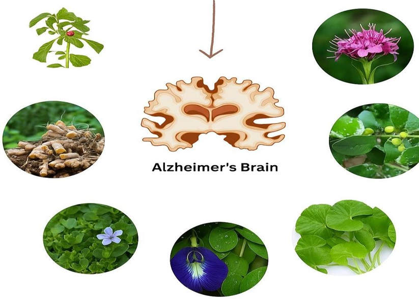
1. Ginkgo biloba (EGb 761)

Mechanisms :  Antioxidant and free-radical scavenging. Anti-inflammatory effects and modulation of microglial activation.Improves cerebral microcirculation and mitochondrial function; may modulate neurotransmission (serotonergic/dopaminergic/cholinergic).

Evidence : Multiple meta-analyses and RCTs report modest cognitive and functional benefits in cognitive impairment and dementia when using the standardized extract EGb 761 at 120–240 mg/day; benefits are most consistent for stabilization or slowing of decline in short-to-medium term (weeks–months) but effect sizes vary.

Safety / Caveats : Generally well tolerated in trials of standardized extracts; bleeding risk and interactions with antiplatelet/anticoagulant drugs remain important concerns. Product variability is a major limitation — only well- standardized extracts (EGb 761) have reproducible evidence.

Research gaps : Need for long-term, biomarker-guided RCTs in well- characterized EOAD populations; combination with standard therapy (and monitoring for interactions) requires study.[45,46]

2. Curcumin (Curcuma longa) and novel formulations

Mechanisms :  Anti-amyloid and anti-tau actions (direct binding/aggregation inhibition), strong antioxidant and anti-inflammatory activities, metal chelation, inhibition of microglial pro-inflammatory signaling, modulation of autophagy.Native curcumin has poor oral bioavailability; nanoformulations and carrier systems (liposomes, ferritin nanocages, polymeric nanoparticles) markedly increase BBB penetration and plasma/tissue exposure.

Evidence : Extensive preclinical evidence for anti-amyloid/tau and neuroprotective effects. Human trials with conventional curcumin formulations were largely negative or inconsistent, but newer pharmacokinetic-enhanced formulations show promising biomarker signals and small clinical improvements in early studies; definitive large RCTs are still lacking. Reviews of nanocarrier strategies highlight translational potential for AD.

Safety / Caveats : Safe at commonly used doses; GI side effects reported rarely. Clinical benefit depends heavily on formulation and dosing trials must report pharmacokinetic data.

Research gaps : Well-powered RCTs using standardized, bioavailability- enhanced curcumin with biomarker endpoints (plasma/CSF p-tau, amyloid, imaging) are needed.[43.44]

3. Bacopa monnieri (Brahmi)

Mechanisms :  Bacosides and other constituents show antioxidant, anti- apoptotic, cholinergic modulation, enhancement of synaptic plasticity and BDNF signaling, and anti-inflammatory effects.

Evidence : Multiple RCTs in healthy older adults and MCI show improvements in memory, attention, and some cognitive domains; systematic reviews/meta-analyses support cognitive benefits but note heterogeneity in extracts, doses, and outcomes. A few longer trials (including an RCT vs donepezil in small cohorts) suggest comparable effects in some measures, but sample sizes are small.

Safety / Caveats : Generally well tolerated; common mild GI complaints. Standardization of bacoside content is necessary for reproducible results.

Research gaps : Larger, longer trials in MCI/early AD (including EOAD subsets) with standardized extracts and biomarker outcomes.[47]

4. Withania somnifera (Ashwagandha)

Mechanisms :  Adaptogenic and anti-inflammatory actions; animal studies indicate reduced amyloid-β, lowered pro-inflammatory cytokines, antioxidant effects, and promotion of neurite outgrowth / synaptic recovery. Clinical mechanisms likely include modulation of neuroinflammation and support of neuroplasticity.

Evidence : Preclinical models (including 5xFAD mice) show improvement in cognition and neuropathology. Small human trials report improvements in memory and attention measures; systematic reviews indicate potential but emphasise small trials and mixed quality. Recent human supplementation studies report benefit on some cognitive endpoints.

Safety / Caveats : Generally safe at typical doses used in trials (e.g., 225– 600 mg standardized extracts), but quality and standardization vary across commercial products.

Research gaps : Need RCTs in early AD/EOAD using standardized extracts, with dose-finding, PK, and biomarker endpoints.[48]

5. Huperzine A

Mechanisms :  Reversible acetylcholinesterase inhibition (cholinergic augmentation) and possible neuroprotective effects (anti-apoptotic, NMDA antagonism reported in preclinical work).

Evidence : Several RCTs (many conducted in China) and meta-analyses report short-term cognitive improvement (8–16 weeks) vs placebo; however, methodological limitations, small sample sizes, and inconsistent longer-term benefits limit conclusions. Larger multicenter trials have reported mixed results.

Safety / Caveats : Side effects overlap with cholinesterase inhibitors (nausea, diarrhea, sweating, bradycardia); interactions with other cholinergic agents must be monitored.

Research gaps : Larger, rigorously designed, longer trials to confirm efficacy and safety; studies in EOAD specifically are lacking.[49]

6. Other candidates & multi-component approaches

Omega-3 fatty acids, resveratrol, citicoline, Panax ginseng, sage and rosemary: mixed evidence; some promising biomarker or small clinical signals but inconsistent across trials. Recent reviews highlight potential of combinations and multi-target strategies rather than single-agent “silver bullets.” [50]

Role of NDDS in Herbal Management of Early-Onset Alzheimer’s Disease

Herbal compounds (curcumin, bacosides, resveratrol, withanolides, huperzine A) have promising anti-Alzheimer’s effects.

Major limitations:

  • Poor solubility and stability.
  • Low oral bioavailability.
  • Rapid metabolism and clearance.
  • Difficulty crossing the blood–brain barrier (BBB).

NDDS Strategies for Herbal EOAD Therapy

1. Nanoparticles : Polymeric (PLGA, chitosan) or lipid-based carriers.Protect herbal actives from degradation and enhance BBB penetration.

Example: Curcumin nanoparticles → improved anti-amyloid activity, antioxidant effect, and memory improvement.[51,53]

2. Liposomes : Phospholipid vesicles encapsulating both hydrophilic & lipophilic herbal molecules.Ligand-modified liposomes target brain receptors (e.g., transferrin-modified liposomes for curcumin).

Example: Huperzine A liposomes → prolonged cholinesterase inhibition.

3. Niosomes & Dendrimers : Niosomes are more stable and cost-effective than liposomes. Dendrimers are branched polymers that carry herbal drugs with high precision.[52]

Example: Resveratrol dendrimers → improved antioxidant neuroprotection.

4. Nanoemulsions & Microspheres : Increase solubility and bioavailability of poorly soluble herbal extracts.

Example: Intranasal curcumin nanoemulsion → rapid brain delivery, enhanced memory protection.[53]

5. Intranasal Delivery Systems : Direct nose-to-brain pathway bypasses BBB.Reduces first-pass metabolism, provides faster action.

Example: Ashwagandha nanoformulation via intranasal route → enhanced cognitive improvement in animal models.

6. Transdermal Patches : Provide controlled and sustained release of herbal actives.Reduce dosing frequency and systemic side effects.

Example: Rivastigmine patch is already used; herbal patches with Bacopa extract are under preclinical study.[51]

CONCLUSION :

Early-onset Alzheimer’s disease (EOAD) is a clinically and genetically heterogeneous form of dementia that differs significantly from the more common late-onset form. Its earlier age of onset, frequent genetic basis, and atypical clinical variants such as posterior cortical atrophy and logopenic progressive aphasia make timely recognition and diagnosis particularly challenging. While conventional treatments like cholinesterase inhibitors and memantine remain the mainstay of management, their benefits are limited and do not halt disease progression. Herbal and natural products including Ginkgo biloba, curcumin, Bacopa monnieri, Withania somnifera, huperzine-A, and others have shown promising neuroprotective mechanisms — ranging from antioxidant and anti-inflammatory effects to cholinesterase inhibition and amyloid modulation. However, evidence from EOAD-specific clinical trials remains scarce, and concerns about safety, herb–drug interactions, and lack of standardization must be carefully addressed.

Integrating herbal therapies as complementary approaches may offer additional symptomatic relief and neuroprotection, but they should be used cautiously under professional supervision. Future research should focus on well-designed, EOAD- targeted studies to establish efficacy, optimize formulations, and provide clearer clinical guidance. Overall, a combined strategy involving early diagnosis, conventional therapy, lifestyle interventions, and evidence-based herbal management holds promise for improving quality of life in individuals with EOAD and their caregivers.[1,5,22,38,45]

REFERENCES

  1. Rossor MN, Fox NC, Mummery CJ, Schott JM, Warren JD. Early-Onset Alzheimer’s Disease: What Is Missing in Research? Curr Neurol Neurosci Rep. 2020;20(5):23.
  2. Young AL, Oxtoby NP, et al. Dissecting the clinical heterogeneity of early-onset Alzheimer’s disease. Mol Psychiatry. 2022;27(6):2812–2820.
  3. Ossenkoppele R, et al. Atypical clinical variants of Alzheimer’s disease. Brain. 2024;147(2):e25.
  4. Bateman RJ, et al. Clinical and biomarker changes in dominantly inherited Alzheimer’s disease. N Engl J Med. 2012;367(9):795–804.
  5. Selkoe DJ, Hardy J. The amyloid hypothesis of Alzheimer’s disease at 25 years. EMBO Mol Med. 2016;8(6):595–608.
  6. Braak H, Braak E. Neuropathological stageing of Alzheimer-related changes. Acta Neuropathol. 1991;82(4):239–259.
  7. Jack CR Jr., et al. NIA-AA Research Framework: Toward a biological definition of Alzheimer’s disease. Alzheimers Dement. 2018;14(4):535–562.
  8. McKhann GM, et al. The diagnosis of dementia due to Alzheimer’s disease: Recommendations. Alzheimers Dement. 2011;7(3):263–269.
  9. Bateman RJ, Xiong C, et al. Clinical and biomarker changes in dominantly inherited Alzheimer’s disease. N Engl J Med. 2012;367(9):795–804.
  10. Cummings JL, Morstorf T, Zhong K. Alzheimer’s disease drug-development pipeline: few candidates, frequent failures. Alzheimers Res Ther. 2014;6(4):37.
  11. World Health Organization (WHO). Global status report on the public health response to dementia 2023.
  12. Van der Flier WM, Scheltens P. Epidemiology and risk factors of dementia. J Alzheimers Dis. 2005;8(3): 85–93.
  13. Harwood DG, et al. Early-onset Alzheimer’s disease: Profile and progression. J Alzheimers Dis. 2010;19(3):665–674.
  14. Kim EJ, et al. Epidemiology and risk factors of early-onset Alzheimer’s disease: A review. Gerontology. 2021;67(4):445–455.
  15. McMurtray A, et al. Early-onset dementias: diagnostic and etiologic features. Alzheimers Res Ther. 2006;2(3):21.
  16. Goldman JS, et al. Genetic counseling and testing in familial Alzheimer’sdisease. Alzheimers Dement. 2011;7(2):212–232.
  17. Vogel JW, et al. Four distinct trajectories of Alzheimer-disease biomarker change. Nat Med. 2021;27(4):512–519.
  18. Bateman RJ, et al. Dominantly inherited Alzheimer network: Delay in biomarker changes. Nat Med. 2016;22(6):653–658.
  19. Gursoy-Ozdemir Y, et al. Pathogenesis of Alzheimer’s disease and environmental factors. Neurochem Int. 2019;133:104671.
  20. Crutch SJ, et al. Posterior cortical atrophy: clinical features and progression. Brain. 2012;135(Pt 11):3100–3112.
  21. Gorno-Tempini ML, et al. Classification of primary progressive aphasia: variants and criteria. Neurology. 2011;76(11):1006–1014.
  22. Ossenkoppele R, et al. Clinical, cognitive, and neuroimaging features of Alzheimer’s variants. J Neurol Neurosurg Psychiatry. 2015;86(9):1016– 1022.
  23. Whitwell JL, et al. Anatomical heterogeneity of Alzheimer disease: structural MRI. Neurology. 2007;69(11):920–926.
  24. Leyton CE, et al. Cognitive variants of Alzheimer’s disease. J Alzheimers Dis. 2017;57(2):321–340.
  25. Frost JL, et al. Behavioral presentation of Alzheimer’s disease: Frontal variant. Neurology. 2015;84(3):284–291.
  26. Richardson K, Mioshi E, et al. Atrophy profiles in EOAD variants: longitudinal changes. Alzheimers Res Ther. 2019;11(1):44.
  27. Jack CR Jr., et al. Imaging-based staging of neurodegeneration in Alzheimer disease. JAMA Neurol. 2018;75(11):1340–1350.
  28. Ossenkoppele R, et al. FDG-PET and MRI in Alzheimer variants. Neurology. 2013;80(11):1047–1055.
  29. McMillan CT, et al. Clinical and neuroimaging characterization of hippocampal-sparing AD. Brain. 2012;135(Pt 8):2427–2438.
  30. Hansson O, et al. CSF biomarkers for Alzheimer’s disease: Clinical use. Lancet Neurol. 2018;17(6):515–526.
  31. Rheaume T, Farhan S. Early-onset Alzheimer disease (EOAD): A brief review of diagnosis and management considerations. Journal of Geriatric Psychiatry and Neurology. 2022;35(3):189-200.
  32. Van der Flier WM, Scheltens P. Early-onset Alzheimer disease: Characteristics and management. Alzheimer’s Research & Therapy. 2018;10(1):59-70.
  33. Smith M, Thompson A. Early-onset dementia: Management strategies and care models. Advances in Psychiatric Treatment. 2016;22(6):389-397.
  34. Janelidze S, et al. Plasma p-tau217 as a biomarker for Alzheimer’s in EOAD. Lancet Neurol. 2020;19(5):400–407.
  35. Thijssen EH, et al. Diagnostic value of plasma p-tau217 and p-tau181. Nat Med. 2022;28(10):2108–2116.
  36. O’Bryant SE, et al. Blood-based biomarkers in Alzheimer’s diagnosis: NIA- AA advancements. Alzheimers Dement. 2020;16(7):1080–1094.
  37. Hansson O, et al. CSF neurofilament light chain as a marker of neuronal damage. JAMA Neurol. 2016;73(6):643–652.
  38. Villegas-Llerena C, et al. TREM2 and microglia in EOAD. Nat Rev Neurol. 2020;16(8):553–564.
  39. Bennett DA, et al. TREM2 and neuroinflammation. Neuron. 2022;108(16):3181–3200.
  40. Hu WT, et al. Novel biomarkers in Alzheimer’s pathology. Brain Pathol. 2023;33(1):e13024.
  41. Villemagne VL, et al. Amyloid and tau imaging in AD: Clinical use. Nat Rev Neurol. 2018;14(11):677–691.
  42. Johnson KA, et al. Tau PET imaging improves diagnosis of EOAD variants. Brain. 2021;144(5):1510–1525.
  43. Salloway S, et al. Lecanemab in early Alzheimer’s: Phase 3 trial. N Engl J Med. 2023;388(1):9–21.
  44. Mintun MA, et al. Donanemab in early Alzheimer’s (Trailblazer-ALZ). N Engl J Med. 2022;387(5):456–468.
  45. Cummings J, et al. Aducanumab controversy overview. Alzheimers Res Ther. 2021;13(1):18.
  46. Zuo Y, Zhao J, et al. Curcumin and nanoformulations in AD: Review. Int J Nanomedicine. 2024;19:567–588.
  47. Birks J, Grimley Evans J. Ginkgo biloba for cognitive impairment and dementia. Cochrane Database Syst Rev. 2009;(1):CD003120.
  48. Pase MP, Kean J, et al. Effects of Ginkgo biloba on cardiovascular disease and hazard ratio. JAMA. 2016;306(22):2449–2453.
  49. Stough C, et al. Bacopa monnieri improves memory: RCT in older adults. Psychopharmacology (Berl). 2008;198(1):127–139.
  50. Choudhary D, Shetty S, et al. A randomized, placebo-controlled trial of Withania somnifera in cognitively impaired adults. Phytother Res. 2017;31(3):363–371.
  51. Curcumin-loaded PLGA nanoparticles induce neurogenesis and reverse cognitive deficits in an Alzheimer's model via Wnt/β-catenin pathway. ACS Nano. 2019;13(6):7355–7369.
  52. Setia A, Vallamkonda B, Challa RR, et al. Herbal theranostics: Controlled, targeted delivery and imaging of herbal molecules. Nanotheranostics. 2024;8(3):344–379.
  53. Kakkar V, Muppu SK, Chopra K, Kaur IP. Curcumin loaded solid lipid nanoparticles: An efficient formulation approach for cerebral ischemic reperfusion injury in rats. Eur J Pharm Biopharm. 2013;85(3):339–345.

Reference

  1. Rossor MN, Fox NC, Mummery CJ, Schott JM, Warren JD. Early-Onset Alzheimer’s Disease: What Is Missing in Research? Curr Neurol Neurosci Rep. 2020;20(5):23.
  2. Young AL, Oxtoby NP, et al. Dissecting the clinical heterogeneity of early-onset Alzheimer’s disease. Mol Psychiatry. 2022;27(6):2812–2820.
  3. Ossenkoppele R, et al. Atypical clinical variants of Alzheimer’s disease. Brain. 2024;147(2):e25.
  4. Bateman RJ, et al. Clinical and biomarker changes in dominantly inherited Alzheimer’s disease. N Engl J Med. 2012;367(9):795–804.
  5. Selkoe DJ, Hardy J. The amyloid hypothesis of Alzheimer’s disease at 25 years. EMBO Mol Med. 2016;8(6):595–608.
  6. Braak H, Braak E. Neuropathological stageing of Alzheimer-related changes. Acta Neuropathol. 1991;82(4):239–259.
  7. Jack CR Jr., et al. NIA-AA Research Framework: Toward a biological definition of Alzheimer’s disease. Alzheimers Dement. 2018;14(4):535–562.
  8. McKhann GM, et al. The diagnosis of dementia due to Alzheimer’s disease: Recommendations. Alzheimers Dement. 2011;7(3):263–269.
  9. Bateman RJ, Xiong C, et al. Clinical and biomarker changes in dominantly inherited Alzheimer’s disease. N Engl J Med. 2012;367(9):795–804.
  10. Cummings JL, Morstorf T, Zhong K. Alzheimer’s disease drug-development pipeline: few candidates, frequent failures. Alzheimers Res Ther. 2014;6(4):37.
  11. World Health Organization (WHO). Global status report on the public health response to dementia 2023.
  12. Van der Flier WM, Scheltens P. Epidemiology and risk factors of dementia. J Alzheimers Dis. 2005;8(3): 85–93.
  13. Harwood DG, et al. Early-onset Alzheimer’s disease: Profile and progression. J Alzheimers Dis. 2010;19(3):665–674.
  14. Kim EJ, et al. Epidemiology and risk factors of early-onset Alzheimer’s disease: A review. Gerontology. 2021;67(4):445–455.
  15. McMurtray A, et al. Early-onset dementias: diagnostic and etiologic features. Alzheimers Res Ther. 2006;2(3):21.
  16. Goldman JS, et al. Genetic counseling and testing in familial Alzheimer’sdisease. Alzheimers Dement. 2011;7(2):212–232.
  17. Vogel JW, et al. Four distinct trajectories of Alzheimer-disease biomarker change. Nat Med. 2021;27(4):512–519.
  18. Bateman RJ, et al. Dominantly inherited Alzheimer network: Delay in biomarker changes. Nat Med. 2016;22(6):653–658.
  19. Gursoy-Ozdemir Y, et al. Pathogenesis of Alzheimer’s disease and environmental factors. Neurochem Int. 2019;133:104671.
  20. Crutch SJ, et al. Posterior cortical atrophy: clinical features and progression. Brain. 2012;135(Pt 11):3100–3112.
  21. Gorno-Tempini ML, et al. Classification of primary progressive aphasia: variants and criteria. Neurology. 2011;76(11):1006–1014.
  22. Ossenkoppele R, et al. Clinical, cognitive, and neuroimaging features of Alzheimer’s variants. J Neurol Neurosurg Psychiatry. 2015;86(9):1016– 1022.
  23. Whitwell JL, et al. Anatomical heterogeneity of Alzheimer disease: structural MRI. Neurology. 2007;69(11):920–926.
  24. Leyton CE, et al. Cognitive variants of Alzheimer’s disease. J Alzheimers Dis. 2017;57(2):321–340.
  25. Frost JL, et al. Behavioral presentation of Alzheimer’s disease: Frontal variant. Neurology. 2015;84(3):284–291.
  26. Richardson K, Mioshi E, et al. Atrophy profiles in EOAD variants: longitudinal changes. Alzheimers Res Ther. 2019;11(1):44.
  27. Jack CR Jr., et al. Imaging-based staging of neurodegeneration in Alzheimer disease. JAMA Neurol. 2018;75(11):1340–1350.
  28. Ossenkoppele R, et al. FDG-PET and MRI in Alzheimer variants. Neurology. 2013;80(11):1047–1055.
  29. McMillan CT, et al. Clinical and neuroimaging characterization of hippocampal-sparing AD. Brain. 2012;135(Pt 8):2427–2438.
  30. Hansson O, et al. CSF biomarkers for Alzheimer’s disease: Clinical use. Lancet Neurol. 2018;17(6):515–526.
  31. Rheaume T, Farhan S. Early-onset Alzheimer disease (EOAD): A brief review of diagnosis and management considerations. Journal of Geriatric Psychiatry and Neurology. 2022;35(3):189-200.
  32. Van der Flier WM, Scheltens P. Early-onset Alzheimer disease: Characteristics and management. Alzheimer’s Research & Therapy. 2018;10(1):59-70.
  33. Smith M, Thompson A. Early-onset dementia: Management strategies and care models. Advances in Psychiatric Treatment. 2016;22(6):389-397.
  34. Janelidze S, et al. Plasma p-tau217 as a biomarker for Alzheimer’s in EOAD. Lancet Neurol. 2020;19(5):400–407.
  35. Thijssen EH, et al. Diagnostic value of plasma p-tau217 and p-tau181. Nat Med. 2022;28(10):2108–2116.
  36. O’Bryant SE, et al. Blood-based biomarkers in Alzheimer’s diagnosis: NIA- AA advancements. Alzheimers Dement. 2020;16(7):1080–1094.
  37. Hansson O, et al. CSF neurofilament light chain as a marker of neuronal damage. JAMA Neurol. 2016;73(6):643–652.
  38. Villegas-Llerena C, et al. TREM2 and microglia in EOAD. Nat Rev Neurol. 2020;16(8):553–564.
  39. Bennett DA, et al. TREM2 and neuroinflammation. Neuron. 2022;108(16):3181–3200.
  40. Hu WT, et al. Novel biomarkers in Alzheimer’s pathology. Brain Pathol. 2023;33(1):e13024.
  41. Villemagne VL, et al. Amyloid and tau imaging in AD: Clinical use. Nat Rev Neurol. 2018;14(11):677–691.
  42. Johnson KA, et al. Tau PET imaging improves diagnosis of EOAD variants. Brain. 2021;144(5):1510–1525.
  43. Salloway S, et al. Lecanemab in early Alzheimer’s: Phase 3 trial. N Engl J Med. 2023;388(1):9–21.
  44. Mintun MA, et al. Donanemab in early Alzheimer’s (Trailblazer-ALZ). N Engl J Med. 2022;387(5):456–468.
  45. Cummings J, et al. Aducanumab controversy overview. Alzheimers Res Ther. 2021;13(1):18.
  46. Zuo Y, Zhao J, et al. Curcumin and nanoformulations in AD: Review. Int J Nanomedicine. 2024;19:567–588.
  47. Birks J, Grimley Evans J. Ginkgo biloba for cognitive impairment and dementia. Cochrane Database Syst Rev. 2009;(1):CD003120.
  48. Pase MP, Kean J, et al. Effects of Ginkgo biloba on cardiovascular disease and hazard ratio. JAMA. 2016;306(22):2449–2453.
  49. Stough C, et al. Bacopa monnieri improves memory: RCT in older adults. Psychopharmacology (Berl). 2008;198(1):127–139.
  50. Choudhary D, Shetty S, et al. A randomized, placebo-controlled trial of Withania somnifera in cognitively impaired adults. Phytother Res. 2017;31(3):363–371.
  51. Curcumin-loaded PLGA nanoparticles induce neurogenesis and reverse cognitive deficits in an Alzheimer's model via Wnt/β-catenin pathway. ACS Nano. 2019;13(6):7355–7369.
  52. Setia A, Vallamkonda B, Challa RR, et al. Herbal theranostics: Controlled, targeted delivery and imaging of herbal molecules. Nanotheranostics. 2024;8(3):344–379.
  53. Kakkar V, Muppu SK, Chopra K, Kaur IP. Curcumin loaded solid lipid nanoparticles: An efficient formulation approach for cerebral ischemic reperfusion injury in rats. Eur J Pharm Biopharm. 2013;85(3):339–345.

Photo
Shahril Raza Khan
Corresponding author

Central India College Of Pharmacy, Lonara, Nagpur

Photo
Tohed Chand Quresh
Co-author

Central India College Of Pharmacy, Lonara, Nagpur

Photo
Mohammad Sufiyan Ansari
Co-author

Central India College Of Pharmacy, Lonara, Nagpur

Photo
Ayanuddin Salimuddin Mulla
Co-author

Central India College Of Pharmacy, Lonara, Nagpur

Photo
Ganesh Pradip Akone
Co-author

Central India College Of Pharmacy, Lonara, Nagpur

Photo
Rubeena Shiekh
Co-author

Assignment Professor: Central India College Of Pharmacy, Lonara, Nagpur

Photo
Dr.Sayyad Sajauddin Gulab
Co-author

Central India College Of Pharmacy, Lonara, Nagpur

Shahril Raza Khan, Tohed Chand Quresh, Mohammad Sufiyan Ansari, Ayanuddin Salimuddin Mulla, Ganesh Pradip Akone, Rubeena Shiekh, Dr. Sayyad Sajauddin Gulab, Early Onset of Alzheimer’s Disease it’s Variant and its Herbal Managerial Tools, Int. J. of Pharm. Sci., 2025, Vol 3, Issue 9, 2252-2264. https://doi.org/10.5281/zenodo.17165676

More related articles
Role of Extraction Process of Herbal Medicine in A...
Naina Sara Sabu, Farsana K. S., Fasila M. V., Jilna John, Jumana ...
Pharmacological Approaches in Alzheimer’s Diseas...
R. Pravallika Sai Sri, K. Sunitha, ...
Impact Of Air Pollution on Chronic Obstructive Pulmonary Disease...
Priyadharshini M., Gnanavi H. R., Harshitha K. , Hilal Ahmad Dass , Jayanti Mandal , Jhanavi L. Redd...
Phytochemical And Pharmacological Potential of Ixora Coccinea L. Against Alzheim...
R. Gayathri, P. Saranya, L. Gopi, Dr. V. Kalvimoorthi, Dr. K. Kaveri, Dr. S. Syed Abdul Jabbar Basha...