1,2NMT Gujarati College of Pharmacy, Indore M.P
3Ujjain Institute of Pharmaceutical Sciences
4The Indore Management Institute (IMI) Pharmaceutical Studies
5Swami Vivekanand College of Pharmacy, Indore
The present investigation aimed to formulate and evaluate in vitro performances of controlled release amoxicillin microspheres for the potential application in the treatment of gastric and duodenal ulcers, which were associated with Helicobacter pylori. Method: Amoxicillin control release microspheres containing as guar gum and xanthan gum as control release polymer were prepared by an external ionotropic gelation method using sodium alginate and calcium chloride. The prepared microspheres were evaluated for their micromeritic properties, drug content and encapsulation efficiency and characterized by Fourier transform infrared spectroscopy, differential scanning calorimetry, X-ray diffraction and scanning electron microscopy. The in vitro release studies were performed by buffer change method using pH 1.2 buffer and pH 6.8 phosphate buffer. Results: The preliminary trials results indicate that quantity of gelling agent, drug-to-polymers ratio, time for stirring and speed of rotation has impact on the characteristics of microspheres. The prepared microspheres were half white, free flowing, spherical in shape with mean particle size in the range of 450 to 779 µm. The drug-loaded microspheres showed 60.42-89.23% of entrapment and in vitro drug release was extended up to 12 hr releasing 18.9 –93.86 % of the total drug from the microspheres. The optimized formulation batch exhibited high drug entrapment efficiency of 83%, the particle size was 450 µm and a controlled drug release was observed for more than 12 hr. The infrared spectra showed stable character of amoxicillin in the drug-loaded microspheres and revealed the absence of drug-polymer interactions. The morphological characteristics of the microspheres were studied under a scanning electron microscopy revealed that the microspheres were spherical and porous in nature. In vitro release studies showed that amoxicillin released bit slightly faster in pH 1.2 gastric fluid than in pH 7.8 phosphate buffer. The release kinetics was achieved with Koresmeyer-Peppas plot followed by zero order with Fickian diffusion mechanism. Conclusions: The prolonged gastrointestinal residence time and enhanced amoxicillin stability resulting from the amoxicillin microspheres might make a contribution to H. pylori complete eradication
The oral route is considered to be most convenient for the administration of drugs to patients. The oral administration of different dosage forms is the most common form of administration due to greater patient compliance and flexibility. Where drug normally dissolves in the gastro-intestinal fluids and is absorbed from these regions of the gastro-intestinal tract, and both process depends upon the physicochemical properties of the drug [1-4]. The dosage form is modified to deliver the drug at the target region or at the disease region is known as targeted drug delivery system [5]. Drugs which show poor absorption from the stomach as intestine including peptide are most suitable for controlled drug delivery system. Novel drug delivery aim is to deliver the drug at a rate directed by the needs of the body during the period of treatment and channel the active entity to the site of action. A number of novel drug delivery systems have emerged encompassing various routes of administration to achieve controlled and targeted drug delivery. Microspheres based drug delivery systems has received considerable attention in recent years. Hence, microspheres of degradable and non-biodegradable polymers have been investigated for sustained or controlled release based on their application [6]. Generally, antibiotics acts against bacterial activity by inhibiting the synthesis of bacterial cell walls. Amoxicillin is a semi-synthetic penicillin and is a simply the hydroxyl analogue of ampicillin. It acts against bacterial activity by inhibiting the synthesis of bacterial cell wall and also specifically stops the cross-linkage between the linear peptidoglycan polymer chains that make up a major component of the bacterial cell wall. It is used in the treatment of bacillary dysentery, stomach ulcers, intestinal ulcers caused by the bacteria H. Pylori and to prevent the ulcers from returning. Amoxicillin is rapidly absorbed after oral administration. Mostly amoxicillin is excreted unchanged in the urine. It binds to blood serum and is approximately 20% protein binding and its plasma elimination half-life is 1 hour, in order to maintain therapeutic plasma levels, the drug must be administered approximately 750-1000 mg daily by oral individual dosage [7-17]. Amoxicillin belongs to BCS class III, its solubility has to be improved. Hence the present research work was aimed to develop amoxicillin microspheres by external ionotropic gelation method.
MATERIALS AND METHODS
Materials: Amoxicillin, guar gum and xanthan gum were purchased from Yarrow Chem products, Mumbai, India. Sodium alginate was purchased from Finar Chemicals, Mumbai, India. Calcium chloride anhydrous was purchased from HiMedia Labs, Pvt. Ltd. Mumbai, India. Hydrochloride and potassium dihydrogen orthophosphate were purchased from Yarrow chem.Products, Mumbai, India. All other chemicals and reagents used in this study were of analytical grade.
METHODS:
Pre-formulation studies: Before design of the dosage forms of a amoxicillin pre-formulation studies such as organoleptic characterization, melting point, UV-Visible spectrophotometer, and fourier transform infrared (FTIR) spectroscopy studies were performed to know the quality and purity of the drug. Drug content estimation, compatibility studies like visual inspection, FTIR spectroscopy, Differential Scanning Calorimetry (DSC) studies.
Compatibility studies: These studies were performed to know the compatibility of drug and excipients by using FTIR, DSC, XRD and SEM.
Method of preparation: The microspheres were prepared by external ionotropic gelation method. Sodium alginate in combination with various polymers such as guar gum and xanthan gum in different ratios were taken. Sodium alginate was dissolved in distilled water to form homogeneous polymer solutions with each polymer. The drug amoxicillin (100 mg) was added to the polymer solution and the resulting dispersion was added manually drop wise into 5 % w/v of calcium chloride solution with stirring at 1000 rpm to complete the curing reaction to form microspheres. The microsphere was collected and dried in hot air oven. The microspheres were washed several times and dried at room temperature for 3 hr. The microspheres were stored in a desiccator containing calcium chloride. The composition of microspheres formulations was given in Table No. 1
Table No. 1: Formulation of amoxicillin microspheres.
|
Formulation Code (mg) |
F1 |
F2 |
F3 |
F4 |
F5 |
F6 |
F7 |
F8 |
F9 |
F10 |
F11 |
F12 |
F13 |
F14 |
F15 |
|
Drug |
100 |
100 |
100 |
100 |
100 |
100 |
100 |
100 |
100 |
100 |
100 |
100 |
100 |
100 |
100 |
|
Sodium alginate |
200 |
200 |
200 |
200 |
200 |
200 |
200 |
200 |
200 |
200 |
200 |
200 |
200 |
200 |
200 |
|
Guar gum |
50 |
75 |
100 |
125 |
150 |
200 |
- |
- |
- |
- |
- |
- |
- |
50 |
75 |
|
Xanthan gum |
- |
- |
- |
- |
- |
- |
50 |
75 |
100 |
125 |
150 |
200 |
250 |
- |
- |
All the formulation contains 5% w/v calcium chloride.
Evaluation of amoxicillin microspheres
Organoleptic properties: Organoleptic properties such as appearance, odour and colour were identified.
Micromeritic properties: Micrometric properties like angle of repose, bulk density, tapped density, Carr’s index and Hausner’s ratio of the prepared microspheres were studied [8, 9].
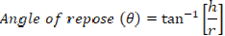
Angle of repose: The flow characteristics of microspheres were assessed by determining the angle of repose. Determination of angle of repose of amoxicillin microspheres were carried out by employing fixed funnel method by using Eq. 1.
(1)
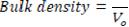
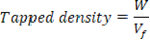
Bulk density and tapped density: Initial volume (Vo) was measured and then, the density apparatus was set for 100 taps and after that, the volume (Vf) was measured. The bulk density and tapped density were calculated by using formulas given in Eq. 2 and Eq. 3 where W- is weight of prepared microspheres.
(2)
(3)
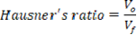
Hausner’s ratio: It measures the ratio of the final volume to initial volume and can be calculated by using Eq. 4.
(4)
Carr’s index: To analyze the flow ability, the Carr’s index is calculated on the basis of the bulk density and tapped density. It can be calculated by using Eq. 5.
Percentage yield: The percentage yield was calculated by using Eq. 6.

Drug content estimation: Accurately weighed quantity (equivalent to 100 mg of drug) of microspheres was crushed into powder and added to 100 ml of pH 6.8 phosphate buffer. The resulting mixture was kept stirring for 2 hours and kept it a side over night. The solution was diluted using pH 6.8 phosphate buffer and analyzed spectrophotometrically at 230 nm to get practical drug content [10].
Drug entrapment or encapsulation efficiency: The drug entrapment efficiency was calculated by Eq. 7.

Particle size analysis: The particle size can be identified with the help of optical microscope in the range of 0.2 to 500 µm. The particle size was measured using an optical microscope and the mean microsphere diameter was calculated by measuring 50 particles with the help of a calibrated ocular micrometer.
Swelling studies: A known weight ≠50 mg of microspheres was placed in a glass vial containing 10 ml of distilled water at 37 ± 0.5 °C in incubator with occasional shaking. The microspheres were periodically removed, bottled with filter paper and their changes in weights were measured during the swelling until equilibrium was attained. Finally, the weight of the swollen microspheres was recorded after a period of 3 hours and the swelling ration (SR) was then calculated from the formula given in Eq. 8. The studies were carried out in triplicate.
(8)
Fourier Transform Infrared Spectroscopy (FTIR) studies: The FTIR spectra of the amoxicillin, Optimized formulation, guar gum, xanthan gum using KBr pellet technique, samples were scanned over the range of 400-4000 cm-1 at the spectral resolution of 4 cm-1 [11].
Differential Scanning Calorimetry (DSC): The DSC analysis of indomethacin and optimized formulation were carried out to evaluate any possible drug polymer interaction. The DSC analysis was performed in the range of 20 ? C to 300 ? C temperature at a rate of 10 ? C/min with 25 ml/min of nitrogen flow [12].
X-ray Powder Diffractometry (XRD): The XRD studies of indomethacin and optimized formulation were recorded using Philips X-ray diffractometry (Uitima-III, Rigaku, Japan) with copper target to investigate the effect of microencapsulation on crystallinity of drug [13].
Scanning Electron Microscopy (SEM) studies: The SEM analysis of indomethacin and optimized formulation microspheres powder were taken using a scanning electron microscope (JSM 6360, Joel make, UK). Samples were fixed on an aluminum stub with conductive double-side adhesive tape.
In vitro drug release studies: Dissolution studies were carried out for 12 hours using basket type dissolution apparatus (USP-XXIII Electro lab, containing 900 mL of dissolution medium maintained at 37 ±0.5 ? C at 100 rpm speed. The dissolution was carried out first 2 hours in 1.2 pH buffer and followed by pH 6.8 phosphate buffers for next 10 hours with the respective sampling intervals 5 ml of aliquots were withdrawn and replaced with an equal volume of fresh prewarmed dissolution medium. After suitable dilutions, the samples were analyzed spectrophotometrically at 230 nm.
Release kinetics studies: Quantitative analysis of the values obtained in dissolution studies was used in mathematical formulae to express the dissolution results as function of dosage form characteristics.
RESULTS AND DISCUSSION
Organoleptic properties: The organoleptic properties of amoxicillin were identified the appearance is as fine powder, odour is penicillin-type and white in color.
Micromeritic properties: The micromeritic properties of microspheres of all the batches F1-F15 were shown in Table No. 2. The angle of repose of prepared microspheres was found to be in the range of 9.64 to 16.47°. The bulk density of prepared microspheres was found to be between 0.32 to 0.50 gm/cm3, tapped density was 0.42 to 0.59 gm/cm3, Hausner’s ratio 1.03 to 1.18 and Carr’s index was found to be between 5.21 to 10.35 %. The flow properties of different batches of microcapsules were excellent as the angle of repose values were found to be less than 25°, compressibility index less than 12 % and Hausner’s ratio less than 1.18 in all the batches.
Table No. 2: Micromeritic properties of amoxicillin microspheres.
|
S. No. |
Formulation code |
Angle of repose ( θ 0) |
Bulk density (g/cm3) |
Tapped density (g/cm3) |
Hausner’s ratio |
Carr’s index% |
|
1. |
F1 |
10.75±0.25 |
0.32±0.13 |
0.57±0.22 |
1.11 |
9.98 |
|
2. |
F2 |
9.64±0.01 |
0.45±0.03 |
0.48±0.09 |
1.15 |
10.35 |
|
3. |
F3 |
11.14±0.22 |
0.49±0.04 |
0.53±0.02 |
1.10 |
9.83 |
|
4. |
F4 |
14.09±0.15 |
0.48±0.05 |
0.52±0.03 |
1.08 |
7.60 |
|
5. |
F5 |
11.06±0.08 |
0.46±0.08 |
0.49±0.09 |
1.06 |
7.62 |
|
6. |
F6 |
12.84±0.06 |
0.49±0.09 |
0.52±0.08 |
1.09 |
9.02 |
|
7. |
F7 |
16.17±0.12 |
0.47±0.07 |
0.51±0.05 |
1.10 |
9.79 |
|
8. |
F8 |
12.13±0.05 |
0.42±0.13 |
0.50±0.01 |
1.12 |
9.13 |
|
9. |
F9 |
14.28±0.01 |
0.45±0.02 |
0.52±0.03 |
1.11 |
10.31 |
|
10. |
F10 |
16.18±0.09 |
0.47±0.12 |
0.55±0.01 |
1.18 |
9.4 |
|
11. |
F11 |
12.44±0.03 |
0.49±0.15 |
0.52±0.92 |
1.09 |
8.7 |
|
12. |
F12 |
15.65±0.05 |
0.42±0.05 |
0.46±0.05 |
1.05 |
5.21 |
|
13. |
F13 |
16.47±0.12 |
0.50±0.14 |
0.59±0.01 |
1.03 |
7.05 |
|
14. |
F14 |
11.89±0.23 |
0.46±0.03 |
0.43±0.01 |
1.04 |
9.57 |
|
15. |
F15 |
13.62±0.78 |
0.49±0.04 |
0.42±0.05 |
1.07 |
8.47 |
Where all the values are expressed as mean ± S.D., n=3.
Percentage yield, drug entrapment eficacy and drug content estimation: The percentage yield, drug entrapment efficacy and drug content of all prepared microspheres of different batches F1-F15 were given in the Table No. 3. Percentage yield and entrapment efficiency of different batches were found to be in the range of 62.46 to 94.27 % and 60.428 % to 89.23 % respectively. The drug content of microspheres of different batches F1 –F10 were found to be in the range of 64.18 to 91.42 % respectively.
Table No. 3: Percentage yield, drug entrapment efficiency and drug content of amoxicillin microspheres.
|
S. No. |
Formulation code |
Theoretical yield (g) |
Practical yield (g)a |
Percentage yield (%)a |
Entrapment efficiencya |
Drug contenta |
|
1. |
F1 |
2.43 |
2.1±0.13 |
86.41±0.21 |
79.26±0.18 |
73.51±0.52 |
|
2. |
F2 |
1.25 |
1.1±0.04 |
88.07±0.12 |
73.64±0.16 |
76.89±0.84 |
|
3. |
F3 |
1.68 |
1.15±0.12 |
68.24±0.13 |
75.58±0.13 |
79.64±0.45 |
|
4. |
F4 |
2.65 |
2.37±0.09 |
86.66±0.16 |
83.68±0.08 |
84.18±0.16 |
|
5. |
F5 |
2.25 |
1.94±0.11 |
85.08±0.11 |
82.23±0.11 |
65.42±0.75 |
|
6. |
F6 |
2.18 |
1.56±0.14 |
71.88±0.09 |
60.42±0.12 |
76.92±0.91 |
|
7. |
F7 |
2.85 |
2.15±0.10 |
87.65±0.10 |
72.31±0.09 |
88.45±0.41 |
|
8. |
F8 |
3.84 |
2.86±0.08 |
74.09±0.12 |
61.32±0.13 |
64.18±0.28 |
|
9. |
F9 |
2.22 |
1.95±0.16 |
88.28±0.14 |
82.31±0.07 |
83.64±0.34 |
|
10. |
F10 |
3.39 |
2.23±0.07 |
65.59±0.13 |
83.57±0.21 |
85.42±0.19 |
|
11. |
F11 |
3.72 |
2.25±0.09 |
62.46±0.11 |
84.46±0.12 |
86.92±0.32 |
|
12. |
F12 |
2.35 |
2.19±0.16 |
93.19±0.06 |
88.20±0.05 |
75.42±0.44 |
|
13. |
F13 |
2.28 |
2.15±0.08 |
94.27±0.12 |
89.23±0.09 |
89.64±0.45 |
|
14. |
F14 |
3.56 |
3.35±0.11 |
86.59±0.08 |
81.57±0.14 |
74.18±0.55 |
|
15. |
F15 |
2.35 |
1.89±0.05 |
82.17±0.17 |
86.41±0.18 |
91.42±0.75 |
Where all the values are expressed as mean ± S.D., a=3.
Particle size (µm): The average particle size range is 450±1.25 µm to 779.5±1.25 µm. The major fraction of the microspheres (50-55%) of all batches were within a range from 450µm mesh size. The data was represented in Table 4.
Swelling Index (SI): swelling property of microspheres was prepared by using different concentration of natural gums like guar gum, xanthan gum. The swelling index range of all the prepared microspheres were in the range from 87% to 222%, values were given in Table 4.
Table No. 4: Particle size and swelling index of amoxicillin microspheres.
|
S. No. |
Formulation code |
Particle size (µm) |
Swelling Index (%) |
|
1. |
F1 |
450±1.25 |
98±0.11 |
|
2. |
F2 |
472±1.30 |
102±0.8 |
|
3. |
F3 |
493±0.96 |
132±0.15 |
|
4. |
F4 |
540±2.65 |
90±0.14 |
|
5. |
F5 |
554±1.54 |
170±0.23 |
|
6. |
F6 |
581±1.17 |
168±0.7 |
|
7. |
F7 |
602±0.97 |
222±0.04 |
|
8. |
F8 |
624±0.35 |
193±0.11 |
|
9. |
F9 |
665±1.77 |
87±0.06 |
|
10. |
F10 |
692±0.22 |
157±0.11 |
|
11. |
F11 |
713±2.67 |
121±0.17 |
|
12. |
F12 |
719±0.57 |
97±0.13 |
|
13. |
F13 |
779±1.80 |
201±0.11 |
|
14. |
F14 |
588±1.27 |
95±0.08 |
|
15. |
F15 |
692±0.65 |
150±0.12 |
Where all the values are expressed as mean ± S.D., n=3.
Fourier Transform Infrared Spectroscopy (FTIR) studies: FTIR analysis describes about the drug:polymer interaction. Figure 1, shows IR spectrum of amoxicillin, xanthan gum and optimized formulation F13. The FTIR spectrum of amoxicillin showed the characteristic peaks at 3440 cm-1 (N-H Stretching), 1576 cm-1 (aromatic C=C Stretching), 2968cm-1 (C-H Stretching), 1772.3cm-1 (C=O Stretching), 1177.8 cm-1 CH3 directly attached to the ring, 1246.6 cm-1 C-O-C in COOH. The similar drug result were reported by Kultida S et al. therefore, it was identified as amoxicillin in Figure 1a. The FTIR spectrum of xanthan gum showed the absorption band nearly at 3268 cm-1 (O-H), 1718.3 cm-1 (C=O) and 2911.1 cm-1 (C-OH) stretching vibration. The same peaks were also reported in drug loaded microcapsules in optimized formulation F13, prepared using sodium alginate and its combination with xanthan gum. There was no change or shifting of characteristics peaks of amoxicillin in drug loaded microcapsules, suggested that there was no significant drug polymer interaction which indicated the stable nature of drug inside the formulation. This conformed the stability of the formulation.
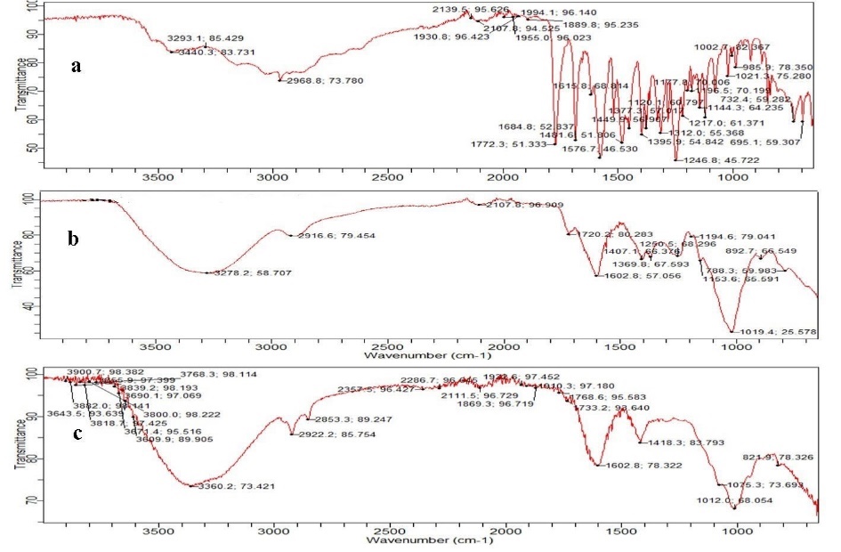
Figure 1: FTIR spectrum of a) Amoxicillin, b) Xanthan gum and c) Formulation SF13.
Differential scanning Calorimetry (DSC): DSC thermogram of amoxicillin showed a sharp endothermic peak at 202.83 ?C and onset 196.19 ?C corresponding to its melting point and shown in Figure 2. The obtained value matched with the value given in literature. The melting peak of sodium alginate was observed at 77.34 ?C. And the melting peak of guar gum was observed at 160.27 ?C. Optimised formulation F13 was prepared by ion gelation method. Melting peak of formulation was shited to a higher temperature 199.73 ?C. The change in melting point endotherm of drug may be attributed to mixiing process which lowers purity of each component. Therefore, it could be concluded that drug in microspheres was in amorphous phase of molecular dispersion or solid solution state in polymer matrix. However, in the interaction studied the drug was shown to be fairly stable and inert with the excipients used in the microsphers formulation.
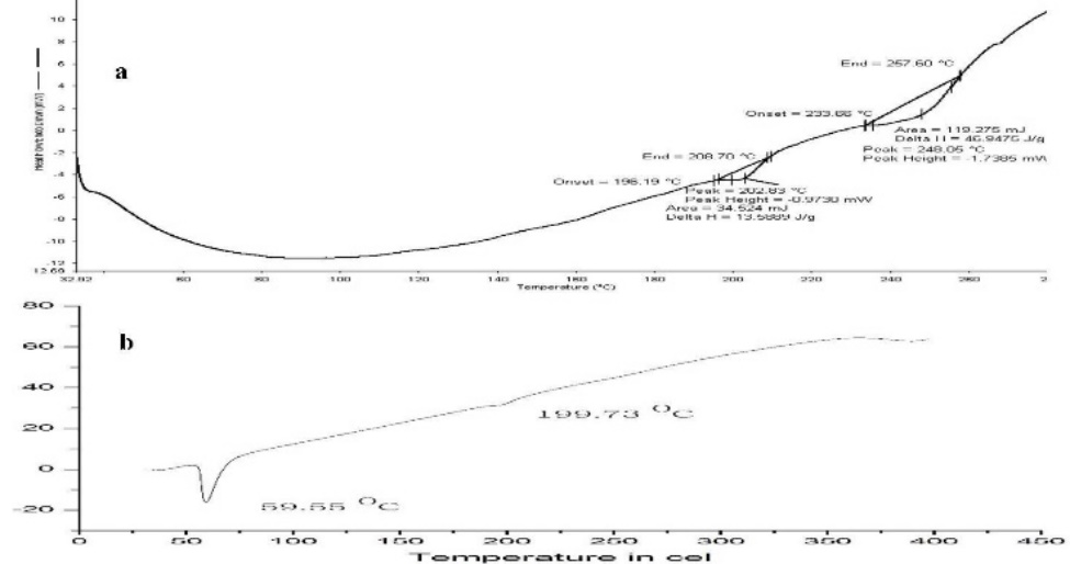
Figure 2: DSC thermogram of a) Amoxicillin and b) Fomualtion F13.
X-ray Powder diffractometry (XRD): The XRD Patterns of Amoxicillin and microspheres formulation F13 are shown in Figure 3. Amoxicillin exhibited characteristics sharp and intense diffraction peak at 200 equivalent to 12.2130, 15.3060, 16.3110, 18.2890, 19.1120, 26.6480and 28.8960 this signifies the amorphous in nature of amoxicillin. The XRD of optimized formulation F13 shown less intense peak 200 equivalent to 28.7690. It demonstrates the homogeneous dispersion of drug in microsphere as well as partial transition from crystalline amoxicillin to amorphous state. Transition may be a dilution effect by the gum. It doesn’t affect the therapeutic efficacy of the drug as all the all function groups are present.
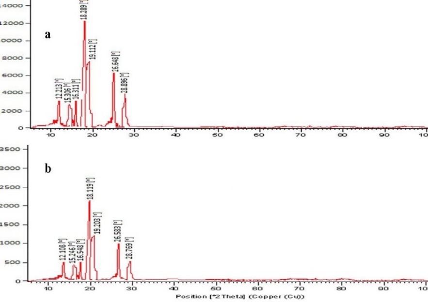
Figure 3: XRD of a) Amoxicillin and b) Optimized formulation F13.
Scanning Electron Microscopy (SEM) studies: The microphotography of the microsphere was shown Figure 4. It suggests the prepared microspheres were free flowing nature. It was distinct, multinucleate and monolithic with even geometry. Microphotograph also revealed that microspheres were round and totally wrapped with gums. Roughness of the microsphere surface indicated the presence of the drug.
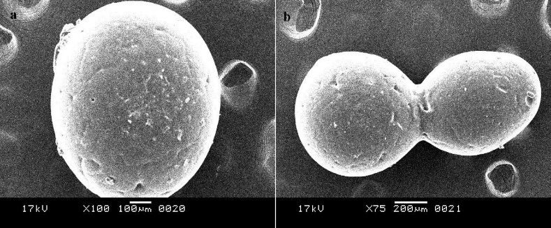
Figure 4: SEM of a) Amoxicillin and b) Optimized formulation F13.
In vitro drug release studies: The % cumulative in vitro release studies were performed in triplicate (n=3) for all the prepared formulation F1 to F15 with different concentration of polymer in each formulation in 1.2 pH buffer for 12 hrs the samples were spectrophotometrically analyzed at 230 nm. Guar gum used microspheres containing amoxicillin release profile shown in Figure 5a. The formulations of guargum showed in complete drug release within 12 hrs of dissolution. As the concentration of guargum was increased the drug release pattern was decreased (F2). The formulation F3, F4, F5 showed similar drug release profile was F2 formulation. The formulation F1 to F6 contains similar quantities of sodium alginate 2 g. the amoxicillin release was less due to the more concentration of sodium alginate. The sodium alginate forms high viscosity solutions where the amoxicillin was entrapped. Therefore, the amoxicillin release was less. The sodium alginate guar gum (2g) good matrix solution where the amoxicillin entrapped and showed higher release. The F14 to F15 formulation showed incomplete drug release. The formulation F7 to F13 was prepared by using various concentration of Xanthan gum withvarying concentration of sodium alginate. The formulation F7 to F13 contains 2 g of sodium alginate with increasing concentration of xanthan gum. The F12 formulation showed approximately 93% of drug release within 12 hrs of dissolution were shown in Figure 5b. The F13 formulation showed a complete drug release and good release pattern within 3hrs dissolution showed approximately 45% of drug release whereas at 6 hrs of dissolution it showed 70% of drug release. The formulation F14 to F15 in complete drug release (approximately 91 %). Among all the formulation of xanthan gum used amoxicillin microspheres, F13 formulation (0.250 g of xanthan gum, 1g of sodium alginate). Showed better release profile over a period of 12 hrs. this may be due to increased concentration of xanthan gum and decrease concentration of sodium alginate. The combination of sodium alginate, xanthan gum maintained as appropriate gel barrier and tortosity. Therefore, F13 formulation was selected as optimized formulation.
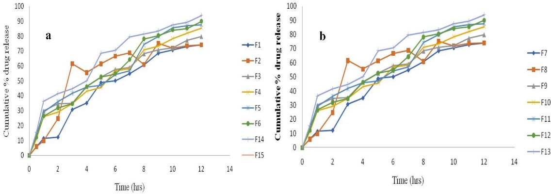
Figure 4.14: In vitro drug release profile of a) Guar gum formulation F1 – F6, F14, F15 and b) Xanthan gum formulation F7 – F13.
Drug release kinetic study:
CONCLUSION
The microspheres were evaluated for pre-compression parameters like angle of repose, bulk density, tapped density and Carr’s index. The result obtained were found to be satisfactory and within specified limits. The drug entrapment efficiency and percentage yield increased progressively with increasing the concentration of polymer. The in vitro amoxicillin release studies were performed 0.1 N HCl buffer. In this investigation it was observed that out of all formulation (F1 to F15), the F13 (0.1 g drug, 0.250 g xanthan gum, 1g sodium alginate) shown a promising and effective controlled release of drug over a period of 12 hrs. The XRD and SEM supported the study. Hence F13 was considered as best formulation. Analysis of release data as per different kinetic models indicated that amoxicillin release from these microspheres was best fit towards zero order kinetics. Correlation coefficient values in the zero order models were higher than those first order, indicating the drug release was independent of drug concentration. Correlation coefficient (R2) values in Higuchi model suggest that the release of drug was following diffusion type of mechanism. When the release data of optimized batch F13 was analyzed as per Korsemeyer – Peppas equation, as the release exponent ‘n’ indicating the Fickian diffusion mechanism. From the results we can conclude that design of microspheres by employing natural polymers as release retardants by external ionotropic gelation technique was widely used to encapsulate a broad range of drugs to achieve controlled delivery of drug. Natural polymer (xanthan gum) was a suitable release retardant polymer. It is showed its best along with another natural polymer, sodium alginate. The combination of sodium alginate and xanthan gum was suitable to develop microsphere, which showed better amoxicillin release over a period of 12 hrs. the external inotropic gelation method was a suitable method for the preparation of microsphere. The controlled amoxicillin microspheres were capable of maintaining constant plasma concentration up to 12 hrs. so this can expected to decrease the drug intake eventually and increasing the patient compliance which was the major drawback of conventional amoxicillin formulations. This combination of sodium alginate and xanthan gum can be used develop microspheres for other drugs.
REFERENCES


Afreen Khan*, Arpita Verma, Ashutosh Rajak, Dhawal Pal, Rajat Pawar, Development of Controlled Release Microspheres of amoxicillin by using Natural Gums, Int. J. of Pharm. Sci., 2025, Vol 3, Issue 3, 1577-1587. https://doi.org/10.5281/zenodo.15041886
 10.5281/zenodo.15041886
