Shree Chhatrapati Shahu Maharaj Shikshan Sanstha , Institute of Pharmacy, Maregaon
Carbon dots (C-dots), the youngest members of the carbon nanomaterials family, were first discovered in 2004 during the purification of carbon nanotubes and have since emerged as versatile and innovative nanomaterials. These particles, typically smaller than 10 nm, are distinguished by their exceptional photoluminescence, biocompatibility, chemical stability, and low toxicity, making them ideal for a wide range of applications. Their highly tunable optical properties and ease of surface functionalization enable their use in bioimaging, drug delivery, and advanced cancer therapies such as photothermal therapy (PTT) and photodynamic therapy (PDT). Synthesized through top-down methods like laser ablation and chemical oxidation or bottom-up approaches like hydrothermal and green synthesis, C-dots can also be produced sustainably from plant-based precursors. Furthermore, their applications extend to biosensing, where they demonstrate high sensitivity in detecting biomolecules and toxic heavy metal ions. This review explores the synthesis, properties, and biomedical applications of C-dots, emphasizing their role as multifunctional nanomaterials in diagnostics, therapeutics, and emerging fields such as cancer theranostics and nanomedicine.
Carbon dots
(C-dots) are a novel type of carbon-based nanomaterial discovered by Xiaoyou Xu’s in 2004. C dots were accidentally discovered during the separation and purification of single-walled carbon nanotubes, showing that the carbon size is less than 10 nm. (1) Sun Ping Presented a synthetic approach to making C-dots with improved fluorescence emissions via surface passivation in 2006 and termed them carbon quantum dots (CQ-dots), carbon nanoparticles (CNPs), or carbon nanodots (CN-dots).(2) C-dots are studied and are of interest to scientists all over the world compared to 2D carbon materials due to their unique, novel properties, great photostability, ease of production, strong biocompatibility, high quantum yield, small size, low toxicity, low cost, weak interactions with proteins, and good water solubility, providing important applications in many fields including chemistry, engineering, and biological science along with their respective applications, particularly in imaging and therapeutics. (3,5) In the last ten years, the biological and biomedical application of C-dots has become widespread due to their small size (110 nm), unique optical properties, easy functionalization, and bioactivity. Because of their small size, they can cross numerous natural biological barriers in vivo, including ion channels, the blood– brain barrier (BBB), and the glomerular filtration barrier. Owing to their tunable functional properties, they are suitable nanocapsules and nanocarriers for loading and transporting drugs and genes to specific sites in vivo. In addition, the optical qualities of some C-dots open up possibilities for biomedical applications. (6,7) C-dots exhibit significant absorption in the near-infrared spectral range, which enables in situ photothermal effects in photoacoustic imaging and photothermal therapy. C-dots are also used as fluorescent contrast agents for deep tissue fluorescence imaging. (8) CDs have been acknowledged as wise choices for the diagnosis of cancer.(9) CDs have been investigated for cancer diagnosis by PA imaging, MR imaging, and others in addition to using FL imaging.(10) Some CDs also naturally possess anticancer characteristics, which take part in PDT and PTT.(11) PDT typically involves the use of photosensitizers.(12) to produce reactive oxygen species (ROSs) under the right lighting conditions, which are then used to cause oxidation reactions with biological macromolecules in cancer cells, leading to cell toxicity.(13)
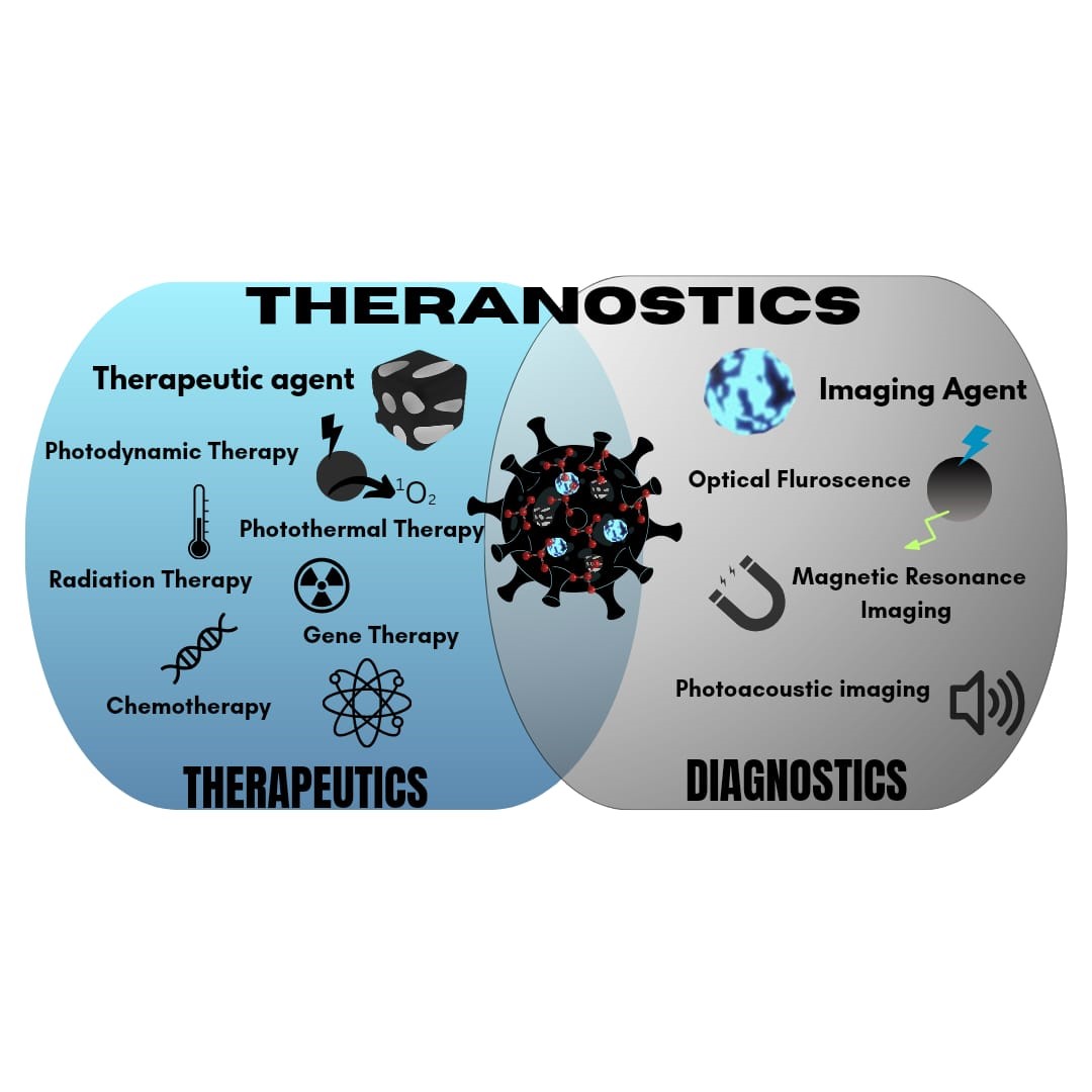
Figure 1. Schematic representation of therapeutics, diagnostics, and theranostics of Carbon Dots.
1.2 Cancer
Cancer has become a leading global health crisis due to its high incidence and mortality rates. Despite significant advancements in treatment, the overall5-year rate for cancer patients remains low. In 2018 alone, approximately 9.6 million cancer-related deaths and 18 million new cases were reported with projections suggesting an alarming rise to 22 million annuals cases within the next two decades. Lung cancer, the deadliest form, currently claims over 350 lives per day, which is 2.5 times more than colorectal cancer (CRC)—the second leading cause of cancer death—and exceeds fatalities from breast, prostate, and pancreatic cancers combined. Of the estimated 130,180 lung cancer deaths anticipated in 2022, roughly 81% (105,840) will be linked to smoking, with an additional 3,650 deaths attributed to second-hand smoke exposure. Notably, the remaining 20,700 non-smoking related lung cancer deaths would rank as the ninth most common cause of Cancer death for both sexes if counted separately. Despite advances in early detection and treatments such as surgery, chemotherapy, and immunotherapy, cancer remains a significant global health challenge with ongoing research focused on improving outcomes and finding more effective cures. (14)
2.Classification of carbon dots:-
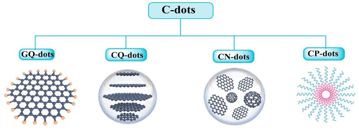
Figure 2. Structure of different types dots: graphene quantum dots (GQ dots), carbon quantum dots (CQ-dots), carbon nanodots (CN-dots), and carbonized polymer dots (CP-dots). (15)
Classification of carbon dots: -
2.1. Graphene quantum dots- Graphene quantum dots (GQDs) are considered tiny chunks of graphene and are zero-dimensional fragments of bulk carbon materials with properties of both carbon dots and graphene. (16) An ideal GQD will have only one atomic layer of carbon atoms although the lateral sizes may be large. (17) Graphene quantum dots (GQDs) are nanometre-sized fragments of graphene with a 2D planar structure composed of sp?2;hybridized carbon atoms arranged in a hexagonal lattice. Typically, 2–20 nm in diameter, GQDs exhibit quantum confinement and edge effects, resulting in unique photoluminescence properties. The presence of oxygen-containing functional groups at their edges enhances water solubility and chemical reactivity. Due to their excellent conductivity, photostability, and biocompatibility, GQDs are widely used in bioimaging, sensors, and optoelectronic applications. However, most of the synthesized GQDs have multiple atomic layers with sizes less than 10 nm and also contain functional groups like oxygen and hydrogen. (18)
2.2. Carbon quantum dots-Carbon quantum dots (CQDs) are a new type of zero-dimensional carbon nanomaterials that exhibit similar luminescence performance and small size characteristics as traditional quantum dots. Additionally, they possess good water solubility, low biotoxicity, and good electrical conductivity. As a result, they have garnered significant attention in the fields of biomedicine, sensors, optoelectronics, and light emitting diodes. Carbon quantum dots (CQDs) are spherical nanoparticles generally under 10 nm, made up of amorphous or crystalline carbon with sp?2; and sp?3; hybridization. They possess a core-shell structure, with the core being carbon-rich and the surface bearing various functional groups like carboxyl’s and hydroxyl’s. CQDs display tunable photoluminescence attributed to quantum confinement and surface state emissions, along with high chemical stability and water dispersibility. Their non-toxic nature and versatile optical properties make them ideal for applications in bioimaging, drug delivery, and photovoltaic technologies. (19,20)
2.3. Carbon nanodots- Carbon nanodots (CDs), discovered in the mid-2000s are one of the protagonists of carbon nanoscience. CDs are nanoparticles smaller than?10 nm typically composed by carbon, oxygen, nitrogen and hydrogen. Carbon nanodots (CNDs) are carbon-based nanoparticles typically less than 10 nm in size with a more amorphous structure compared to CQDs. They consist of a mix of sp?2; and sp?3; hybridized carbon regions, and their surface is often enriched with functional groups that enhance solubility and reactivity. CNDs exhibit luminescence primarily due to surface state emissions, rather than intrinsic quantum confinement, making their optical properties dependent on surface chemistry. Due to their ease of functionalization, low toxicity, and photoluminescence, CNDs are used in bioimaging and as fluorescent markers. (21,22)
2.4. Carbonized polymer dot- Carbonized polymer dots (CPDs) are novel emerging fluorescent nanomaterials, which are composed of organic polymer chains and a carbon core. Carbonized polymer dots (CPDs) are formed through the controlled thermal carbonization of organic polymers or small molecules, incorporating carbon and other heteroatoms like nitrogen or sulphur. CPDs are typically 2–10 nm in diameter, with a mixed sp?2;/sp?3; carbon network and a less crystalline structure than GQDs. Their photoluminescence is highly tunable, influenced by the carbonization process and surface groups. CPDs have high quantum yields and can be doped to modify properties, making them useful for bioimaging, drug delivery, and light-emitting applications such as LEDs. (23,24)
3. Structural data of carbon dots: -
C-dots are commonly described in terms of a carbogenic core consisting of amorphous and crystalline parts with surface functional groups. As-prepared fluorescent carbon materials always consist of sp2/sp3 carbon, oxygen/ nitrogen-based groups, and postmodified chemical groups. Carbon materials, which include graphite, diamond, fullerenes, carbon nanotubes (CNTs), and graphene, have been well known for many years. To make these materials fluorescent, their size and surface chemical groups should be carefully modulated. C-dots always possess at least one dimension less than 10 nm in size and fluorescence as their intrinsic properties. Nuclear magnetic resonance data indicates that the inner part of C-dots mainly con sists of sp2 hybridized carbon atoms, while outer part is composed of sp3 hybridized carbon atoms. Both sp2-and sp3-hybridized C atoms found in the C-dot. (8)

Figure 3 Structure of Carbon Dots.
Table 1: Properties of Carbon dots along with description
|
Property |
Description |
|
Chemical composition |
Carbon(C), oxygen (o) Hydrogen (H), Nitrogen (N) (depending on synthesis method) |
|
Core structure |
Graphene -like carbon core, often amorphous or containing sp2 and sp3 hybridized carbons. |
|
Size |
Typically, 1-10 nm in diameter. |
|
Surface functional groups |
Hydroxyl (-OH), carboxyl (-COOH), amino(-NH2), aldehyde (-CHO), and other oxygen/nitrogen – containing groups |
|
Optical properties |
Fluorescence emission, quantum yield (QY) typically ranging from 1080%, excitation-dependent emission spectra. |
|
Surface charge |
Zeta potential values can vary from negative to positive, depending on surface group and ph. |
|
Crystal structure |
Amorphous or crystalline, depending on the preparation method (e.g., hydrothermal, laser ablation, etc.). |
|
Photoluminescence (PL) Behaviour |
Excitation- dependent PL, tunable emission wavelength, broad absorption range (200-400 nm). |
|
Synthetic method |
Hydrothermal synthesis, microwave- assisted synthesis, laser ablation, solvothermal synthesis, etc. |
4. Synthesis processes of carbon dots: -
4.1 The Chemical synthesis method of carbon dots (CDs):
Synthesis methods can be roughly divided into two methods, including the top-down method and the bottom-up method. In the top-down process, macromolecular carbon materials are broken down into small molecular nanoparticles using specific physical and chemical methods. The bottom-up method refers to the synthesis of large carbon nanoparticles from small molecule materials by certain physical and chemical methods.
4.1.1. Synthesis of CDs by the Top- down method: -
The top-down method includes the arc discharge method, laser ablation method, electrochemical method, and chemical oxidation method.
1.Arc discharge method: -The arc discharge method is that the continuous discharge between the cathode and the anode leads to high temperature, making the anode gradually consume and then form CDs. In 2004, Xiaoyou Xu and colleagues prepared the first carbon nanoparticles using the arc discharge method. Since the arc discharge method synthesizes carbon nanomaterials like nanotubes and fullerenes by creating an electric arc between graphite electrodes in an inert gas atmosphere. The high temperatures vaporize carbon, which then cools and condenses into nanomaterials. Factors such as current, gas pressure, and catalysts influence the material's quality. While this method produces high-quality products, it requires significant energy and control and often results in mixed outputs needing purification. hen, the arc discharge method has been popularized as a rapid preparation method.
2.Laser ablation method: -The laser ablation method uses a high-energy laser as a light source to prepare CDs by irradiating carbon materials. Carbon nanoparticles were first prepared using laser ablation in 2006 by Sun Ping and colleagues.The laser ablation method is a synthesis technique where a high-powered laser beam is focused on a solid carbon target in an inert gas atmosphere (e.g., argon or helium) at elevated temperatures. The laser's energy causes rapid vaporization of the target, forming a plume of carbon atoms and clusters that cool and condense into nanomaterials such as carbon nanotubes and quantum dots. By adjusting factors like laser power, wavelength, and gas type, the properties of the produced materials can be finely tuned. This method yields high-purity, low-defect nanomaterials but requires costly equipment and significant energy, limiting its scalability.
3.Electrochemical method: -The electrochemical method uses graphite rods as a carbon source to prepare CDs through electrochemical treatment. In 2012, Dhanraj Shinde and colleagues found that the electrochemical device can control the size of CDs by adjusting the potential, electrolyte concentration, reaction temperature and other factors. By manipulating parameters such as voltage, electrolyte composition, and reaction duration, the size, surface properties, and optical characteristics of the CDs can be tuned. This technique is cost-efficient, environmentally safe, and works under mild conditions, though it may need further optimization to ensure uniformity and quality for particular uses.
4.Chemical oxidation method: -The chemical oxidation method uses strong oxidants to prepare CDs. In 2007, Liu and colleagues used oxidizing acid to treat soot from candle burning to obtain CDs. The chemical oxidation method for making carbon dots (CDs) involves treating a carbon precursor with strong oxidizing agents like nitric acid or hydrogen peroxide. This process breaks down the precursor into smaller fragments, forming nanoparticles with controllable size, surface properties, and optical characteristics. It is a simple, cost-effective method suitable for large-scale production, though it requires careful handling of chemicals.
4.1.2. Synthesis of CDs by the Bottom-up method: -
The bottom-up method includes the microwave method, hydrothermal method, and pyrolysis method.
1.Microwave method: - The microwave method can be used for the preparation of CDs from different precursor sources, so it is widely used. By microwave Treatment of citric acid (CA) and urea. The microwave method for synthesizing carbon dots (CDs) uses microwave radiation to heat a carbon precursor, like citric acid or glucose, in a solvent. This rapid heating causes carbonization and fragmentation, forming nanoparticles with controlled size and properties. It is a fast, efficient, and scalable method, producing high-quality CDs with good uniformity, although optimization may be needed for consistent results.
2.Hydrothermal method:-The hydrothermal method is the mildest reaction method to prepare CDs by putting the precursor in the liquid phase and keeping it in a constant temperature environment for a certain time. The hydrothermal method for synthesizing carbon dots (CDs) involves heating a carbon precursor in water or solvent under high temperature and pressure in a sealed container. This process breaks down the precursor to form carbon nanoparticles with controlled size, surface properties, and optical characteristics. It is a simple, cost-effective, and scalable method, though optimization is needed for consistent product quality.
3.Pyrolysis method: -The pyrolysis method is to place the precursor in an inert gas to prepare CDs at a high temperature. In 2020, Lidong Yu and colleagues used naphthalene for liquid phase pyrolysis to obtain CDs. The pyrolysis method for synthesizing carbon dots (CDs) involves heating organic precursors like sugars or citric acid at high temperatures (300–800°C) in the absence of oxygen. This process decomposes the precursor, forming carbon dots with controllable size and properties. The method produces high-quality, fluorescent CDs but requires precise control of temperature and time to ensure uniformity and avoid excessive carbonization. (25)
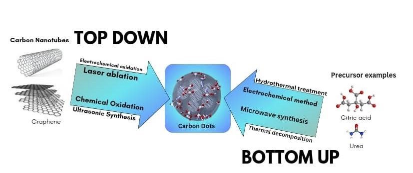
Figure 4. Top-down and bottom-up approaches for the synthesis of C-dots.
4.2. Synthesis of CDs by Green synthesis: -
Green synthesis of CDs mainly utilizes biomass. Biomass synthesis makes use of natural raw materials (organisms, waste material, protein products, or natural polymers), instead of reaction precursors usually used in the traditional methods, and also Requires external energy supply. Using diverse raw materials, CDs with different structures and properties can be obtained. Usually, a temperature of 100–200 °C is required, which is Much lower than that required in the traditional methods, and the synthesis is carried out in aqueous media. Generally, hydrothermal or solvothermal treatments, ultrasonication, microwave Irradiations, and microwave-assisted hydrothermal/pyrolysis are Used in the green synthesis of CDs. (26) Hydrothermal methods Convert the raw material into carbonized matter. Although relatively simple, the procedure takes several hours. Microwave irradiation, in contrast, provides homogenous and effective Heating and speed up the reaction to merely a few minutes. Hence, this approach is considered the fastest and simplest Amongst the synthesis methodologies and has become widely Used. The “top-down” approach involves breaking down bulky carbonaceous materials, such as carbon fibres, carbohydrates, proteins, and carbon soot, through chemical or physical methods. The carbon containing material is oxidized and broken down Into CDs using oxidants such as sulfuric acid and nitric acid. As Green methods are limited regarding the raw materials, the “top down” method is not very common in green approaches. (27,28) The “bottom-up” method consist of carbonization of smaller Organic molecules. This method basically involves four phases, that is, condensation of the molecules followed by polymerization, carbonization, and passivation. Small molecules are Condensed into intermediate chains and then polymerized into Clusters of carbonaceous material. Carbonization of this material at elevated temperatures leads to the formation of carbon Cores. The residual groups on the surface act as surface-passivating agents and can be manipulated to ameliorate surface luminescence properties. Biomass is rich in small organic Compounds suitable for carbonization at elevated temperature And, hence, “bottom-up” approaches are extensively used for the green synthesis of CDs. (29)
4.3 Synthesis of CDs by Plant source: -
Synthesis of CDs from plant-based sources has the potential to Be scaled up and comes with a number of benefits, including Reduced chemical exposure, cost-effectiveness, renewability of Sources, waste reduction, and ample source availability. It is Thus environmentally friendly and advantageous. (30,31) Plant Parts such as roots, stem, leaves, fruits, flowers, and seeds have Been used for the production of CDs. Several low-value plant Materials can also be converted into functional materials with Excellent biocompatibility by manufacturing CDs from these Plant components. Plant-based precursors that contain Heteroatoms (nitrogen and sulphur) are preferred over carbon sources that demand supplementary heteroatoms for the synthesis of CDs (32).
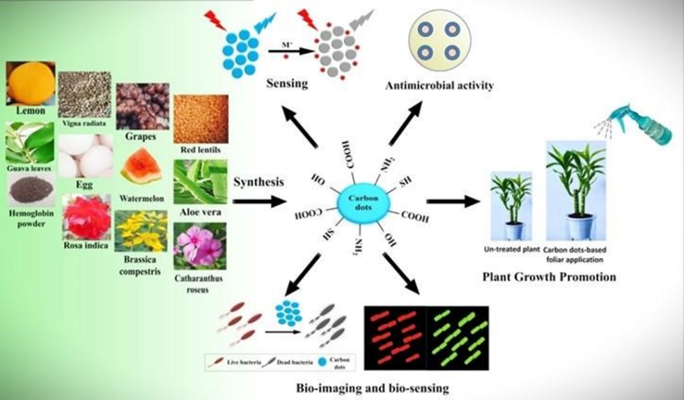
Figure 5. Illustration of the environmentally friendly sources employed for the synthesis of CDs and their applications from 2015–2022. (33)
5. Special properties of Carbon dot’s: -
C-dots exhibit unique properties that enable them to be used in various applications such as sensors, optronics and electrochemical luminescence. These properties can be tailored by controlling the size, shape, heteroatom doping or functionalization with biomolecules. When compared to organic dyes and traditional QDs, C-dots are advantageous in their photostability against photobleaching and blinking. Photobleaching is a major set-back for many fluorophores, in which the PL degrades over time with or without continuous excitation, limiting the shelf life of the fluorophores and long-term imaging capacity. (34) C-dots also tend to have lower toxicity, be chemically inert and have better biocompatibility when compared to inorganic semiconductor quantum dots. This allows for C-dots to be used as effective carriers for drug delivery or bioimaging, and thus is key to their potential in the theranostics area. Optical Properties: - The optical properties of C-dots are perhaps their most important and most studied property. C-dots possess highly tunable photoluminescence (PL) from deep ultraviolet to near-infrared (NIR). Despite the diverse structures of C-dots, prepared from different approaches with different precursors, C-dots usually share some similar optical properties in terms of absorption and fluorescence characteristics. A characteristic property of CDs is their ability to fluorescence under UV irradiation. However, the CD fluorescence mechanism remains a topic of debate and is suspected to vary according to the synthesis parameters. The CD surface proper ties (e.g., degree of oxidation, decoration with functional groups), size-dependant quantum confinement effect, and the incorporation of fluorescent molecules at the CD core or surface can all impact the mechanism. (35)
6.Chemical Composition: -
Doping CD’s with a heteroatom such as nitrogen is common practice since it is known to enhance QY by modifying the electronic properties and surface chemistry of the CDs.(36) Moreover, while the presence of oxygen in green-synthesized CD’s is common due to the abundance of oxygen in renewable raw materials and also many organic compounds, they can also impact the QY by affecting the degree of oxidation of the CD’s.(37)
Renewable raw materials are a source of heteroatoms such as nitrogen, sulphur, and phosphorus. X-ray photoelectron spectroscopy (XPS)is commonly used to measure the CD elemental composition on anatomic basis. In the green CD literature ,CDs have an oxygen-to-carbon (O:C) ratio of 0.07–1.12, and a nitrogen-to-carbon (N:C) ratio of 0.02–0.43 .CDs synthesized from Allium fistulosum exhibit a sulphur-to-carbon (S:C) ratio of 0.11 while those prepared from Eleocharis dulcis contained traces of phosphorus resulting in a phosphorus-to carbon(P:C) ratio of 0.004.The elemental diversity of renewable raw material scan be advantageous when synthesizing hetero-doped CDs, but complicate our ability to tune the chemical composition of the CDs. In such cases, renewable refined compound should be used. (38) Despite being synthesized from a wide variety of precursors, half of the CD’s have an O:C ratio in the narrow 0.31- 0.39 region. Cellulose is a major plant component, but has an O:C ratio of 0.83, well above the range indicated. Thermal reduction has been observed in complex compounds similar to CDs. For example, Chen et al, were able to thermally reduce graphene oxide with an O:C ratio of 0.48 to partially reduced graphene oxide with an O:C ratio of 0.18, as measured by XPS, using microwave irradiation. (39) Therefore, it is possible that the cellulose in plants, along with other biomolecules, is thermally reduced to a similar extent in reported studies, leading to similar O:C ratios in the resulting CDs. Another possible explanation for this convergence to a common O:C ratio could be that the CD is more likely to forma chemically stable structure in this region. (40)
7.Mechanism of Carbon Dots in Cancer Therapy: -
7.1 Theranostics approach in cancer treatment
In cancer treatment, chemotherapy, radiation, and surgery have certain limitations because one treatment pathway cannot address all types of cancer. (41) Tumour theranostics focuses on developing new structures that are biocompatible and biodegradable and able to perform efficient target therapy.317 Currently, several types of theranostics nanoparticles have been developed for treating cancer, however not all are efficient and safe. (42) In this respect, carbon dots are considered potential candidates for developing tumour theranostics due to the physicochemical and optical properties described above. Indeed, compared with other existing theranostics agents such as and metal–organic framework (MOF)-based nanomedicine, liposomes and polyester micelles, CDs possess a series of advantages that include low cytotoxicity, biocompatibility and stable photoluminescence. (43,44)
7.2 Targeted Cancer Therapy :
As CDs accumulate specifically in tumours, they are ideal for further use in targeted cancer therapy. (45) Meanwhile, with CDs' excellent performance in various bioimaging techniques, it is possible to develop nanotheranostic strategies that can reveal simultaneous diagnosis and treatment of various cancers. At an early stage, CDs are demonstrated to be excellent nanocarriers to reunite with different drugs to enhance targeted therapeutic effect. As nanomaterials develop, CDs have also been used as a new class of nanocarriers for creating nanocomposite with a series of functional nanoparticles such as Au NPs, Fe3O4 NPs, and some inorganic quantum dots, endowing them with additional characteristics, such as oxidative stress amplifier, magnetic functions and radioactivity, for advanced therapeutic applications. In addition, the special photophysical properties, such as strong absorbance of NIR light and/or excellent photothermal/photodynamic character, endow various CDs for light-active nanotheranostic of cancer. Compared with the traditional chemotherapy, the light-active nanotheranostic have received more and more attention due to its non-invasive and stimuli-responsive features and the promising characters to over drug-fast. In addition, several kinds of CDs have been observed distinctive cytotoxicity with novel metabolic pathway in cell growth and death, enabling their direct application for cancer therapy as nanomedicines. Finally, with the development of nanotechnologies, CDs also play important role in various advanced technologies and exhibit excellent applications in several kinds of cancer to overcome the limitation of common medicine. (46)
7.3 Gene therapy
Although the delivery of small molecule drugs has been extensively studied using C-dots, there are now reports of C-dots being used as gene carriers. Gene therapy has seen several advancements as a therapeutic approach to cure many debilitating diseases. Until now, viral vectors are the most efficient vector in delivery of genes and ~70% of clinical gene therapy trials use viral vectors. (47) However, there are major problems related to viral vectors such as severe immunogenicity response, random genomic integration, very limited capacity to accommodate long nucleic acids and high pro duction costs. (48) Yu et al. designed a C-dot based multifunctional delivery system that can deliver both drugs and genes. In the design of their vector, they synthesized amphiphilic C-dots by attaching alkyl epoxide to the amino groups on the surface of PEI derived C-dots. They then used green fluorescence protein (GFP) expression and flow cytometry data to confirm that the prepared C dots had higher transfection efficacy than a conventional vector (Lipofectamine-2000) in cancerous A549 cells. (49)
7.4 Therapeutic mechanisms of CDs in PDT
The three key elements of PDT are light source, PS and O2. The mechanism of action is that PS absorbs energy from the ground state to the excited state when irradiated by a light source and reacts to produce ROS, which causes cytotoxicity leading to cell death. (50) The PS receives light and absorbs energy to change from the ground state to the excited state, which can result in two reactions: the first is an electron transfer between the excited state of PS and intracellular substrates (such as nucleic acids, proteins and lipids) to form free radicals, which then interact with O2 to produce hydrogen per oxide, superoxide anions and hydroxyl radicals (H2O2, O2 ? and •OH), i.e. Type I reaction [Fig.6.] (51) The second is the direct transfer of energy from the excited PSs to oxy gen molecules through an ‘inter-system crossing’ pro cess, resulting in the formation of singlet oxygen (1O2), i.e. a Type II reaction [Fig.6.] (52) The Type III reaction mechanism is that Type III PSs selectively target biomolecules, such as nucleic acids, proteins and other macromolecules, with the result that biological target molecules can be directly and effectively destroyed. Due to the stringent requirements of Type III reaction on PSs, research on the mechanism of Type III reaction is still relatively scarce. (53)

Figure 6. Schematic illustration of photodynamic reactions (either Type I or Type II) and cell death pathways in the process of PDT. (54)
Analytical and Biomedical Applications:
8.1. Analytical applications (Diagnostic): C-dots have been used for a variety of biomedical applications due to their unique optical properties, their large surface area and the flexible ability for surface functionalization. Although, there are still currently some biosafety concerns about the use of C-dots, up until now no acute toxicity or morpho logical changes have been found from in vitro cytotoxicity studies of numerous series of cell lines.
(55)
8.1.1. In vitro and in vivo toxicity studies:
Although in vivo toxicity studies of CDs remain a critical issue, the low cytotoxicity of these nanoparticles could be the main reason for scarcity of studies in animal models. Wang et al. evaluated the genotoxicity, the acute toxicity and subacute toxicity of CDs fabricated from raw soot by nitric acid treatment and PEG passivation. The results showed no genotoxic or toxic effects in the mice model used. (56) On the other hand, many recent reviews have described several studies on the cytotoxic effect of CDs against different cell line models including, breast adenocarcinoma SKBR3, mouse embryonic fibroblast NIH-3T3. (57) Human renal epithelial cells 293T, normal breast epithelial cells MCF-10A, normal human liver cells HL-7702 (L-02), normal human skin fibro blasts NSFbs, (58) human lung adenocarcinoma cells A549, human colorectal adenocarcinoma HCT-15 and others (59) To date, several of these cytotoxic studies have shown that CDs did not alter cell viability. Therefore, according to various researchers, the immense variety of synthesized CDs with different physicochemical properties is the next challenge in exploring the in vitro and in vivo toxicity of CDs. In fact, CD toxicity can be reduced by surface-engineering techniques that use surface passivation molecules or biocompatible polymers at a minimum concentration. (60)
8.1.2. Bio-imaging :
C-dots tend to be superior to the current organic dyes and semiconductor QDs due to their distinctive advantages marked by multicolour emission profile, small sizes, low cytotoxicity, good biocompatibility, and excellent photostability, as C-dots do not tend to photo bleach unlike most of currently used fluorescence tracking dyes. (61) Synthesis methods of CDs are an essential way to prepare multifunctional CDs with imaging ability. Bhunia et al. synthesized a series of fluorescent CDs from several carbohydrates including sucrose, glucosamine, glucose, and dextran of different molecular weights, ascorbic acid and cellulose. The synthesized nanoparticles were then incubated with HeLa cells for 3–6 hours. The CDs showed very low binding properties to the cells due to their low surface charge and small size. Moreover, the particles emitted different fluorescence spectra based on their size. (62)
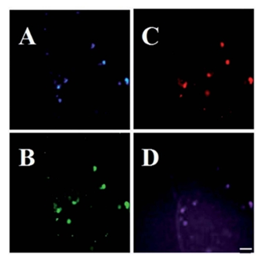
Figure 7.Exosomes isolated from the cell culture medium of MDA-MB-231cellstreated with CNP’s (2mg mL-1). Images were acquired with different excitation filter sets:(A) 350nm, (B) 490 nm, (C) 550 nm and (D) 630 nm. Scale bar: 20mm. (64)
Yang et al. were the first to use carbon dots (CDs) as contrast agents in live mice. They synthesized CDs and ZnS-doped CDs via laser ablation, then modified them with PEG1500N for in vivo optical imaging. After injection into mice, the carbon dots diffused slowly, and fluorescence faded within 24 hours. (63)
CDs were intravenously injected into mice for whole-body circulation, with fluorescence detected in the bladder, kidneys, and liver. The injected CDs were primarily excreted through urine. However, it was noted that blue and green emitting CDs are not ideal for in vivo imaging, as their short excitation wavelengths could potentially harm living cells and biological systems. (65,66)
8.1.3. Biosensing
C-dots have been employed by researchers as a bio- and chemical-sensing materials due to their unique properties like excitation-dependent emission, higher photostability, low cytotoxicity and aqueous solubility. (67) This sensing usually occurs by a change in their fluorescence properties that can take place via different mechanisms, such as resonance energy transfer, inner filter effect, and photo induced electron and charge transfer. C-dots can be used for sensing of several biological molecules and intracellular metal ions, such as hydrogen peroxide (H2O2), Fe3þ, glucose, vitamin B12, L-cysteine, and galactose, etc. Several researchers have developed hydrogen peroxide (H2O2) sensing, Wu et al. (68) Finally, Shan et al. used Boron-doped C-dots for selective detection of H2O2, and the detection limit was 8.0 mm. These C-dots could also be used for detection of glucose in the presence of glucose oxidase which produces H2O2 via oxidation of glucose. (69)
8.1.4. Detection of Metal Ion
C-dots have been employed for the detection of the highly toxic heavy metal ion mercury (Hg2-) on several occasions. Zhou et al. demonstrated the use of unmodified C-dots for the detection of Hg2- and biothiols (glutathione, cysteine, and homocysteine) with higher selectivity and sensitivity. They observed that the addition of Hg2- to C-dots caused fluorescence quenching. However, subsequent addition of biothiols to the Hg2-/C-dots recovered the fluorescence via the removal of Hg2- ions, which has a high affinity towards the thiol (-SH) groups. The time-evolution UV-vis ab sorption and photoluminescence (PL) spectra were recorded to investigate the formation of C-dots. (70)
8.2. Biomedical Applications (Therapeutic):
8.2.1. Gene transfer.
Gene therapy is a promising alternative for the treatment of many pathological disorders. This experimental technique requires an efficient and safe carrier for the delivery of biocompatible therapeutic loads with fluorescence properties. (71) In this context, the properties of CDs make them efficient emerging carriers for cellular uptake monitoring and gene delivery. Gene therapy includes the use of rDNA, mRNA, and noncoding RNAs, which can be internalized by endocytosis (72). Yu et al. designed a C-dot based multifunctional delivery system that can deliver both drugs and genes. In the design of their vector, they synthesized amphiphilic C-dots by attaching alkyl epoxide to the amino groups on the surface of PEI derived C-dots. They then used green fluorescence protein (GFP) expression and flowcytometry data to confirm that the prepared C dots had higher transfection efficacy than a conventional vector (Lipofectamine-2000) in cancerous A549 cells. (73)
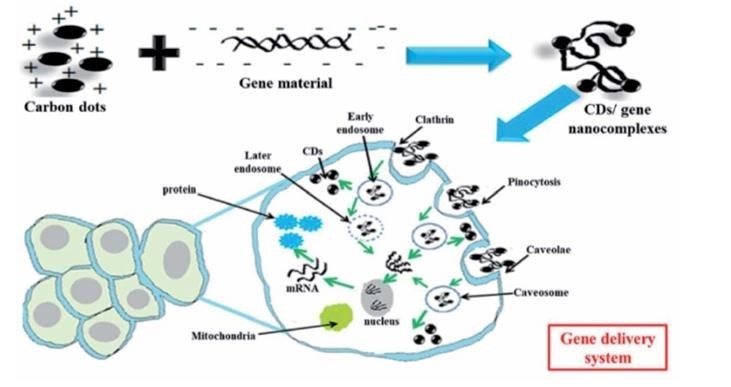
Figure 8. General mechanisms of gene delivery via CDs: here, CDs bind gene materials via electrostatic interactions, enter the cells via endo cytosis and release the payloads into the nucleus. (74)
8.2.3. Biomedicine
CDs show promise in biomedical applications, in part due to the myriads of functional groups that decorate their surface, which allows for active targeting. For instance, Wang et al. used bovine serum albumin to develop a 6.8 nm hollow CD (HCD) loaded with doxorubicin. After 90 minutes of incubation with the doxorubicin–HCD complex, red fluorescence stemming from the drug was observed in the nucleus of A549 cells. The authors proposed a mechanism whereby the doxorubicin–HCD complex enters the cell through endocytosis and upon entering the lower pH lysosome, the complex releases the doxorubicin which then enters the nucleus. (75) CDs have also been used in antimicrobial applications. For instance, CDs synthesized from henna leaves were found to be much more effective antimicrobial agents than the bulk henna leaves against both Gram-positive Staphylococcus aureus and Gram-negative Escherichia coli. (76)
8.2.4. Photodynamic therapy (PDT) and photothermal therapy (PTT)
PTT and PDT are a new class of therapeutic strategy which involves laser light for treatment of disease. In case of PTT, the photo absorber should have a strong absorbance in the NIR region to convert the absorbed energy into heat. The generated local heat from photons leads to the thermal ablation of the target cells and subsequent cell death. Compared with other traditional therapeutic approaches for cancer treatment, like aggressive surgery, chemotherapy or radiotherapy, PTT offers distinctive advantages; such as being non-invasive, highly specific and having precise temporal selectivity. (77)
In recent years, many research groups have been investigating the therapeutic potential of photothermal therapy (PTT) for cancer treatment. PTT can directly target and destroy cancer cells at the primary tumour site or local metastasis in nearby lymph nodes, helping to combat the early stages of cancer spread. The effectiveness of PTT largely depends on the ability of photothermal agents, particularly nanomaterials, to convert light into heat. The unique structure of carbon dots (C-dots), with their high number of pi electrons and strong electron-electron interactions, enhances their ability to convert absorbed light into heat through non-radiative pathways. In addition to PTT, C-dot-based photodynamic therapy (PDT) has also gained attention. PDT involves a photosensitizer, a light source, and reactive oxygen species (ROS) that are generated when the photosensitizer is excited by light. These ROS, including singlet oxygen (?1;O?), superoxide (O??), and hydroxyl radicals (OH), are highly cytotoxic and can damage cancer cells. (78)
C-dots derived from chitosan and diketopyrrolopyrrole are capable of generating singlet oxygen species (?1;O?) upon single laser irradiation. These C-dots demonstrated good hydrophilicity and biocompatibility in both in vitro and in vivo tests. The study showed that the C-dots were non-toxic to HepG2 cells at concentrations up to 200 µg/mL without laser exposure, but with laser irradiation at 540 nm, a concentration of 100 µg/mL was sufficient to kill 50% of the cells. In another example, Hua et al. synthesized C-dots through a one-step hydrothermal process involving chitosan, ethylenediamine, and mercaptosuccinic acid. These C-dots were conjugated with the photosensitizer rose bengal (RB), resulting in efficient cellular uptake and targeted accumulation in mitochondria, enabling mitochondria-targeted photodynamic therapy. (79)
9.C0NCLUSION AND FUTURE PROSPECTS:
Carbon dots (C-dots or CDs) have garnered significant attention across multiple fields due to their exceptional photoluminescence, good water dispersibility, biocompatibility, and low toxicity. These properties make them ideal for applications such as cellular imaging, biosensing, catalysis, electronics, drug delivery, and advanced biomedical uses. In cancer theranostics, CDs stand out as promising tools, leveraging their small size and surface functionalization to penetrate biological barriers and target tumours effectively. They enhance traditional therapies like photodynamic therapy (PDT) and photothermal therapy (PTT) by converting absorbed light into reactive oxygen species or heat, enabling precise cancer cell destruction with minimal side effects. Additionally, CDs serve as efficient nanocarriers for drug and gene delivery, improving treatment specificity and reducing off-target effects. Their role in advanced imaging techniques, such as fluorescence and photoacoustic imaging, facilitates early cancer detection and real-time therapeutic monitoring. Future prospects for CDs include optimizing their synthesis for enhanced biocompatibility, exploring their potential in targeting metastatic cancers, and integrating them into multifunctional platforms that combine diagnostics and treatment. These advancements aim to overcome current limitations, paving the way for innovative, non-invasive approaches to cancer management and expanding their applications in tackling other conditions like infections, inflammation, neurodegenerative disorders, and cardiovascular diseases. With continued research and development, CDs have the potential to revolutionize the field of nano-theranostics and modern medicine.
REFERENCES


Chaitanya Bopate*, Minal Paneri, Aakanksha Panajwar, Dr. Nilesh Chachda, Carbon Dots: A Review on Nowel Type of Carbon-Based Nanomaterial Used Treat Cancer, Int. J. of Pharm. Sci., 2024, Vol 2, Issue 12, 1001-1019. https://doi.org/10.5281/zenodo.14330230
 10.5281/zenodo.14330230
