Bharat School of Pharmacy, Mangalpally, Ranga Reddy, Telangana, India, 501510.
Calvarial abnormalities compromise neurological protection and cranial integrity. These cranial abnormalities are defined by the partial or whole absence of cranial vault bones. These abnormalities provide serious functional and cosmetic issues. Some small errors remain asymptomatic, larger problems needed to be surgically treated to restore the skull form. These abnormalities can be aroused from variety of infections, neoplasms, trauma, congenital abnormalities, and surgical resections. CT and MRI are the mostly employed diagnostic techniques for appropriate treatment. The Surgical method of restoration of calvarial abnormalities focuses to restore both function, structure, and to enhance cerebrospinal fluid dynamics.
Calvarial abnormalities are comprising of both appearance and neurological protection. This abnormality includes partial or complete lack of cranial vault bones. Smaller errors will not show any type of symptoms and larger abnormalities are to be corrected only by surgery. 3D printing and biomaterials are the recent advancements which improved calvarial repair outcomes (1,2,3).
Definition:
Calvarial defect is the malformation or bone loss in the calvarium, the top part of skull. As skull shields the brain and other vital organs of head, these abnormalities show a maximum impact on the patients’ health (4).
Etiology:
Some of the causes of calvarial defects include infections, neoplasms, congenital malformations, trauma, and surgical removal of tumors or other diseases (5).
Diagnosis:
In order to diagnose cavarial defects, doctors often use imaging techniques including computed tomography (CT), magnetic resonance imaging (MRI), and x-rays.
Management Of Calvarial Defects:
Cranial deformities should not be treated conservatively; instead, surgical intervention is necessary.By restoring a barrier between the cranium and the outside world, calvarial defect reconstruction attempts to improve CSF dynamics and lessen headaches, exhaustion, lightheadedness, and psychological discomfort (5). Neurosurgeons frequently perform cranioplasty to fix calvarial and scalp abnormalities. Primary wound healing, dead space obliteration, sterile region closure, normal barrier restoration, producing a long-lasting or permanent reconstruction utilizing biologically inert materials, and cosmetic restoration are the key goals (6). Autologous and synthetic materials are among the materials used in cranioplasty. Strong, pliable, light, and able to blend in with the skull are the best qualities. It should be long-lasting, infection-resistant, and enable imaging assessment without artifacts (5).
Autologous Reconstruction:
The gold standard for fixing skull abnormalities is autologous reconstruction, which uses spare bone from the split-thickness calvarium, ribs, iliac crest, pelvis, and tibia. Benefits include reduced infection risk, osteogenesis, and safe usage in young patients. On the other hand, drawbacks include possible bone resorption, donor-site morbidity, and challenges in molding donor bone. Autologous ribs are appropriate for juvenile patients, although they can induce instability in the chest wall and have size restrictions. In adulthood, the iliac crest is more effective in replicating the calvarium curvature (5).
Alloplastic Reconstruction:
Because of its accuracy, durability, and donor-site morbidity, alloplastic implants are the gold standard for calvarial abnormalities, replacing autologous bone. PMMA, hydroxylapatite, PEEK, and titanium mesh are examples of common synthetics (5).
Reconstruction Materials:
Although it is technically difficult, it is possible to rebuild a skull defect using artificial or organic tissue. The perfect cranioplasty replacement needs to be biocompatible, robust, lightweight, pliable, nonmagnetic, radiolucent, nonferromagnetic, easily accessible, affordable, and safe. Because of its strength, durability over time, and biocompatibility, natural bone is the recommended option. Although they are accessible, alloplastic implants are prone to exposure and infection. Autologous bone can be used to handle partial or no graft loss and has a decreased rate of graft loss (6).
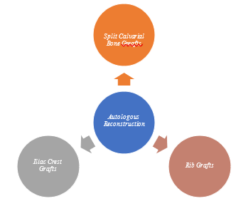
Figure 1. Autologous Reconstruction Materials
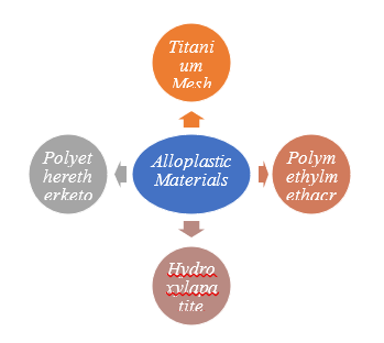
Figure 2. Alloplastic Reconstruction Materials
ADVANTAGES:
Reconstructions of scalp defects are accomplished with calvarial bone transplants; however problems are few. Research indicates that upon repair, there is no resorption or loss of calvarial transplants. Autogenous calvarium is superior to the majority of alloplastic materials in terms of quality and properties. Because of its mechanical qualities, anatomicofunctional fusion, and histocompatibility, fresh autologous bone can be used to restore cranial defects. Excellent outcomes are obtained when nearby bone merges with live tissue. Additionally, autologous bone transplants exhibit bone repair processes and have a low rate of infection (6).
DISADVANTAGES:
When repairing cranial defects, choosing the appropriate material is essential. Although synthetic bone and dura substitutes are frequently employed, they are not as strong as biological materials and can result in infection and inflammation. Limitations of calvarial transplants include their small size and potential for inner table violations. It takes skill to split the bone, and it might result in problems such dural rips, subarachnoid hemorrhage, intracerebral hematoma, and CSF leaks (6).
Case Discussion:
A 64 years old female patient was admitted in the neurosurgery ward with chief complaints of post op left frontal ICH left FTP DC evacuation on 7/1/2025. Patient had lipto spirosis antibiotic treatment received at rehabilitation. Patient don’t have any fresh complaints. Patient is tracheostomised. No h/o fever, seizures, headache. Admitted for bone flap replacement. Her past history was FTP decompression-on Jan 2015. On examination the patient has spontaneous eye opening+ Tracheostomy+ insitu, E4VTM3-4, pupils B/L ERTL, sunken flap+ in abdomen + weakness on all over limbs+. Patient was presented with above mentioned complaints evaluated and submitted. Patient underwent surgery on 26/4/2025. The laboratory investigations of Hb were found to be 8.3gm% (N.R=12 -15.0) Findings; After removing subgalel flap little tense, so ventricle tap done – 40to 50ml csf drain implant- stryker 2 hole (2) 4 hole (1). Patient improved clinically. Vitals of this patient are stable with E4VTM3-4 daily physio was done patient improved and discharged stably.
Drug Interactions-
Drug - Drug Interactions.
1)Cefpodoxime - Pantoprazole
You shouldn't use cefpodoxime and pantoprazole at the same time. The anti-infective efficacy of cefpodoxime is diminished when pantoprazole lowers stomach acid levels and thus cefpodoxime absorption and blood levels. Cefpodoxime may interact negatively with pantoprazole, therefore your doctor may recommend switching antibiotics or advising you to stop taking pantoprazole temporarily.
2)Metaxalone - Levetiracetam
Levetiracetam and metaxalone combined may amplify the symptoms of vertigo, lethargy, disorientation, and inability to focus. Cognitive, judicial, and motor impairments are also possible, particularly in the elderly. While using these drugs, you should not drink alcohol or drink very little. Until you know how the prescriptions impact you, stay away from driving or operating dangerous equipment, both of which need mental awareness. In case you have any inquiries or worries, you should speak with the doctor.
Drug - Food Interactions.
Cefpodoxime - Food
1) Consuming food might increase the body's absorption of cefpodoxime pills. To maximize the body's absorption of cefpodoxime, take the pills with meals. Taking the cefpodoxime suspension with or without meals is not a problem. Cefpodoxime is best taken daily at the same time(s).
2)Metaxalone - Food
Alcohol may amplify the effects of metaxalone on the neurological system, making it more difficult to focus, sleepy, and dizzy. Mental fogginess or impaired decision-making may also affect certain individuals. While using metaxalone, it is best to abstain from alcohol or drink in moderation. Be cautious not to drive or operate heavy equipment until you have a better idea of how metaxalone affects you, and do not take more than the prescribed dosage. Feel free to see your physician or pharmacist with any inquiries or concerns you may have.
3)Levetiracetam - Food
The effects of levetiracetam on the neurological system, including lightheadedness, lethargy, and trouble focusing, may be exacerbated by alcohol. Mental fogginess or impaired decision-making may also affect certain individuals. While using levetiracetam, you should cut down or abstain from alcohol altogether. Do not drive or operate heavy equipment until you have determined how levetiracetam affects you; also, do not exceed the dosage suggested for this drug. Feel free to see your physician or pharmacist with any inquiries or concerns you may have.
CONCLUSION:
Calvarial anomalies necessitate precise diagnosis and surgical treatment since they can jeopardize both cerebral protection and attractiveness. Although synthetic materials and 3D-printed implants are good substitutes, autologous bone transplants are still the recommended method. The case study demonstrates a steady recovery following a successful bone flap replacement. Patient outcomes are continuously improved by ongoing advancements in reconstructive procedures.
REFERENCES


Dornala Manoj*, Mattam Pavani, Safa Hussain, Calvarial Defect- A Case Report, Int. J. of Pharm. Sci., 2025, Vol 3, Issue 8, 14-19. https://doi.org/10.5281/zenodo.16671138
 10.5281/zenodo.16671138
