Kasturi Shikshan Sanstha Collage of Pharmacy, Shikrapur, Pune, India 412208
Nanotube-based technologies have emerged as powerful tools in tissue engineering and regenerative medicine due to their unique structural, mechanical, and biological properties. Their high surface-area-to-volume ratio, tunable chemistry, and ability to mimic native extracellular matrix features enable enhanced cell adhesion, proliferation, and differentiation across a range of tissue types. Carbon nanotubes, titanium dioxide nanotubes, and peptide-based nanotubes are among the most widely studied, each offering distinct advantages for scaffold reinforcement, drug delivery, and bioactive signaling. Recent research highlights their potential to improve electrical conductivity in cardiac and neural constructs, guide stem-cell fate through nanoscale cues, and deliver therapeutic molecules with controlled release kinetics. Despite these promising outcomes, challenges remain related to long-term biocompatibility, degradation behavior, and potential cytotoxicity, particularly at higher concentrations or with improper functionalization. This review summarizes current advancements, applications, and safety considerations of nanotube-integrated systems, emphasizing their transformative potential while outlining the critical gaps that must be addressed for successful clinical translation.
Tissue engineering and regenerative medicine represent innovative approaches aimed at restoring, repairing, or replacing damaged tissues and organs. the development of effective therapies in these fields relies heavily on the design and application of advanced biomaterials that can mimic the natural extracellular matrix provide structural support, and promote cellular functions such as adhesion, proliferation, and differentiation. among emerging nanomaterials, nanotubes including carbon nanotubes boron nitride nanotubes and other tubular nanostructures have attracted significant attention due to their unique mechanical, chemical, and electrical properties.
nanotubes exhibit high surface area, exceptional mechanical strength, and tunable surface chemistry, making them ideal candidates for creating scaffolds that support tissue regeneration. their tubular structure enables the incorporation and controlled release of bioactive molecules, such as growth factors, genes, and therapeutic drugs, which can enhance cellular responses and accelerate tissue repair. additionally, surface functionalization of nanotubes can improve their compatibility with biological systems, reduce cytotoxicity, and tailor them for specific regenerative applications.
these nanostructures have been explored in a wide range of tissue engineering applications, including bone, cartilage, neural, and cardiovascular tissue regeneration. for instance, incorporating nanotubes into polymeric or ceramic scaffolds can enhance mechanical stability while simultaneously promoting cellular growth and differentiation. despite these promising properties, challenges remain, including potential toxicity, immune responses, and concerns about long-term biodegradability and clinical translation.
this review aims to provide a comprehensive analysis of the role of nanotubes in tissue engineering and regenerative medicine. it discusses their structural and functional properties, applications in various tissue types, strategies for improving biocompatibility, and potential challenges. by summarizing recent advances and future prospects, this review seeks to guide researchers in harnessing the full potential of nanotubes for the development of next-generation regenerative therapies.
TYPES OF NANOTUBES IN TISSUE ENGINEERING AND REGENERATIVE MEDICINE :
1. Carbon Nanotubes (CNTs)
Strong, conductive, and high surface area; used to reinforce scaffolds and support neural or cardiac tissue growth. Carbon nanotubes (CNTs) are strong, conductive, carbon-based nanostructures that enhance the mechanical and electrical properties of scaffolds used in tissue engineering. Their unique surface features support better cell attachment, alignment, and communication, which is especially important for regenerating bone, cartilage, nerve, and muscle tissues. CNTs can also be functionalized to deliver therapeutic molecules directly to the repair site, although their long-term safety depends on proper purification and biocompatible modification.[5]
Example: Single-walled (SWCNTs), Multi-walled (MWCNTs).
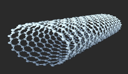
Fig1: Carbon Nanotube
2. Titanium Dioxide Nanotubes (TiO?-NTs)
Biocompatible and osteoconductive; commonly applied in bone and dental implants. Titanium dioxide nanotubes are biocompatible nanostructures that enhance cell adhesion, proliferation, and osteoblast activity, making them ideal for bone tissue engineering and implant surface modification. Their porous tubular architecture also allows for controlled delivery of therapeutic agents, such as growth factors or antibiotics, supporting tissue regeneration and improving implant integration. [11]
Example: Anodized TiO? nanotubes on titanium implants.
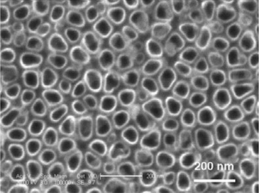
Fig2: Titanium Dioxide
3. Halloysite Nanotube
Hollow, biocompatible, and capable of carrying drugs; used in contolled release and wound healing. Hallosite nanotube enhance scaffolds in tissue engineering by improving mechanical strength, biocompatibility, and controlled drug delivery, while also promoting cell adhesion, proliferation, and tissue regeneration. Their hollow structure allows sustained release of growth factors or drugs, making them ideal for bone, soft tissue applications. [11]
Example: Naturally occurring aluminosilicate clay nanotubes.
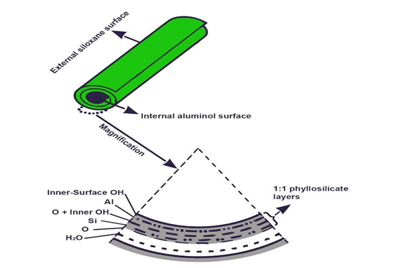
Fig3: Halloysite Nanotube
4. Peptide/Protein-Based Nanotubes
Biodegradable and bioactive; mimic extracellular matrix to guide cell adhesion and differentiation. Protein-based nanotubes improve tissue engineering scaffolds by enhancing biocompatibility, supporting cell adhesion, and enabling controlled delivery of bioactive molecules. Their tunable structure and natural bioactivity make them suitable for promoting tissue regeneration, wound healing, and targeted therapeutic release. [7]
Example: Self-assembling RADA16 peptide nanotubes.
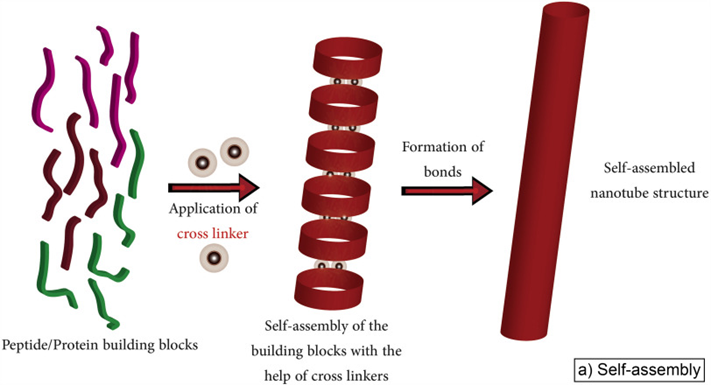
Fig4: Protein Based Nanotube
5. Boron Nitride Nanotubes (BNNTs)
Mechanically strong and thermally stable; useful in bone/cartilage scaffolds and neural tissue engineering. Boron nitride nanotubes (BNNTs) enhance tissue engineering scaffolds by providing excellent mechanical strength, chemical stability, and biocompatibility, while supporting cell growth and differentiation. Their high surface area and ability to carry bioactive molecules make them promising for drug delivery, bone regeneration, and advanced regenerative medicine applications.[13]
Example: Hexagonal boron nitride nanotubes.
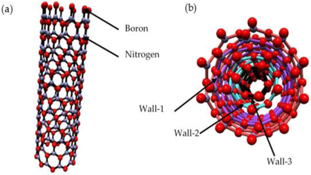
Fig5: Boron Nitride Nanotube
6. Hybrid/Functionalized Nanotubes
Enhanced biocompatibility and tailored functionality; applied in smart scaffolds and multifunctional tissue engineering. Hybrid nanotubes improve tissue engineering scaffolds by combining the advantages of different nanomaterials, such as enhanced mechanical strength, biocompatibility, and bioactive molecule delivery. Their multifunctional properties support cell adhesion, proliferation, and tissue regeneration, making them ideal for advanced regenerative medicine applications. [9]
Example: CNT-polymer composites, graphene-CNT hybrids.
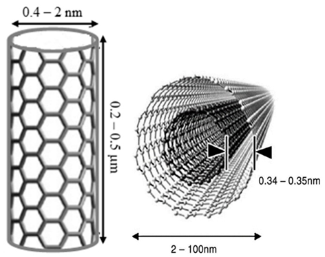
Fig6: Hybrid Nanotube
PHYSICOCHEMICAL PROPERTIES RELEVANT :
In tissue engineering and regenerative medicine, the physicochemical characteristics of biomaterials are critical for their functionality and interaction with biological systems. These properties influence cell behaviour, scaffold performance, and ultimately the success of tissue regeneration.
1. Surface Chemistry
The chemical composition and functional groups on a biomaterial’s surface dictate protein adsorption, cell adhesion, and signaling pathways. Surface modification can enhance biocompatibility or provide sites for targeted cell attachment [17]
2. Hydrophilicity and Hydrophobicity
The balance between hydrophilic and hydrophobic domains affects cell attachment, proliferation, and nutrient diffusion. Hydrophilic surfaces often favor cell adhesion and spreading, while excessive hydrophobicity can hinder biological interactions.
3. Topography and Roughness
Micro and nanoscale surface features influence cell morphology, migration, and differentiation. For example, nanoscale roughness can mimic the natural extracellular matrix (ECM) and guide stem cell fate. [8]
4. Mechanical Properties
Biomaterial stiffness, elasticity, and tensile strength must match the native tissue’s mechanical environment. Mechanical mismatch can lead to implant failure or poor tissue integration. Dynamic properties such as viscoelasticity also impact cellular responses.
5. Degradation Rate and Stability
Biodegradable scaffolds must maintain structural integrity long enough to support tissue regeneration while degrading into non-toxic byproducts. The degradation kinetics should be tailored to the specific tissue type. [18]
6. Porosity and Pore Interconnectivity
Adequate porosity facilitates nutrient transport, waste removal, and vascularization. Interconnected pores are essential for cell infiltration and tissue ingrowth.
7. Swelling and Water Uptake
Hydrogels and other soft biomaterials absorb water, which affects nutrient diffusion, mechanical behavior, and scaffold volume. Controlled swelling can improve tissue integration. [8]
8. Electrical and Thermal Properties
Conductive biomaterials may enhance nerve or cardiac tissue engineering, while thermal stability is relevant for sterilization and processing.
FABRICATION METHODS AND SCAFFOLD INTEGRATION OF NANOTUBES :
Nanotubes can be combined with scaffolds to improve their strength, bioactivity, and support for cell growth. Several methods are commonly used:
1. Electrospinning:
Nanotubes are mixed with a polymer solution and stretched into thin fibers using an electric field. The resulting scaffold mimics natural tissue structure and helps cells grow, especially in nerve and heart tissues.
2. 3D Bioprinting:
Nanotube-containing bio inks are printed layer by layer to make scaffolds with precise shapes. This method allows control over the scaffold structure and improves both strength and conductivity.
3. Freeze-Drying and Pyrogen Leaching:
Scaffolds are made porous by freezing and drying a mixture of polymer and nanotubes or by removing temporary particles. Porous scaffolds allow nutrients and cells to move through easily.
4. Sol–Gel and Hydrothermal Methods:
Inorganic nanotubes like titanium dioxide or halloysite can be added chemically to scaffolds or grown directly on surfaces. These methods improve bone attachment and can carry drugs or growth factors. [17]
5. Surface Modification:
Nanotubes can be coated or chemically altered to mix evenly with scaffolds, reduce toxicity, and help cells attach and develop properly.
6. Composite Scaffolds:
Nanotubes are often combined with biodegradable polymers or gels to create hybrid scaffolds that are strong, safe for the body, and encourage tissue regeneration.
APPLICATIONS [13]:
1. Bone Tissue Engineering
Nanotubes, particularly carbon nanotubes (CNTs), are used to create scaffolds for bone regeneration. Their high mechanical strength and flexibility make them ideal for supporting osteoblasts (bone-forming cells). CNT-based scaffolds help mimic the extracellular matrix (ECM), enhancing cell adhesion and promoting osteogenesis. Surface functionalization with osteo inductive molecules further stimulates bone growth and mineralization.
2. Nerve Regeneration
CNTs are employed in nerve tissue engineering to promote the regeneration of damaged neurons. Their electrical conductivity mimics the natural electrical properties of nerve tissue, helping to guide nerve growth and regeneration after injury. CNT-based scaffolds facilitate the alignment of neurons and enhance nerve fiber regeneration by conducting electrical signals, which is crucial for restoring neural function in cases like spinal cord injury or peripheral nerve damage.
3. Cardiovascular Tissue Engineering
In cardiovascular applications, CNTs are integrated into scaffolds for vascular and cardiac tissue regeneration. Their electrical conductivity supports the synchronized contraction of cardiomyocytes (heart muscle cells), essential for cardiac tissue repair. CNT-based vascular grafts and stents provide structural support for blood vessels, while their flexibility and mechanical properties allow them to function effectively in blood vessel repair and heart tissue engineering.
4. Cartilage Regeneration
CNTs are used to create scaffolds for cartilage tissue engineering, promoting the growth and differentiation of chondrocytes (cartilage cells). Their mechanical properties mimic the compressive and elastic nature of cartilage, which is essential for joint function. CNTs help enhance chondrocyte proliferation and matrix production, and their electrical conductivity may also support stem cell differentiation into cartilage-producing cells. These scaffolds are useful in treating cartilage defects or diseases like osteoarthritis.
5. Wound Healing and Skin Regeneration
CNTs are explored in wound healing and skin regeneration, particularly for chronic wounds or burns. CNT-based scaffolds help promote cell migration and proliferation, facilitating skin regeneration at the wound site. These scaffolds can also be functionalized with antimicrobial agents to reduce infection risks. Additionally, CNTs can serve as carriers for growth factors like epidermal growth factor (EGF), which accelerates skin cell proliferation and wound closure.
6. Drug Delivery and Therapeutic Systems
CNTs are utilized in drug delivery systems for controlled and targeted release of therapeutic agents. Their large surface area allows for encapsulation of a variety of drugs, growth factors, or genetic materials. CNTs can deliver these agents directly to the site of injury or disease, ensuring more efficient and localized treatment. This application is valuable in regenerative medicine, where targeted delivery of growth factors or stem cells can significantly enhance tissue healing and regeneration
7. Muscle Tissue Engineering
CNTs are used in muscle tissue engineering to support the growth and differentiation of muscle cells (myoblasts). Their mechanical properties help mimic muscle tissue, while their electrical conductivity aids in synchronized contraction. CNT-based scaffolds promote muscle fiber formation, making them useful for repairing skeletal and cardiac muscle, including conditions like muscular dystrophy and heart injury.
8. Pancreatic Tissue Regeneration
In pancreatic tissue regeneration, CNTs are explored to support the regeneration of insulin-producing beta cells in the pancreas. CNT-based scaffolds promote cell proliferation and differentiation, aiding the restoration of functional pancreatic tissue. This approach holds promise for developing treatments for type 1 diabetes by potentially restoring insulin production.
ADVANTAGE [21] :
1. High Surface Area Promotes Cell Adhesion and Growth
Nanotubes feature an extremely high surface-to-volume ratio, which allows for efficient adsorption of proteins, signaling molecules, and growth factors. This nanostructured surface mimics the natural extracellular matrix, creating an environment that encourages cells to attach, spread, and proliferate. By providing nanoscale topographical cues, nanotubes can also guide stem cell differentiation, supporting regeneration of tissues such as bone, cartilage, and neural structures.
2. Customizable Physical and Chemical Properties
The size, shape, surface chemistry, and functionalization of nanotubes can be precisely tailored to meet specific tissue engineering requirements. Functional groups or bioactive molecules can be attached to the nanotube surface to enhance cellular interactions or stimulate particular biological pathways. This adaptability allows scaffolds to be optimized for mechanical strength, degradation rate, and cellular response, making them highly versatile for diverse regenerative application
3. Electrical Conductivity for Nerve and Muscle Regeneration
The electrical conductivity of carbon nanotubes (CNTs) makes them highly effective for promoting nerve regeneration by conducting electrical signals, which can stimulate the growth and differentiation of neurons. Similarly, they aid in muscle tissue regeneration by supporting electrical stimulation necessary for muscle function.
4. Biocompatibility and Cell Interaction
Functionalized nanotubes show excellent biocompatibility, reducing immune responses when used in vivo. They can be modified to enhance cell adhesion, promote proliferation, and even guide stem cell differentiation, making them ideal for regenerative applications.
5. 3D Scaffolds for Tissue Growth
Nanotubes can be used to create highly porous, 3D scaffolds that mimic the extracellular matrix (ECM), providing a suitable environment for cell growth and tissue formation. These scaffolds can be tailored to suit the specific needs of different tissues, from bone to cartilage.
6. Controlled Drug and Growth Factor Delivery
Nanotubes can encapsulate and release bioactive molecules, such as growth factors or drugs, in a controlled manner. This ability enables precise, localized treatment for tissue regeneration and accelerates healing by promoting cell proliferation and differentiation at the injury site.
7. Antimicrobial Properties
The antimicrobial properties of nanotubes help reduce the risk of infection in tissue-engineered implants. Surface functionalization can further enhance their antibacterial effects, ensuring that implanted scaffolds remain sterile and effective in promoting tissue repair.
LIMITATION [15] :
1. Cytotoxicity and Biocompatibility Concerns
Although many nanotubes are designed to be biocompatible, certain types—especially carbon nanotubes—can cause cytotoxic effects depending on their size, length, concentration, and surface properties. Unmodified nanotubes may induce oxidative stress, inflammation, or DNA damage in cells, which poses challenges for their safe use in clinical applications
2. Poor Biodegradability
Most nanotubes are highly stable and resistant to natural degradation in physiological environments. This can result in long-term accumulation within tissues or organs, potentially causing adverse effects. Developing nanotubes that are both mechanically strong and biodegradable remains a significant challenge in regenerative medicine. Limitations
3. Difficulty in Achieving Uniform Dispersion
Nanotubes have a strong tendency to aggregate due to van der Waals forces, making it difficult to distribute them evenly in scaffolds or composite biomaterials. Poor dispersion can compromise mechanical properties, reduce bioactivity, and limit cell-scaffold interactions, reducing overall regenerative efficiency.
4. Complex Functionalization and Manufacturing
Functionalizing nanotubes with bioactive molecules or surface coatings often involves complicated chemical processes. These procedures can alter the nanotubes’ intrinsic properties or reduce the effectiveness of incorporated biomolecules. Scaling up production while maintaining consistent quality and functionality is still a challenge for clinical applications.
5. Limited Long-Term In Vivo Data
Most studies on nanotube-based scaffolds are performed in vitro or in small animal models. There is limited information regarding their long-term behavior, immune response, or potential toxicity in larger animals and humans, making translation to clinical use more cautious.
SAFETY, TOXICITY AND REGULATORY CONSIDERATIONS IN (TERM) [25] :
1. Safety Considerations in TERM
Patient Factors: Age, health status, and tissue sensitivity affect safety. Older patients or those with comorbidities may face higher risks of side effects, and sensitive tissues (e.g., skin) require caution.
Treatment Parameters: Energy intensity and correct targeting are critical. Excessive energy can lead to thermal damage, while imprecise targeting risks harming surrounding tissues.
Device Safety: Devices must undergo strict quality assurance testing. Proper operator training and built-in safety features (e.g., automatic shutoffs) are essential to prevent user errors.
2. Toxicity Concerns in TERM
Cellular Toxicity: High-energy treatments can cause heat-induced cell damage, while non-thermal modalities might disrupt cellular processes, leading to inflammation or apoptosis.
Long-Term Effects: Repeated or prolonged use of TERM therapies may cause cumulative damage, scarring, or even increase the risk of cancer in some cases, especially with ionizing radiation or high-energy lasers.
Biocompatibility: Interaction with implants or biomaterials can cause localized toxicity, alter material properties, or lead to adverse tissue responses.
3. Regulatory Considerations
Regulatory Guidelines: Regulatory bodies like the FDA (U.S.) and EMA (EU) assess TERM therapies based on risk classifications, requiring rigorous preclinical and clinical data before approval.
Clinical Trials: Proper trials are essential to monitor both short-term and long-term safety and efficacy. They evaluate potential adverse events, including toxicity and side effects.
Post-Market Surveillance: After approval, continuous monitoring is necessary to detect and report adverse effects. If safety issues arise, regulatory actions such as recalls or modifications may be implemented.
FUTURE PERSPECTIVES:
Nanotubes hold significant promise for advancing tissue engineering and regenerative
medicine due to their exceptional mechanical, electrical, and surface properties. Future research will likely prioritize the development of biocompatible and biodegradable nanotube-based scaffolds that support tissue growth while minimizing cytotoxicity and long-term accumulation in the body. Functionalization techniques, including bioactive molecule conjugation and stimuli-responsive coatings, may allow scaffolds to actively guide cell behavior, promote differentiation, and control therapeutic release. [19]
Integration of nanotubes with 3D bioprinting and additive manufacturing is expected to enable precise fabrication of complex tissue constructs with controlled architecture, mechanical strength, and electrical conductivity. This is especially relevant for neural, cardiac, and muscular tissues, where aligned and conductive scaffolds can enhance cell organization, signaling, and functional integration.
The development of hybrid nanomaterials, combining nanotubes with polymers, graphene, or nanocellulose, offers opportunities to achieve multifunctional scaffolds that combine strength, bioactivity, and responsiveness. Additionally, nanotube-based scaffolds may serve as platforms for electrical stimulation and bioelectronic interfaces, allowing real-time monitoring and modulation of tissue regeneration. [20]
To realize clinical translation, research must address long-term safety, biodegradability, and standardization, along with scalable fabrication methods that maintain uniform properties. By combining material innovation with biological insight, nanotube-based scaffolds are poised to become versatile tools for repairing and regenerating a wide range of tissues, potentially transforming regenerative medicine in the coming decade.
CONCLUSION :
Nanotubes are becoming very useful in tissue engineering and regenerative medicine because of their special size and structure. They can support cells, help them grow, and guide them to form new tissues. Different types of nanotubes, like carbon and halloysite nanotubes, can improve the strength of scaffolds, help deliver drugs, and even provide signals that encourage cells to heal damaged tissues such as bone, nerve, and heart.
However, there are still some concerns, especially about their safety, long-term effects, and how the body reacts to them. More studies are needed to make sure they are completely safe and to improve their design.
In general, nanotubes have great potential to improve future medical treatments. With more research and better technology, they may become an important part of creating new, healthy tissues in the body.
ACKNOWLEDGEMENT:
The author would like to express sincere gratitude to the faculty members of the Department of Pharmacy for their continuous guidance, valuable suggestions, and academic support throughout the preparation of this review article. Their encouragement and critical insights played a significant role in shaping the structure and scientific content of this work.
The author is also thankful to the institution for providing access to academic resources, including journals, online databases, and library facilities, which were essential for the literature survey and compilation of this review. Special appreciation is extended to all researchers and authors whose published studies contributed to the understanding and development of this topic.
Finally, the author gratefully acknowledges the support and motivation received from family and peers, which helped in the successful completion of this manuscript.
REFERENCES


Rutuja Chavan, Vijaykumar Kale, Mahesh Thakare, Vaibhav Narawade, Mamta Pavale, An Overview of Nanotube in Tissue Engineering and Regenerative Medicine, Int. J. of Pharm. Sci., 2025, Vol 3, Issue 12, 4053-4064. https://doi.org/10.5281/zenodo.18096499
 10.5281/zenodo.18096499
