P.S.V College Of Pharmaceutical Science And Research,Orappam (Village), Krishnagiri (Dt), Tamilnadu – 635 108.
Caesalpinia bonducella L. (family Caesalpiniaceae), commonly known as fever nut, Bonduc nut, or Nicker nut, is an important medicinal shrub widely used in Ayurveda, Siddha, and Unani systems of medicine. All parts of the plant—roots, leaves, seeds, bark, and stem—are traditionally used for the treatment of diverse ailments. Accurate identification and quality control of plant materials are essential to ensure the safety, efficacy, and reproducibility of herbal drugs. Pharmacognostic evaluation involving macroscopic and microscopic studies, physicochemical parameters and modern analytical techniques such as TLC form the basis of standardization and UV-visible spectroscopy, as recommended by WHO. Literature reveals that C.bonducella contains important phytoconstituents including bonducin, bonducellin, phytosterols, ?-sitosterol, flavonoids, furanoditerpenes, fatty acids, amino acids, and alkaloids. The present study therefore emphasizes systematic pharmacognostical and phytochemical evaluation of C. bonducella to support its standardization and quality assessment as a medicinal plant.
Caesalpinia bonducella L, also known as fever nut, Bonduc nut and Nicker nut belongs to the family of Caesalpiniaceae. It is widely used in medicine of Ayurveda, Siddha, Unani. (C. bonducella) is an Indian herb also found in tropical and subtropical parts of Asia such as SriLanka, Vietnam, China, Myanmar and Bangladesh. Name ‘Bonducella’ of the species is derived from the Arabic word “Bonduce” which means a “little ball” that indicates the globular shape of the seed. Genus of Caesalpinia is widespread in 500 species that has medicinal benefits based on their pharmacological activity. It is a common shurb that is scadent prickly is often grown as a hedge plant. All parts of the plant – roots, leaves, seed, bark and stem are used as herbal medicine for different diseases. These investigations aid in the correct identification and authentication of plant parts. The precise identification and quality control of the plant materials is pivotal to guaranteeing the reproducible quality of herbal-based drugs, which will finally ensure their efficacy and safety. The primary steps in the pharmacognostic evaluation of medicinal plants are macroscopic and microscopic characteristics, physicochemical studies on ash values, extractive values, and modern analytical technics inpresence of heavy metals like iron, zinc, candium and TLC, UV-visible spectroscopy. The WHO also states that the macroscopical and microscopical accounts of a medicinal plant are the primary steps in determining its identity and degree of purity. Review of the literature reveals the most part of the plant has been studied in the presence of some imperative phytoconstituents such as bonducellin, phytosterinin, β- sitosterol, furanoditerpenes, flavonoids, aspartic acid, arginine, citrulline, β-carotene. The Limited study on aqueous extract of caesalpinia bonducella have evaluated Pharmacogonostic properties. Physiochemical parameters requird for quality control of the crude drug materials. Stepwise pharmacognostical investigations of the raw materials can help with the standardization process. The present study focuses on the pharmacognostical and phytochemical parameters of the plant of C. bonduc.
2. PLANT DESCRIPTION
c.bonducella, a sprawling shrub or extensive climber with dark gray branches covered in hard yellow prickles, which grows naturally in the hotter part of India and in topical countries of the world. It produces small yellow flowers in dense, spiny racemes and has oblong, spiny pods containing 2 to 3 smooth, hard, greenish or gray seeds that resemble an eyeball.
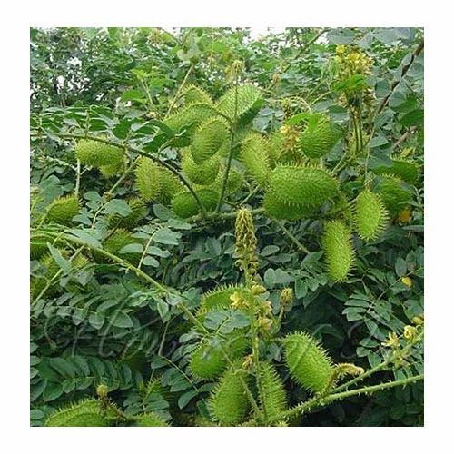
Figure: 1 caesalpinia bonducella
2.1 Taxonomical classification of c.bonducella
2.2 Vernacular Name
2.3 LEAVES:
Each leaves has about 7 pairs of smaller pinnae with 3-8 leaflets on each one. Leaves are 30-45 cm rachis with recurved prickles, pinnae 6-9 pairs, opposite stipules deciduous, large, leaf like, usually lobed, lobes to 2 cm, leaflets 6-12 pairs, oblong 1.5-4 × 1.2-2 cm, membranous both surfaces pubescent, base oblique, apex rounded to acute, mucronate taste - Bitter & astringent properties and they are “Characteristic” of which dark green on the top surface and a lighter more or less pubscent (hairy) gray – green on the underside.

Figure: 2 Leaves
2.4 FLOWER:
The c.bonducella flower is a yellow with five petals and grows in dense racemes at end of its branches in lower part. Bracts: caducous at anthesis, reflexed, subulate, 6-8 mm, pubescent. Pedicel 3-5 mm, Sepals 5-8 mm, both sides ferruginous hairy and these petels are yellowish standard tinged with red spots, oblanceolate, clawed Stamens Filaments are short, hairy in basal part with bitter taste. They are Unpleasant and nauseating odour and yellow or pale yellow in colour.
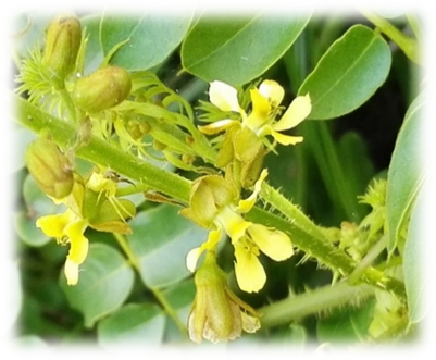
Figure 3 Flower
2.5 Seed:
They are typically grey, hard and shiny & have a varity of purpose of medicinal properties. They are globular in shape with typically 1.5 to 2cm long, taste - very bitter, pungent & astringent properties. Glossy seed dicot odour with 2 or 3, greyish, shiny, ovoid to globule. These seeds were cleaned to remove any adhered foreign material, then washed several times with tap water, air-dried, further homogenized to powder.
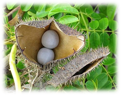
Figure 4: seed
4. MATERIALS AND METHODS:
4.1 Soxhlet Extraction (Successive Solvent Extraction)
The Soxhlet extraction method is used for continuous extraction of a component (usually a solid–liquid extraction) when the desired compound has limited solubility in a solvent and the impurities are insoluble.
Procedure
Dry and powder plant material (50–200 g) and Load into a Soxhlet extractor. The material is extracted successively with solvents of increasing polarity such as Petroleum ether (non-polar), Chloroform, Ethyl acetate, Methanol/Ethanol (polar), Water (highly polar). Each extraction runs for 6–8 hours until siphoning becomes colourless. Concentrate extracts with a rotary evaporator and store at 4°C.
Purpose
Separates phytochemicals based on polarity and gives high-quality phytochemical-rich extracts.
4.2 Cold Maceration (Methanolic or Aqueous)
Cold maceration extraction is used for extracting heat-sensitive (thermolabile) compounds from plant or animal materials without applying heat.
Procedure
Mix 50 g powdered plant material with 500 ml methanol/water and Shake occasionally and keep at room temperature for 48–72 hours. Filter through muslin cloth + Whatman No. 1 filter paper. Evaporate solvent on a water bath to obtain the extract.
Purpose
Suitable for heat-sensitive compounds
Good for flavonoids, phenolics, alkaloids
4.3 Hot Continuous Percolation (Soxhlet Alternative)
Hot continuous percolation extraction, also known as the Soxhlet-type percolation or Hot Percolation (Modified Soxhlet) Method, is used for exhaustive extraction of constituents from plant materials using hot solvent in a continuous cycle.
Procedure
Place powdered plant material in a percolator and pass hot solvent (ethanol/methanol) continuously over the material then collect extract until it becomes clear. Concentrate under reduced pressure.
Purpose
Faster than maceration
Higher extraction efficiency than cold methods
4.4 Aqueous Decoction (Traditional Method)
Aqueous decoction extraction is used for extracting water-soluble and heat-stable constituents from hard or coarse plant materials by boiling them in water.
Procedure
Boil crushed seeds/roots/leaves in water (1:10 w/v) for 30–60 min. Allow the mixture to cool and then filter it. Concentrate under mild heat.
Purpose
Used in Ayurvedic preparations of C.bonducella.
Extracts tannins, saponins, polysaccharides.
4.5 Microwave-Assisted Extraction (MAE)
Microwave-assisted extraction (MAE) is used for rapid and efficient extraction of bioactive compounds from plant or other solid materials using microwave energy to heat the solvent and sample.
Procedure
Mix plant powder with the solvent and heat in microwave at controlled power (300–600 W) for 5–10 minutes. Then Cool and filter the mixture.
Purpose
Efficient extraction of phenolics, flavonoids
Faster and uses less solvent
5. PHARMACOGNOSTIC EVALUATION 5.1 Macroscopic studies:
Morphological studies were done using simple microscope. The shape, apex, base, margin, taste and odour. They are somewhat flattened on one side due to the close pressing of adjacent seeds. Hilum and micropyle are seen close together. About 40nm in diameter, a dark circle surrounds the hilum mostly with a pale remnant to the funicular and the microply at the periphery of dark area. The weights of a single seed was approximately 2gm.
Figure: 6 morphology of c. bonduc seed
Seeds globous or rounded, smooth, shiny, 1.2-2.5 cm in diameter which is Slightly flattened on one side due to close pressing of adjacent seed and hilum and micropyle close together hilum surrounded by a dark area around 4 mm in diameter, usually with a whitish remnat to funiculus Micropyle near the periphery of dark area, Seeds coat greenish - gray to bluish gray, lineate, shiny100 seeds weigh from 150 to 250 g. The stem bark pieces was observed as they were thin, double or compound quilled, 1-2 mm thick, scaly, externally blackish brown with intermittent creamish brown patches and inner surface was smooth, pale brown. Stem is woody, hard and brownish grey in appearance. Surface rough with spins on younger branches. Leaves are very large about 30-40 cm long and composed of 2 or 3 round segments, rachis rout cylindrical, pubescent, mani rachis 5-7 inches hooked usually in pair, 6-8 pairs of pinnae, 1.5 – 2.5 inches. The leaflets are 2.5 – 4.5cm long and it oblique to elliptically oblong, rounded at base bluntly pointed and with a sharp micro at the apex, entire, pubescent at the margin and on the midrib or all over beneath.
5.2 Microscopic studies:
The free hand transverse section of leaves and stem were taken and stained differential staining technique and mounted in Dibutylphthalate polystyrene xylene. The cellular and anatomical illustration was photographs were taken with the help of digital camera. The leaf is peeled off for the study of stomata in upper epidermis is very few stomata present generally 0-10mm² and lower epidermis 80-150mm² and trichomes of upper is 0-5mm² and lower is 10-25mm². The seed powder was treated with phloroglucinol and hydrochloric acid for the detection of liguin. As a part of quantitative microscopy, stomatal number, stomatal index, vein islet number and vein termination number were determined by using fresh leaves of the plant. Section of testa demonstrates an outermost compact and single row arrangement of very narrow, translucent, radially elongated arranged cells which forms a palisade layer (Malpighian layer). In surface view, these cells appear hexagonal containing pectin rich thick walls. 2 or 3 layers sub-epidermis of consists of thick walled bearer cells, followed by multilayered osteosclereids. The size of osteosclereids cells gradually increases, elongate laterally; intercellular spaces toward the inner side also increases. A brown substance was observed in the outer few osteosclereids layers; laterally elongated vascular elements tissues were observed in the lower part of this zone.
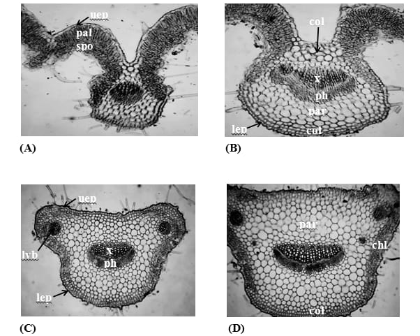
Figure 7: Microscopy of Caesalpinia bonduc leaf
(A and C): Control plant; (B and D): Plant treated with nano zeolite loaded nitrogen , (chl) chlorenchyma, (col) collenchyma, (lep) lower epidermis, (lvb) lateral vascular bundle,(pal) palisade tissue, (par) parenchyma, (ph) phloem ,(spo) spongy tissue, (uep) upper epidermis, (x) xylem.
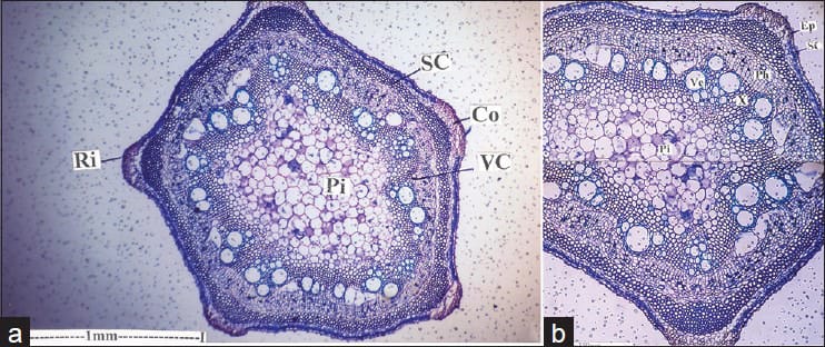
Figure 8: Microscopy of Caesalpinia bonduc stem
(a) Entire view of transverse section of stem. (b)Enlarged view of transverse section of stem. (Co) Cortex, (Ri): Ridges, (SC) Sclerenchyma, (VC) Vascular cylinder, (Ep) Epidermis, (Ph) Phloem, (Pi) Pith, (SC) Sclerenchyma, (Ve) Vessels, (X) Xylem.
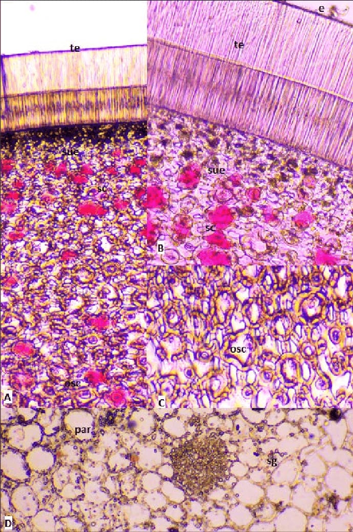
Figure 9: Microscopy of Caesalpinia bonduc seed
(A) Transverse section of seed, ( B & C) A portion enlarged, (D) Transverse section of cotyledon; (e) Epidermis, (osc) Osteosclereids, (par) Parenchyma cells, (sc) Sclereids, (sg) Starch grains, (sue) Sub-epidermis, (te) Testa.
In the transverse section, the stem bark shows, outer most cork 40-45 layered cells rectangular, tangentially elongated, measuring 17.5 - 70 μm (31.5) long and 3.5 - 21μm (7) wide; walls slightly thick, contents slightly with golden yellow coloured, interspersed with stone cells, isolated or in groups, mostly rectangular, measuring 21 - 42 μm (35) long and 7 - 17.5 μm (10.5) wide walls thick contents scanty. The transverse section of the seed reveals an outmost compact and single row arrangement of very narrow, translucent, radially & longated cells. Which form the palisade or in other words the malpighian layer. In normal view, these cells will appear as hexagonal shape and have thick walls containing a rich quantity of pectin. Subsequently 2 or 3 layers of sub epidermis consisting of thick walled bearer cells which were stemmed by multi layered osteosclerosis. The size osteosclerosis cells slowly increase and elongated laterally and the intrercellular space towards the inner side also increases. A brown substance was detected in the outer few osteosclerosis layer and laterally elongated vascular elements in the tissue were also recorded in the lower part of this zone. There are two types of sclerosis present in this region. Some are small, oval to circular shaped osteosclerosis bone or barred shaped elongated sclerosis dilated at their ends and that filled with brown gradually compact and round towards the inner margin. The cotyledon usually display a single outer layer of the epidermis containing small diameter cells. Interestingly the inner parenchymatous ground tissue cells are with fixed oil and are seen with uniformly distributed empty cavities. Cotyledon are also rich in starch grains.
6. PHYTOCHEMICAL EVALUATION
6.1 Preliminary (qualitative) screening:
6.1.1 Phytochemical screening
Phytochemical screening is a fundamental laboratory process to rapidly identify major groups of natural compounds (phytochemicals) in plants, like alkaloids, flavonoids, tannins, saponins, phenols, using simple chemical tests and colour changes, revealing potential medicinal properties and guiding further research into specific bioactive agents for health products or drug development.
6.1.2 Test for alkaloids
To 1 ml of plant extract, add 1 ml mayer’s reagent appearance of deep yellow or white precipitate indicates presence of alkaloids.
To 2ml of plant extract, add 1 ml of Dragendorff’s reagent along the side of test tube. Appearance of orange or orange reddish brown precipitate indicates presence of alkaloids.
c) Hanger’s test:
To 1 ml of plant extract, add few drops of hanger’s reagent. Appearance of yellow colour precipitate indicates the presence of alkaloids.
d) wagner’s test:
To 1 ml of plant extract, add 2 ml of wagners reagent appearance of reddish brown colour precipitate indicates the presence of alkaloids.
6.1.3 Test for carbohydrates:
Plant extract was treated with 2-napthol and add few drops of sulphuric acid from the side of the test tube. Formation of violet ring between the junction indicates the presence of carbohydrates.
b) Fehling’s test:
To the sample, equal volume of fehlings solution A&B are added &heat in water bath for few minutes and formation of reddish colour precipitate.
c) Benedicts test:
To the sample, add Benedicts reagent and heat for few minutes in boiling water bath appearance of orange red colour precipitate indicates the presence of carbohydrates.
6.1.4 Test for tannin & phenols:
To 1 ml of extract, add 3 ml distilled water then followed by 10% aqueous ferric chloride solution formation of blue or green colour indicates the presence tannins & phenols.
To the extract, add 1% gelatin solution containing sodium chloride solution in appearance of white precipitate indicates the presence tannins & phenol compound.
6.1.5 Test for flavonoids:
To 2 ml of extract add 1 % ammonia solution in appearance of yellow colour indicates tannins & phenol compound.
6.1.6 Test for protein and amino acids:
To the extract, add few drops of ninhydrin reagent in appearance of purple colour which indicates the presence of protein and amino acid.
To the extract, add millions reagent gently heat turns red colour precipitate indicates presence of protein.
6.1.7 Test for glycosides:
5ml of extract, add 2ml glacial acetic acid containing one drop of ferric chloride solution. This undelayed with 1ml of conc. sulphuric acid. Browning of interface indicates deoxy-sugar characteristic of carotenoids.A violet ring appears below the brown ring while in the acetic acid layer, a greenish ring may form just gradually throughout thin layers
To the sample add 1ml pyridine and few drops of sodium nitro- pursside solution and then it is made alkaline with sodium hydroxide solution. Appearance of pink colour indicates presence of glycoside.
6.1.8 Test for saponins:
To 2ml of extract, dilute with 5ml of distilled water in test tube shake vigorously add few drops of olive oil formation of stable foam indicates saponins.
6.1.9 Test for fixed oil and fats:
Take few drops of 0.5N potassium hydroxide and add few drops of phenolphthalein added to the extract this is heated in water bath for about 1-2hrs formation of soap indicates the presence of fixed oil and fats.
Small amount of extract is allowed to pass through the filter papers appearance of oil stain indicates presence of fixed oil and fats.
6.1.10 Test for terpenoids:
a) Chloroform test
5ml of extract add few drops of Chloroform and conc. sulphuric acid was added from side of test tube apperance of reddish brown colour indicates terpenoids..
Table 1: (preliminary qualitative screening) Summary of reported preliminary phytochemical screening of different parts of Caesalpinia bonducella
|
Phytochemical Class |
Test Used |
Leaves |
Seeds |
Stem Bark |
Roots |
|
Alkaloids |
Dragendorff’s / Mayer’s test |
+ |
+ |
+ |
– |
|
Flavonoids |
Shinoda / Alkaline reagent |
+ |
+ |
– |
– |
|
Tannins |
Ferric chloride test |
+ |
+ |
+ |
+ |
|
Phenols |
Ferric chloride test |
+ |
+ |
+ |
+ |
|
Saponins |
Foam test |
– |
+ |
– |
– |
|
Glycosides |
Keller–Killiani test |
– |
+ |
+ |
– |
|
Terpenoids |
Salkowski test |
+ |
+ |
+ |
+ |
|
Steroids |
Liebermann–Burchard test |
– |
+ |
+ |
– |
|
Carbohydrates |
Molisch test |
+ |
+ |
+ |
+ |
|
Proteins |
Biuret test |
– |
+ |
– |
– |
|
Amino acids |
Ninhydrin test |
– |
+ |
– |
– |
|
Fixed oils & fats |
Spot test |
– |
+ |
– |
– |
Note: (+) indicates presence and (–) indicates absence of phytoconstituents. Results may vary with extraction solvent, plant origin, and test method.
6.2 Preliminary (Quantitative) screening
6.2.1 Determination of total phenolic content:
TPC is measured folin ciocalteu method in a 96 well microplate formate 25 ml of diluted c.boundc extract was mixed 100ml of distilled folin ciocalteu reagent then shaken after 4 mins, 75 ml of sodium carbonate solution 100g/L was added & shaken again. The mixture was kept at room temperature for 2hrs.Absorbance was then read at 765nm using a microplate reader. A blank using ethanol instead of all extract was used for correction. Gallic acid (10-200mg/l) was used at the standard and results were expressed as milligrams of gallic acid equivalents (mg GAE) per gram of plant extract.
Table 2: (Total phenolic content)
|
Plant Part |
Total Phenolic Content (mg GAE/g extract) |
|
Root |
89.81 ± 3.00 |
|
Stem |
144.42 ± 16.05 |
|
Leaf |
146.64 ± 3.94 |
|
Seed kernel |
70.34 ± 10.59 |
6.2.2 Determination total flavonoids content:
Total flavonoids content was estimated using aluminium chloride colorimetric method. Quercetin was used at the standard in different concentration (30-100µg/ml) prepared in 96% ethanol. In a 96 well plate, 50ml of plant extract (1mg/ml) or standard solution was mixed with 10ml of 10% aluminium chloride, 105ml of 96% ethanol and 10ml of 1M sodium acetate. The mixture was incubated for 40mins at room temperature in dark after maceration, the absorbance was measured at 45nm using a microplate reader.The TFC was calculated and expressed as mg of quercetin equivalents (mg QE) per gram of plant extract.
Table 3: (Total flavonoid content)
|
Plant Part |
Total Flavonoid Content (mg QE/g extract) |
|
Root |
12.55 ± 0.08 |
|
Stem |
21.82 ± 0.46 |
|
Leaf |
31.05 ± 0.35 |
|
Seed kernel |
13.21 ± 1.35 |
Studies have reported highest amount of phenolic compounds was present in leaf (146.64 mg of GAE/g of crude extract) and the lowest was in seed kernel extract (70.34 mg of GAE/g). Leaf also have highest total flavonoid content (31.05 ± 0.35 mg QE/g) while the lowest was in root (12.55 ± 0.08 mg QE/g).
7. PHYSICOCHEMICAL EVALUATION
In the present study, we have attempted to evaluate the various physicochemical parameters on the seed of C. bonduc.
7.1 Foreign Matter
Foreign matter refers to any unwanted substances such as soil, sand, stones, insects, or other plant parts mixed with the crude drug. It is used to determine the purity of plant material, detect adulteration, and ensure the herbal drug is clean and safe for further processing.
Method:
About 100 g of dried plant material was spread on a clean tray and examined visually. Extraneous substances such as stones, soil particles, sand, damaged seeds, and other plant parts were removed manually. The separated foreign matter was weighed and expressed as a percentage of the total sample.
Observation / Result:
The foreign matter was found to be less than 1%, indicating a clean and good-quality crude drug.
7.2 Moisture Content (loss on drying)
Moisture content is the amount of water or volatile components present in the plant material, measured by drying it to constant weight. It is used to assess the stability of the drug, prevent microbial growth or spoilage, and ensure the material remains suitable for long-term storage.
Method:
Approximately 2–5 g of powdered sample was accurately weighed and placed in a hot-air oven at 105 ± 2°C. Drying was continued until a constant weight was achieved (3–5 hours). The moisture content was calculated based on the difference in weight before and after drying.
Observation / Result:
The moisture content was in the range of 4–8%, showing low moisture and good storage stability.
7.3 Ash Values
Ash value is the total amount of inorganic residue left after incinerating the plant drug. It is used to detect contamination with inorganic matter, evaluate purity, identify adulteration with sand or soil, and ensure the mineral content stays within acceptable limits.
Total Ash
Method:
Two grams of powdered drug was taken in a silica crucible and incinerated at 450–600°C until white or grey ash was obtained. After cooling in a desiccator, the residue was weighed.
Result:
Total ash was found to be 2–5%, reflecting normal inorganic content.
7.4 Extractive Values
Extractive value refers to the amount of active constituents extracted from the plant using different solvents like water or alcohol. It is used to estimate the presence of phytochemicals, select the best extraction solvent, and standardize the quality and potency of herbal drugs.
Method:
Five grams of coarse powder was macerated with 100 mL of solvent (ethanol, water, petroleum ether) for 24 hours with intermittent shaking. After filtration, 25 mL of the filtrate was evaporated to dryness, and the extractive value (%) was calculated from the dried residue.
Observation / Result:
Alcohol extract: 6–13% (rich in flavonoids, glycosides, alkaloids)
Water extract: 9–16% (contains tannins, phenolics, sugars)
Petroleum ether extract: 22–27% (contains fixed oils and fats)
7.5 pH of Extract
The pH of an extract indicates its acidity or alkalinity when dissolved in water. It is used to ensure chemical stability, determine compatibility with formulations, monitor microbial safety, and maintain proper storage conditions for herbal preparations.
Method:
A 1% w/v aqueous extract was prepared. The pH was measured using a calibrated digital pH meter standardized with pH 4.0 and 7.0 buffers.
Observation / Result:
The extract showed a pH of 5.2–6.0, indicating slightly acidic nature.
7.6 Powder Flow Properties
Powder flow properties describe how easily powdered plant material can move, pack, and compress, measured through parameters like angle of repose and bulk density. They are used to evaluate flowability for tablet/capsule preparation, ensure smooth processing, and optimize handling during manufacturing.
Method:
Bulk density was obtained by pouring the powder into a graduated cylinder and calculating mass/volume. Tapped density was measured after tapping the cylinder 100 times. The angle of repose was determined by allowing powder to flow through a funnel, measuring the height and radius of the formed cone, and applying the formula.
Observation / Result:
Bulk density: 0.43–0.55 g/mL
Tapped density: 0.62–0.75 g/mL
Angle of repose: 28–32° (indicates good flow)
7.7 Foaming Index
The foaming index measures the ability of an aqueous extract to produce foam, indicating the presence of saponins. It is used to estimate saponin content, identify saponin-rich drugs, and support the standardization and quality evaluation of herbal materials.
Method:
A 1% decoction was prepared and added to 10 test tubes in serial dilution. Each tube was shaken for 15 seconds and left undisturbed for 15 minutes. Foam height was recorded.
Observation / Result:
Foaming index was < 100, indicating very low saponin content.
7.8 Swelling Index
The swelling index represents the increase in volume of plant material when soaked in water, reflecting the content of mucilage, gums, or pectins. It is used to evaluate mucilage-rich drugs, assess quality for demulcent or laxative use, and standardize formulations requiring swelling action.
Method:
One gram of plant powder was placed in a 25 mL graduated cylinder with water. It was shaken periodically and left for 24 hours, after which the final swollen volume was measured.
Observation / Result:
Swelling index was below 1 mL, indicating low mucilage content.
7.9 Fluorescence Analysis
Fluorescence analysis involves observing the color changes of plant material under UV light after treatment with different reagents. It is used for identification and authentication of crude drugs, detecting adulteration, and establishing characteristic fluorescence fingerprints for quality control.
Method:
Powdered drug was treated with different reagents (NaOH, HCl, Methanol) and observed under visible light and UV light (254 nm & 366 nm) to detect characteristic fluorescence.
Observation / Result:
The sample showed yellowish-green fluorescence under UV, particularly with alkaline reagents, confirming authenticity and presence of specific phytochemicals.
8. MODERN ANALYTICAL TECHNIQUES
8.1 Thin layer chromatography
Thin-layer chromatography (TLC) is a chromatography technique that separates components in non-volatile mixtures. Thin-layer chromatography. Separation of black ink on a TLC plate.
Procedure:
Take a TLC plate and with the help of pencil draw two straight lines on the white surface of the plate, one 2 cm from the bottom of the plate and another 1 cm from top of the plate. Never use a pen as dyes (used in ink) may interfere with the results by developing spots on the plate. Then mark 10 equidistant points on the bottom line for loading of amino acids samples and test sample. While marking the lines and points do not make a trough with the pencil. Allow all the amino acid samples and test sample to come to room temperature. Then spot 1 μl of each amino acid and test sample along the bottom line on the TLC plate. While spotting use separate tips for each sample. Allow the plates to air dry (~ 10 minutes). Further drying should be done by keeping the TLC plate at 70?C in a hot air oven or incubator for 2 – 3 minutes. Take 10 ml of solvent system in the TLC chamber (with lid) and keep for 10 minutes at room temperature. Place the TLC plate inside the chamber with clean forceps. While keeping the plate make sure that the spotted samples are near the solvent. Furthermore, the TLC plate should be in a straight position so that the solvent phase can move uniformly along the plate. Allow the solvent front to reach the top line of the plate. After that take it out with the help of clean forceps and air dry the plate for 15 – 20 minutes. Keep the plates at 70?C for 2 minutes for further drying. Add 1 ml of the Developing Reagent on the plate and swirl the plate very carefully. Look for the development of the coloured spots of different amino acids and the test sample.
Rf value
The retention factor, or Rf, is defined as the distance traveled by the compound divided by the distance traveled by the solvent.

Developing the TLC profile
The spots appeared in both short (254nm) and (366nm) UV after the TLC profile was developed. The produced TLC of the raw extract showed three layer spots and several minor spots at 245nm with an RF value range of 0.16-0.9. Four spots with RF value between 0.12 and 0.89 were found at 366nm.While eight major spots with RF value between 0.02 and 0.08 were found in derivatized sample. But the derivatized sample in visible light detected five major spots with an Rf value range of 0.05-0.80. Preliminary phytochemical were further confirm using TLC. Aluminum sheet with silica gel 60 F254 plate was used as stationary phase and solvent system (toluene: ethyl acetate: methanol: formic acid [5:4.5:0.5:0.5]) used as a mobile phase for the development of chromatogram.
Figure 10: TLC profile of c.bonduc plant extract
8.2 UV-Visible spectroscopy Analysis
UV-Vis spectroscopy is an analytical technique that measures the amount of discrete wavelengths of UV-visible light that are absorbed by or transmitted through a sample in comparison to a reference or blank sample.
Calibration procedure:
Switch on the instruments and operate the instrument as per the respective current version of operating procedure.
Parameters;
Control of wavelength
Weigh 120mg of holmium oxide in a clean and dry test tube. Dissolve contents in 3.0ml of 1.4M perchloric acid solution. Verify the wavelengths with above solution using 1.4M perchloric acid as blank (or) standard holmium filter. Record the spectrum holmium perchlorate solution from 200nm to 600nm using 1.4M perchloric acid as reference solution.
Control of absorbance
Dry the potassium dichromate AR grade material to a constant mass at 130oC.
Calculation:
A (1%, 1cm) = absorbance x 100
weight in gm x 100
Base line stability with pair cells
Fill the quartz cells with distilled water and back correct the instruments from the 900nm to 190nm. Now scan and record the spectrum from 900nm to 190nm. Criteria, baseline absorbance value: ±0.001A.
Resolution power (For second order derivative spectrum):
Record the second order derivative spectrum of a 0.02%V/V solution of toluene in methanol. The spectrum shows a small negative extremum located between 2 large negative extrema at 261nm and 268nm. Criteria about the ratioA/B is not less than 0.2.
UV analysis of caesalpinia bounduc plant
The qualitative UV/VIS profile of plant extract of C.bonducella was analysed using the wavelength of 200 nm to 1100nm. The spectrum showed the peaks at 218.05, 585.10, 675.75, 762.80, 860.05, 989.50 and 1036.80nm with the absorption 4.0000, 0.6608, 0.6432, 0.6315, 0.5880, 0.5678 and 0.5748 as shown in below table.
Table 4: UV- Visible Analysis of caesalpinia bonduc plant
|
S.NO |
Wave length (nm) |
Absorbance (A 1%,1cm) |
Acceptance criteria |
|
1. |
218.05 |
4.0000 |
3.0000 - 6.0000 |
|
2. |
585.10 |
0.6608 |
0.5608 - 0.7608 |
|
3. |
675.75 |
0.6432 |
0.5432 - 0.7432 |
|
4. |
762.80 |
0.6315 |
0.5315 - 0.7315 |
|
5. |
860.05 |
0.5880 |
0.4880 - 0.6880 |
|
6. |
989.50 |
0.5678 |
0.4678 - 0.6678 |
|
7. |
1,036.80 |
0.5748 |
0.4748 – 0.6748 |
Absorption bands observed that related to UV-Vis spectrum of C. bonducella plant extract was displayed in (Figure 8.3.1). Absorption spectrum of Caesalpinia bonducella plant extract was almost transparent in the wavelength region of 200 to 1100nm. However, the appearance of peak in the region from 200 to 400 nm showed the presence of unsaturated and heteroatomic groups. The spectrum also confirms the presence of organic chromophores within the plant extract. Two absorption maxima in the ranges of 230–285 nm and 300–350 nm made up the flavonoids' spectra. Key details about the makeup of flavonoids can be cleaned from the exact location and relative strengths of these spectra. Due to the difficulty in identifying the absorption peaks with any ingredient, this spectrum is limited.
Figure 11: UV-Visible spectrum of plant seed extract of Caesalpinia bonducella
Acceptance criteria
The ratio of absorbance at the maximum at 269nm to that at the minimum at 266nm is not less than 1.5
CONCLUSION
In the present work, detailed Pharmacognostical (macro and microscopical), Physico-Chemical, Phytochemical properties and Modern analytical techniques of Caesalpinia bonduc has been reported. Several valuable phytochemical have been elicited from different parts of the C .bonducella plant. Comparison of macroscopic and microscopic characters resulted in the documentation of diagnostic features, which could aid in correct identification of the plant. The review has reiterated the fact that different parts of C.bonducella contain a number of active metabolites which have high potential to treat a number of ailments. The purity, quality and stability of the crude drug have been ascertained by physicochemical parameters including ash value, moisture content extractive values and flow properties. Preliminary phytochemical screening also showed the inclusions of significant bioactive metabolites such as alkaloids, flavonoids, tannins, phenolics, glycosides, fixed oils and terpenoids validating its worth and additional traditional medicinal uses. The characteristic profiles established by the TLC, and UV–Vis techniques were suitable for standardisation and quality verification. Overall conclusion, confirmed that C. bonducella is a good medicinal plant and would provide benchmark information for identification and potential pharmacological or formulation studies.
REFERENCES


K. Sojarna*, V. hileep, J. Niroshkumar, S. Pavithra, B. Sandhiya, D. Vinorithika, A Review on Pharamacognostic and Phytochemical Evaluation of Caesalpinia Bonduc Plant, Int. J. of Pharm. Sci., 2026, Vol 4, Issue 2, 2051-2068. https://doi.org/10.5281/zenodo.18628563
 10.5281/zenodo.18628563
