Narasaraopet Institute of Pharmaceutical Sciences, Narasaraopet, Andhra Pradesh 522601
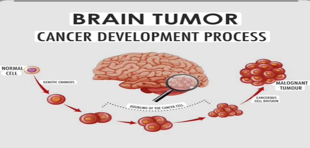
Brain cancer also refers as brain tumour. Brain tumours are caused by uncontrolled and rapid growth of cell. Detecting brain cancer is vitally important for saving lives. Brain tumour can be classified based on their origin, pace of development and stage of progression which helps in choosing the right treatment. Brain tumour segmentation aims to delineate accurately the areas of brain tumours. Brain tumours can be malignant (or) benign. Only about one-third of brain tumours are cancerous. But whether they are cancerous or not, they can impact brain functions. Brain tumours are common requiring general medical providers to have a basic understanding of their diagnosis and management. The most prevalent brain tumours are intra cranial metastases from systemic cancers, meningiomas, gliomas, specifically glioblastoma. Brain tumours are the most primitive, invasive and malignant in humans with poor survival. In recent years numerous studies have been carried out to identify new therapeutic protocols and tumour molecular markers capable to predict survival and response to treatment, the life expectancy of neuro-oncological patient is still very limited (24-36 months). About 33% of all brain tumours are gliomas, about 80% of the total malignant central nervous system (CNS) tumours in adult. Recently scientists showed that differentiated tumour cells may have the ability to differentiate acquiring to stem-like phenotype in response to microenvironment stresses such as hypoxia. In addition, the inter and intra-patient tumour heterogeneity causes several obstacles, limiting the improvement of an early diagnosis and treatment protocols.
A brain tumour is an abnormal growth or mass of cells in or around your brain. Together spinal tumour and brain tumours are called central nervous system tumours (CNS). Tumours that develop in your brain are called primary tumour, tumour that spread to your brain after forming in different parts of body are called secondary tumours or metastatic brain tumour.
Health care providers categorize primary tumours are glial composed of glial cells of brain which surround and assist nerve cells. Approximately 78% of cancerous brain tumours are gliomas.
Types of Brain Tumours:
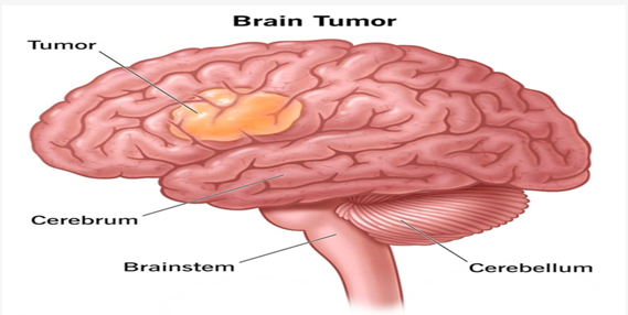
The main three parts of the brain are brainstem, cerebrum and cerebellum. Cerebellum is the second largest component of the brain and manages body motor activities including balance, posture, walking and general coordination of movements. It is positioned behind the brain and connected to the brain stem. The brain stem links to the spinal cord. A brain tumour is the medical term for an unexpected growth of brain cells.
|
Types of tumours Based on |
Type |
Comment |
|
Nature |
Benign |
Less aggressive and grows slowly. |
|
Malignant |
Life-threatening and rapidly expanding. |
|
|
origin |
Primary tumour |
Originates in the brain directly |
|
Secondary tumour |
This tumour develops in another area of body like lung and breast before migrating to the brain. |
|
|
Grading |
Grade I |
Regular in shape and they develop slowly. |
|
Grade II |
Appear strange to the view and grow more slowly. |
|
|
Grade III |
Tumours grow more quickly than grade II cancers. |
|
|
Grade IV |
Reproduced with greater rate |
|
|
Progression state |
Stage 0 |
Malignant but do not invade neighbouring cells |
|
Stage 1 |
Malignant and quickly spreading |
|
|
Stage 2 |
||
|
Stage 3 |
||
|
Stage 4 |
The malignancy invades every part of the body |
Recent Advances:
For many years, the detection of brain abnormalities has involved the use of several medical imaging methods. The two brain imaging approaches are structural and functional scanning.
Techniques including CT, MRI, SPECT (single photon emission computed tomography), PET (positron emission tomography), and ultrasound (US) are utilized to localize brain tumours for their size, location as well as shape and other characteristics.
MRI is non-invasive procedure that utilizes to display 3D anatomical structure of brain where PET is a nuclear medicine technique that analyses the metabolic activity of biological tissues. Fluorodeoxyglucose (FDG) is a popular PET agent for imaging the brain.
Ultrasound uses high-frequency sound waves to create real-time images of the inside of the body. No radiation is used.
Future Perspectives
Patient with have a high symptom burden throughout their disease trajectory and especially in terminal phase of illness. The main applications of CADx systems are in educating and training. The performance metrics outlined in this study provide a helpful and necessary baseline because they all are so dependent. The fact that the image formats utilized to train the models were those characteristics of the AI research field rather those of radiology field is noteworthy.
CONCLUSION
Brain tumours in adults are a rare disease from which survival is generally poor compared with many other cancers. Brain cancer is the leading cause of death from cancer in children. The molecular events that are crucial for normal development and functions are similar between individuals. However, in brain cancer genetic and epigenetic alterations result in cascades of deregulated molecular events which leads to genetically complex highly individual tumours. Numerous new therapies hold great promise for the treatment of patient with brain cancer. Instead of the present model of population risk assessment and empirical treatment, we will move to one of the predictive individualized cares based on molecular classification and targeted therapy for brain cancer.
REFERENCES


Ramya Teja Medarametla, Dr. J. N. Suresh Kumar, A. Lakshmi Prasanna, G. Naga Lakshmi, N. Ravi Teja, U. Srilekha, Y. Bhuvaneswari, A Review on Brain Cancer, Int. J. of Pharm. Sci., 2025, Vol 3, Issue 10, 762-764. https://doi.org/10.5281/zenodo.17295220
 10.5281/zenodo.17295220
