Department of Pharmaceutics, Shri Sarvajanik Pharmacy College, Mehsana
Liposome was first discovered by Alec Douglas Bangham, a British hematologist in 1961 at the Babraham Institute, in Cambridge, England. He published his work in 1964. They were discovered when A.D. Bangham and R.W. Horne was testing a new electron microscope in the institute with a dry phospholipid and gram-negative stain. When a self-forming enclosed lipid bi-layer was hydrated, the liposome or lipid vesicle was discovered. Liposome drug delivery systems have played a key role in the development of powerful drugs that have improved therapies Liposome formulations have recently been aimed at reducing toxicity and increasing accumulation at the target site The suppression of rapid liposome clearance by regulating particle size, charge, and surface hydration is one of several new liposome manufacturing methods based on lipid drug interactions and liposome disposition mechanisms. Liposome is a nanoparticle (size-100nm). Nanoscale drug delivery system using liposome as well as nanoparticle. This technology is for "Rational delivery of chemotherapeutic" drug treatment of cancer. Liposome is use as to study the cell membrane and cell organelles. The advantages of liposome formation using microfluidic approach for bulk-mixing approaches are discussed.
Liposomes are the most common and well investigated nanocarriers for targeted drug delivery. They have improved therapies for a range of biomedical applications by stabilizing therapeutic compounds, overcoming obstacles to cellular and tissue uptake, and improving biodistribution of compounds to target sites in vivo. Liposomes are defined as phospholipid vesicles consisting of one or more concentric lipid bilayers enclosing discrete aqueous spaces. The unique ability of liposomal systems to entrap both lipophilic and hydrophilic compound enables a diverse range of drugs to be encapsulated by these vesicles. Hydrophobic molecules are inserted into the bilayer membrane and hydrophilic molecule can be entrapped in the aqueous center. Liposomal formulation is characterized by properties such as particle size, charge, number of lamellae, lipid composition, and surface modification with polymers and ligands-these all govern their stability in vitro and in vivo Encapsulation within liposomes protect compound from early inactivation, degradation and dilution in the circulation. Liposomes are generally considered to be pharmacologically inactive with minimal toxicity, as they tend to be composed of natural phospholipids. The size of liposomes ranges from 30 nm to the micrometer scale, with the phospholipid bilayer being 4–5 nm thick. liposomes have been widely investigated as delivery vehicles for small molecular drugs, protein, nucleic acid, and imaging agents. Different administration routes, such as parenteral, pulmonary, oral, transdermal, ophthalmic, and nasal routes, have been developed to improve therapeutic efficacy and patient compliance. In addition, liposomes have been widely applied in the fields of food and cosmetics. Besides the specific medicines, liposomes stand as an excellent technique for drug delivery. However, only 14 types of liposomal products are available on the market, which means the advantages of liposomes have not been fully exploited. Therefore, in this review, we summarized the knowledge about commercial liposomal products approved by the FDA and EMA. Attention is paid to the composition and manufacturing technologies adopted in commercial products. In addition, the CQAs of liposomes, the current regulatory environment, and future perspectives are introduced. The purpose of this review is to provide important reference information to accelerate the development of liposomes. The water-soluble drugs are present in aqueous compartments whereas lipid soluble drugs and amphilhilic drugs insert themselves in phospholipids bilayer. The liposomes containing drugs can be administrated by many routes like intervenous, oral inhalation, ocular and local application, used in the treatment of many diseases. It is satisfactory and advanced carrier and has capability to encapsulate hydrophilic as well as lipophilic drugs and shield them from degradation. In general, they are more effective and less toxic than conventional dosage forms due to the bilayer composition and structure.
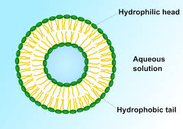
Figure 1: Liposomes
Liposomes are usually applied to the skin as liquids or gels and hydrophilic polymers are considered to be suitable thickening agents. Liposomes as a carrier are biocompatible, biodegradable, targeting, and stimulus-responsive. Local anesthetics are also encapsulated into liposomes have longer duration of action, decrease in circulating plasma levels, decrease central nervous system toxicity and cardiovascular toxicity. Liposomes are useful because they act as carriers for a variety of drugs and have potential therapeutic or other properties. Various carriers such as nanoparticles, microparticles, polysaccharides, lectins, and liposomes can be used to target drug to a specific site. Liposomal drug delivery is gaining interest due to its contribution to various areas like drug delivery, cosmetics, and biological membrane structure. A liposome is a tiny bubble (vesicle), with a membrane composed of a phospholipid bilayer. Membranes are usually made of phospholipids like phosphatidylet- hanolamine and phosphatidylcholine. Phospholipids are amphiphilic with its polar head as hydrophilic and hydrocarbon tail as hydrophobic.
History of Liposomes: [4]
Liposomes were first discovered in the mid-1960s by British hematologist Dr. Alec D. Bangham. He was studying the structure of cell membranes and stumbled upon the liposome while using an electron microscope. Liposomes are tiny spherical structures made up of lipid bilayers, similar to the structure of cell membranes. Dr. Bangham's groundbreaking work laid the foundation for understanding and developing liposomes for various applications. In the following decades, liposomes gained recognition for their potential in drug delivery. Their ability to encapsulate drugs and transport them to specific sites in the body revolutionized the field of pharmacology. Liposomal drug delivery systems allowed for controlled release and reduced side effects of many medications. Since then, liposomes have found applications not only in drug delivery but also in cosmetics, food technology, and gene therapy. Researchers have developed various types of liposomes with different sizes, compositions, and surface modifications to optimize their performance for specific applications. Today, liposomes continue to be an essential tool in the fields of medicine and biotechnology, with ongoing research aimed at improving their effectiveness and versatility in delivering therapeutic agents and other bioactive compounds.
Schematic representation of the different types of liposomal drug delivery system [4]
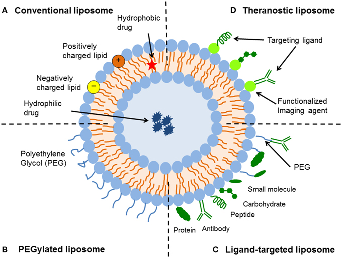
Figure 2: Schematic representation of the different types of liposomal drug delivery system
Structure of Liposome [5]
Lipids are amphiphilic molecules with one water-loving (hydrophilic) and one water-hating (hydrophobic). When lipids come into contact with water, the hydrophobic portions of the molecule establish unfavorable interactions with the solvent, resulting in lipid self-assembly in the form of liposomes. By changing medication absorption, lowering metabolism, prolonging biological half-life, and reducing toxicity, liposomes have been utilized to increase the therapeutic index of new and old pharmaceutical features of the carrier, rather than the physico-chemical characteristics of the pharmacological molecule are therefore used to govern drug distribution. Liposomes can be made from natural or synthetic lipids, and liposome contents are not limited to lipids; new generation liposomes can also be made from polymers (sometimes referred to as polymerases) Liposomes, which are made up of natural or synthetic lipids or polymers, are biocompatible and biodegradable, making them ideal for biomedical research. Liposomes are remarkable in that they have the potential to compartmentalize and solubilize both hydrophilic and hydrophobic molecules. Liposomes are particularly appealing as drug delivery vehicles because of their unique characteristic, which is combined with biocompatibility and biodegradability. Hydrophobic drugs place themselves inside the bilayer of the liposome and hydrophilic drugs are entrapped within the aqueous core or at the bilayer interface. Liposomal formulations enhance the therapeutic efficiency of drugs in preclinical models and in humans compared to conventional formulations due to the alteration of bio-distribution.

Figure 3: Structure of Liposome
Properties of liposomes: [6]
Structural Components of Liposomes: [6]
Phospholipids
Phospholipids are majorly found in the structural composition of biological membranes and are used in the preparation of liposomes extensively. The biocompatibility and Phospholipids are majorly found in the structural composition of biological membranes and are used in the preparation of liposomes extensively. The biocompatibility and Phospholipids are majorly found in the structural composition of biological membranes and are used in the preparation of liposomes extensively. The biocompatibility and Phospholipids are majorly found in the structural composition of biological membranes and are used in the preparation of liposomes extensively. The biocompatibility and The fundamental building blocks of bio membranes are phospholipids. Phosphatidylcholine (PC) is perhaps the most often utilized phospholipid in liposome composition. Phosphatidylcholines derived from natural origins or synthesized using semi-synthetic and synthetic techniques. In terms of bilayer sheet orientation in relation to micellar compositions, phosphatidylcholines vary significantly from other phospholipids. The preparation of liposomes is based on a number of natural and semi-synthetic phospholipids. The molecules of phosphatidylcholine are not miscible in water. They develop a planar bilayer configuration in aqueous environments to decrease undesirable interactions between the bulk aqueous environments as well as in non-esterified fatty acid. The sheets are then folded into enclosed capsules.
Cholesterol
Typically, liposomes prepared by using only phospholipids are not sufficiently rigid primarily because of low phase transition temperature and/or unsaturation in the fatty alkyl chains creating defects in the cell membrane like packaging. During packaging those liposome’s leak, the encapsulated drug. One or two bilayer stabilizers are also used in most liposome formulation in order to avoid such leakage. The additives more widely used are cholesterol and alpha tocopherol. Liposome encapsulation quality varies with variations in the composition of the phospholipid bilayer. Cholesterol is indeed an absolutely vital component of natural lipid bilayers and its presence in bilayer liposomes induces drastic alteration in their characteristics. Cholesterol by itself doesn't develop bilayer complexes, but can be integrated into high concentrations of phospholipid membranes. Rigidity is enhanced due to compact stacking of the bilayers, and permeability of water-soluble molecules is reduced. By reducing bilayer permeability, cholesterol enhances the durability of hydrophilic drugs. Cholesterol reduces the fluidity above the phase transition temperature (Tc) to make the bilayer more ordered. The tricyclic ring is wedged among the first few carbons of the fatty acyl chains, and the hydroxyl group is exposed to the liquid phase, the cholesterol molecule fits in with the phospholipid molecules and orients itself among them. At very significant concentrations, cholesterol to phospholipids ratios of up to 1:1 or even 2:1 can be incorporated into the cellular membrane. Albumin, macroglobulin, and m-transferrin are blood proteins that interact more easily and frequently with cholesterol-free liposomes enabling the vesicle to become unstable as a result its usage as a therapeutic delivery method has declined.
Merits: [6]
Demerits: [6]
Types of Liposomes: [7]
Medium unilamellar vesicles (MUV): size ranges from 40-80 nm. Large unilamellar vesicles (LUV): size ranges from 100nm-1,000 nm
Factors affecting drug release from liposomes[8]
Liposomes are the most successful nanoparticle delivery systems developed to target drugs to the site of action. There has been tremendous development in the field of liposomal drug delivery which has led to various clinically approved formulations that are efficacious, biocompatible and possess improved pharmacokinetics. The drugs loaded in liposomes become bioavailable only when they are released. Therefore, to have optimum therapeutic activity, modifying/controlling the release rate of drug from liposomes is very essential. There are different methods described in literature for improving and optimising rate of drug release from liposomes. Drug release from liposomes is affected by the following factors:
Cholesterol content used in the formation of liposomes:
Liposomes made of phospholipids and cholesterol have demonstrated a decrease in bilayer permeability as the amount of cholesterol increases. The addition of sphingomyelin or cholesterol to liposomes gives the bilayer rigidity and enhances drug retention in liposomes. A study of the impact of cholesterol content on liposome stability and in vitro release has been discussed. The study outlines the potential benefits of cholesterol as a stabilizing agent that helps to give strength to the bilayer, thereby lowering its permeability to electrolyte and non-electrolyte solutes. The study involved loading hydrophilic drug (atenolol) and hydrophobic drug (quinine) into the liposomes. Liposomes were prepared in five different lipid: cholesterol ratios using the thin-film hydration method. Particle size, (%) encapsulation efficiency, and in vitro drug release patterns were assessed for each formulation. According to the results, formulations with high cholesterol levels (up to 50%) had subpar drug release and encapsulation profiles. The optimal encapsulation and medication release profile was demonstrated by the optimized formulation with a 30% cholesterol content. Therefore, achieving high encapsulation and drug loading values during the formulation of drug-loaded liposomes is facilitated by optimizing the cholesterol to lipid ratio. According to studies, cholesterol makes the core part of the membrane bilayer more hydrophobic, which may encourage the incorporation of hydrophobic molecules. Drug encapsulation and loading efficiency are reduced as a result of competition between cholesterol and the hydrophobic drug, both of which prefer to fit into the bilayer. The significance of regulating cholesterol content in assessing the impact of various lipids for figuring out the in vitro release kinetics of liposomes was discussed in another study. The study's objective was to assess the ideal lipid for liposome formulation and the amount of cholesterol needed to enhance the liposomes' overall release kinetics. To mimic medication encapsulation and release, liposomes were created using the sonication method using 14C radiolabeled inulin as a marker. According to the results, 21% cholesterol was chosen in order to maximize the liposomes' stability and release profile.
Nature of encapsulated drug
Another important factor affecting drug release from liposomes is seen in choosing drugs whose physical characteristics favor retention of drug in liposomes. Liposomes possess the ability to entrap both hydrophilic and hydrophobic drug due to their unique structural organization. There are several drugs that have been successfully delivered through liposomes and have been approved by the FDA for clinical applications. Some of the key examples include doxorubicin, vincristine, paclitaxel, bupivacaine, amphotericin B and irinotecan. The physicochemical characteristics of drug of choice and the biocompatibility of lipids are the two main considerations in development of a stable and efficacious liposome system. A study describing the transcutaneous permeation of three drugs: amphotericin B, imiquimod and indole encapsulated by liposomes effectively explained the comparison of the penetration ability of the three drugs through their encapsulation into ultra-flexible liposomes. Among the drugs encapsulated in liposomes for this study, amphotericin B and imiquimod had poor water solubility whereas indole was water soluble. The study stated that formation and encapsulation of the three drugs into ultra-flexible liposomes increased the overall pharmacokinetics of the drugs with the three drugs showing efficient skin penetration. An effective strategy in designing liposomal drug delivery is selection of drugs that can be loaded to retain inside the liposome and selectively release at the site of action to achieve site specific drug delivery.
Membrane composition of liposomes
Membrane composition of liposomes plays a major role in the release of drug from liposomes. Since liposomes can encapsulate both hydrophilic and hydrophobic drugs in their structure it is important to study the composition of liposomes during formulation for effective drug release. A study describing the improved liposomal drug retention was conducted by adding dihydro sphingomyelin (DHSM) in the lipid component during the formulation of liposomes. Vincristine-loaded liposomes were prepared by replacing egg sphingomyelin with DHSM in sphingomyelin: cholesterol (55:45 mol/mol) which resulted in a substantial increase in drug encapsulation and showed sustained drug release. Additionally, there was a three-fold increase in drug release half-life compared to liposomes without DHSM. Studies have also suggested that the formation of molecular complexes within the liposomes has resulted in improved retention of drugs in liposomes. Formation of molecular complexes can help in modulating the release of drug from liposome and could also help in preventing leakage of drug from vesicles. The design of liposomal co-encapsulation of oleanolic acid and doxorubicin for evaluation of antitumor efficacy utilized three different lipid compositions in formulation of liposomes. Liposomes for this study were prepared by ethanolic injection method and composed of the different lipid ratios as predicted by the statistical design. Results showed that the highest encapsulation efficiency and sustained drug release was achieved by controlling the cholesterol content in the formulation.
Application of external stimuli
Studies have shown that in order to obtain controlled release, there are several stimuli-based strategies that allow rapid release of drug at the tumour site. The use of local stimuli strategies for improving drug release from liposomes has been extensively studied. This strategy utilizes the small changes occurring in the tumour microenvironment such as change in pH, difference in temperature or the overexpression of some proteolytic enzymes for improving the rate of drug release. Formulation of thermosensitive liposomes is an effective strategy in controlling the release of drugs from the nano-lipid vesicles. Thermosensitive liposomes have shown tremendous potential when administered with local hyperthermia. There are various studies that have reported the benefits of drug targeting with thermosensitive liposomes. Temperature triggered drug delivery of thermosensitive liposomes using pre and post hyperthermia mechanism has shown promising results. A study describing the formulation of lysolipid containing thermosensitive liposomes in combination with local hyperthermia was used to deliver cytotoxic proteins thereby explaining the mechanism of triggered drug release. Liposomes were prepared by thin film hydration and extrusion technique. Liposomes were prepared with different compositions of lipids. The selected formulation (86:10:4 % mol DPPC: MSPC: DSPE-PEG2000) showed efficient drug release with mild hyperthermia and was thereby considered a promising local tumour delivery strategy for cytotoxic proteins. A study describing the release of doxorubicin based on mild hyperthermia-mediated release from thermosensitive liposomes was explored and studied. Briefly, the study consisted of preparation of thermosensitive liposomes for evaluating the release profile of doxorubicin. Results showed that the injected thermosensitive liposomes loaded with doxorubicin released the drug efficiently with mild hyperthermia at 43 °C and was monitored real time with fibred confocal fluorescence microscopy. Decrease in pH in cellular lysosomes has been successful in improving the rate of drug release in pH sensitive doxorubicin loaded liposomes. The formulation of pH sensitive liposomes was achieved by encapsulating a precursor that had the ability to generate gas bubbles in situ in acidic pH. The bubble generation in acidic pH led to the rapid release of doxorubicin thereby showing antitumor effect at the targeted site. A study involving a novel acid-labile PEGB-Hz-DPPE conjugate in developing dual pH-responsive strategy was part of the preparation of dual pH-sensitive liposomes with enhanced tumour targeted drug delivery. The effectiveness of the study was tested against pancreatic cancer cells. Results showed that as the pH in lysosomes reduced there was efficient release of doxorubicin whose effectiveness increased with the use of ultrasound waves that substantially reduced the viability of cancer cells. Thereby the study concluded by emphasising the potential of pH triggered drug release in liposomes. Thus, there are various techniques/modifications that can be done to improve and control the rate of drug release from liposomes.
Targeting Mechanisms in Liposomal Drug Delivery [9]
Liposomal formulations can utilize both passive and active targeting mechanisms to enhance drug delivery precision. Passive targeting takes advantage of the liposomal structure's natural accumulation in the reticuloendothelial system (RES), making them suitable for targeting specific organs like the liver and spleen. On the other hand, active targeting entails modifying the liposome's surface with specific ligands that can bind to receptors on target cells. This tailored approach allows for a more precise delivery of therapeutic agents, which is particularly beneficial in treatments requiring high specificity, such as cancer therapies.
Mechanism action of Liposome [10,11]
A liposome consists of a region of aqueous solution inside a hydrophobic membrane. Hydrophobic substances can be easily dissolved into the lipid membranes; in this way liposomes are able to carry both hydrophilic and hydrophobic molecules. The extent of location of the drug will depend upon its physiochemical characteristics and composition of lipid. For the release of necessary drug molecules to the site of action, the lipid bilayers fuse with other bilayers of the cell (cell membrane) to release the liposomal content.
Liposome performs their action by four different mechanism,
Evaluation of liposomes: [12, 13]
The liposomal formulation and processing for specified purpose are characterized to ensure their predictable in vitro and in vivo performance. The characterization parameters for purpose of evaluation could be classified into three stapes physical chemical and biological parameters. Physical characterization evaluate parameter includes size, shape, surface, and drug release profile. Chemical characterization includes studies in establish the purity and potency of various lipophilic constituents. Biological characterizations parameters are helpful in establish the safety and suitability of formulation for therapeutic application. Some of parameters are:
Vesicle shape and lamellarity
The Vesicle shape assessed using electron Microscopic Techniques. Lamellarity of vesicles is determined by Freeze Fracture Electron Microscopy and P31 Nuclear Magnetic Resonance Analysis.
Vesicle size and size distribution
The liposome size distribution was determined by the photon correlation spectroscopy.
Microscopic Techniques Optical Microscopy
The microscopic method includes use the Bright Field, phase - Contrast Microscope and Fluorescent Microscope and is useful in evaluating vesicle size of large vesicles.
CRYO-Transmission Electron Microscopy Techniques
(CRYO-TEM)
This technique has been used to elucidate the surface Morphology and size of vesicles.
Diffraction and Scattering Techniques
Laser Light Scattering Photon correlation spectroscopy (PCS)
The analysis of time dependence of intensity fluctuation in scattered laser light due to Brownian motion of particles in solution or suspension since small particles diffuse more than large particles, the rate of fluctuation scattered light intensity varies accordingly. The translational diffusion coefficient (D) can be measured and can be used to determine the mean hydrodynamic radius (RHSS) of particles using the Stoke-Einstein equation using this technique one can measure particles in range of about 3nm.
Hydrodynamic Techniques
This technique includes gel Permeation and Ultracentrifuge. Exclusion chromatography on large pure gels was introduced to separate SUVs from radial MLVs. The large vesicles of 1- 3µmdiameter.usually fail to enter the gel and is retained on top of column. A thin layer chromatography system using agarose beads has been introduced as a convent, fast technique for obtaining a rough estimation of size distribution of liposome preparation. However, it was not reported if this procedure was sensitive to a physical blockage of pores of the agars gel as is the more conventional column chromatography.
Zeta potential determination
The zeta potential was evaluated by the determination of electro mobility of the 90c angle. The measurement was performed in triplicate using the 3000 HS zeta-seizer equipment. The sample was diluted with suitable diluents for the potential determination.
Applications: [14-16]
CONCLUSION:
The liposomal drug delivery is very effective in the delivery of genes and vaccines due to their adjuvant property and tumor targeting ability. Liposomal drug delivery is best tool for targeting of brain. Liposome are made up of lipid bilayer. The ability of liposome to carry both hydrophilic as well as hydrophobic drug for site specific delivery and produce long term effective therapy. Liposome are easily cross 'Blood brain barrier' (BBB) as compared to other dosage form because 'Blood brain barrier is made up of lipid barrier.
REFERENCES


Mitali Oza*, Trusha Patel, Mona Gupta, Dr. Nisha Patel, Sandhya Bodhe, A Comprehensive Review on Liposomes: As Drug Delivery System, Int. J. of Pharm. Sci., 2025, Vol 3, Issue 4, 463-475. https://doi.org/10.5281/zenodo.15133054
 10.5281/zenodo.15133054
