Department Of Pharmaceutics, Modern College of Pharmacy for Ladies, Moshi, Pune.
Centrifugal electrospinning is an advanced technique for the development of nanofibers. It is a vital part of a novel drug delivery system. Oral films are the most convenient dosage form for administration. Hence, oral film is selected as the final dosage form. The study involved the preparation of polymeric solutions such as polyvinyl alcohol and polyvinyl pyrrolidone, and the use of a Design of Experiment for getting trial batches. Furosemide-loaded oral films are prepared by two methods, namely, solvent casting and the centrifugal electrospinning method. The optimized formulation via solvent casting was further utilized for the fabrication of nanofiber oral film using a central electrospinning method. Both oral films were comparatively evaluated by analytical and performance characterization tests, including DSC, FTIR, XRD, in vitro dissolution studies, and disintegration time. The developed nanofiber oral film significantly enhances the solubility of Furosemide, indicating its potential as an effective platform for improving dissolution rate and bioavailability.
Furosemide is widely used as a diuretic and antihypertensive. It is a powerful loop diuretic indicated for the treatment of edematous conditions associated with the heart, liver, and hepatic failure. It is a spironolactone that belongs to the BCS class four (1). The oral bioavailability of the drug is 30 to 50 %. In addition, Furosemide absorption is site-specific, and it occurs only in the upper small intestine. This results in a very narrow absorption window. Furosemide is a poorly soluble and permeable drug. To improve its in vivo performance, improving the drug's aqueous solubility may enhance its uptake in the body. Therefore, hydrophilic polymers and solubilizers are used to improve the drug's solubility. The 10% PVA and 15% PVP solution was used to prepare the oral film. The viscous solution of oral film is made up of distilled water, solubilizers, and polymers (2,3). The solvent casting method forms a mucoadhesive oral film of Furosemide. The hydrophilic polymer Polyvinyl alcohol (film-forming agent ) and Polyvinyl Pyrrolidone are used, glycerol is used as a plasticizer, and sodium starch glycolate is used to improve the disintegration time of the oral film. Sodium starch glycolate shows capillary action when dissolved in water. Hence, drugs easily exit from film and show drug release action within 15 to 20 minutes. (4,5) The optimized oral film was compared with the novel approach called the nanofiber oral web. The needless electrospinning technique is utilized. Needless electrospinning is an alternative method of electrospinning. The centrifugation force plays a vital role in the formation of thread-like nanofibers. The Cotton candy machine is used for centrifugal spinning. This technique is cost-effective compared to needle electrospinning. (6)
Objectives –
1] Improve the solubility of furosemide.
2] Fast drug release will minimize the first-pass metabolism of Furosemide.
3] Improve the bioavailability of the drug.
4] Exploring novel drug delivery systems.
5] Due to the high surface area solubility of the drug will increase.
MATERIAL AND METHODS –
Materials –
The active pharmaceutical, furosemide was provided by (LOBA Chemie Mumbai), for the preparation of various polymeric solution poly(vinyl-pyrrolidone), and polyvinyl alcohol was used, which was provided by (LOBA Chemie Mumbai)Triethanolamine (LOBA Chemie Mumbai), sodium starch glycolate (LOBA Chemie Mumbai) were from Potassium dihydrogen phosphate and sodium hydroxide (Research lab, Mumbai India) were employed for the preparation of the phosphate buffer ( pH 6.8) used for the invitro dissolution test.
Methods –
1] Solvent casting method –
The solvent casting method is most frequently used to create the mouth-dissolving film. In distilled water drug (120 mg) and triethanolamine were added. Then polymeric solution of PVA and PVP, other excipients were added. For the preparation of a homogeneous solution, the beaker was kept on a magnetic stirrer for 1 hr. After cooling down, the solution was cast on a Petri plate. Allowed to dry, then cut into the required number of pieces. (7) The Desice software was used for the application of the design of the experiment. Central composite design (CCD) summarizes the non-electrospinning method [7]. In the polymeric solution, the amount of active ingredient was determined to be 120 mg ( this parameter was kept constant during experiments ). The molar ratios of PVP (W/V%), PVA (W/V%), and SSG (W/W%) were examined at lower and higher levels.
2] Preparation of nanofiber –
Firstly cotton candy machine is cleaned with paper, and this machine by inserting a rotating chamber within it, and the switch is turned on. The rotating chamber is rotated as the voltage is applied. The rotating chamber has one nozzle through which a drop of polymeric solution is entered. A polymeric solution is added to the nozzle of the rotating chamber. The rotatory chamber is continuously rotated as the centrifugal force reaches its maximum level, the polymer starts melting, the solvents are evaporated, and the formation of fibers exits from the rotatory disk and are collected. (8,9) Figure 1 provides images of oral film and the development of nanofibers by the cotton candy machine.
Table 1 – Central composite Design
|
Experimental runs |
PVA (%) |
PVP (%) |
SSG (%) |
|
1 |
8 |
10 |
2 |
|
2 |
10 |
10 |
2 |
|
3 |
8 |
25 |
2 |
|
4 |
10 |
25 |
2 |
|
5 |
8 |
10 |
8 |
|
6 |
10 |
25 |
8 |
|
7 |
8 |
25 |
8 |
|
8 |
8 |
17.5 |
8 |
|
9 |
9 |
17.5 |
5 |
|
10 |
8 |
10 |
5 |
|
11 |
10 |
25 |
5 |
|
12 |
9 |
10 |
5 |
|
13 |
9 |
17.5 |
8 |
|
14 |
9 |
25 |
5 |
As per Table 1 central composite design suggests a total of 14 trial batches of oral film. Every formulation batch is performed by the solvent casting method. Each formulation contains different concentrations of polymer and sodium starch glycolate as a disintegrating agent.
RESULT AND DISCUSSION –
For optimizing best batch of oral film evaluation tests is carried out. The following Table 2 will explain the different characteristics of each film. The characteristics of each film will be compared with standard ranges.
1] Drug Content–
The 2 X 2 cm patch was cut and dissolved into 7.4 pH buffer. This solution was kept on a mechanical shaker for 1 hr. The UV absorbance is checked at 272 nm.
2] Thickness Measurement–
Thickness was measured using a vernier caliper on the three sides of the film.
3] Folding Endurance–
A specific area of the film is repeatedly folded at the same place until it breaks. The number of folds is recorded.
4] Disintegration Time –
A 2X2 cm patch is added to 900 mL of buffer (pH 6.8) in a disintegration apparatus. The disintegration time is noted.
5] Dissolution Test–
A 2X2 cm patch is used in a phosphate buffer (pH 6.8) with USP apparatus 2. The RPM is set at 50 and the temperature at 37°C. The 5 mL sample was collected and filtered, and UV absorbance was measured at 272nm. (11)
Table 2 – Evaluation Test of Oral Film
|
Evaluation Test |
F1 |
F2 |
F3 |
F4 |
F5 |
F6 |
F7 |
F8 |
F9 |
F10 |
F11 |
F12 |
F13 |
F14 |
|
Drug content (%) |
64 |
58 |
23 |
92 |
59 |
87 |
78 |
64 |
89 |
92 |
53 |
60 |
94 |
35 |
|
Thickness (mm) |
0.23 |
0.33 |
0.13 |
0.45 |
0.65 |
0.24 |
0.56 |
0.12 |
0.25 |
0.55 |
0.36 |
0.38 |
0.09 |
0.34 |
|
Folding endurance |
59 |
58 |
83 |
68 |
89 |
78 |
92 |
96 |
88 |
51 |
58 |
65 |
99 |
36 |
|
Disintegration time ( sec) |
40 |
44 |
51 |
56 |
65 |
43 |
56 |
46 |
51 |
39 |
48 |
43 |
36 |
45 |
|
Dissolution test (CDR% % ) |
46 |
52 |
47 |
54 |
64 |
48 |
43 |
31 |
68 |
70 |
59 |
15 |
98 |
75 |
According to the above table, it was proven that the F 13 formulation batch showed show best properties, like a good percentage % cumulative drug release, and a shorter disintegration time. The formula of the F13 batch was used for the fabrication of nanofibers by the central electrospinning technique. The following Figure 1 shows the fabrication of nanofibers by the centrifugal electrospinning technique.
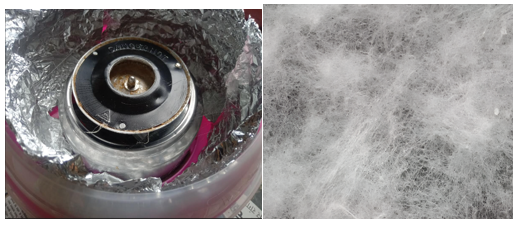
Figure No.1 – Fabrication of nanofiber film by centrifugal electrospinning.
Comparison study of solubility, dissolution, and Ex vivo diffusion between pure drug, oral film, and nanofiber oral web-
Solubility study –
The nanofiber loaded with Furosemide showed that there is an improvement in the solubility of the drug as compared to a fast-dissolving oral film. These could be due to the nanoscale formulation, porous nature of nanofibers, and large surface area of the fiber. Figure 2 explains how the solubility of Furosemide increases.

Figure 2 – Solubility study of pure drug, oral film, and nanofiber
Dissolution study –
Both prepared formulations of furosemide show there is an enhancement in the solubility of furosemide, which also indicates that the oral bioavailability of the drug is improved. Figure 3 explains the % cumulative drug release at a specific time point from the formulation and drug.

Figure 3 – Comparative dissolution study between the drug, oral film, and Nanofiber film
Ex Vivo diffusion study: Nanofibers have a short diffusion pathway, allowing them to quickly diffuse from the fibers. The following Figure 4 explains the % drug that diffuses at a specific time point from the formulation and drug.
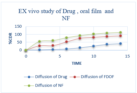
Figure 4- Ex vivo diffusion study of pure drug, oral film, and nanofiber oral web
SEM image of nanofiber oral web - The diameter of the nanofiber is between 82 to 98 nm. It is indicated that fibers are nanosized. Figure 5 shows the diameter of nanofibers. (12,13)

Figure 5 - SEM image of Furosemide-loaded Nanofiber
Characterisation Studies of Pure drug, Oral film and Nanofiber film
DSC - a broad endothermic peak was observed in a nanofiber formulation, which indicates the drug changes crystalline to an amorphous form. Figure 6 explains the DSC of the pure drug, mucoadhesive oral film, and Nanofiber oral web.
FTIR - The peak observed in the FTIR spectrum for furosemide, physical mixture, and formulation indicates that there is no interaction between the drug and excipients. Figure 7 shows the FTIR study of the pure drug, physical mixture, and nanofiber oral film.
XRD - The high-intensity peaks of Furosemide represent its crystalline nature, while the optimized batch of nanofiber oral film showed low-intensity peaks representing the amorphous nature of the drug. Figure 8 shows how the crystalline nature of Furosemide is converted into an amorphous form. (14)

Figure 6 - DSC study of the pure drug, Mucoadhesive oral film, and Nanofiber oral web (C)
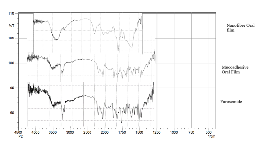
Figure 7 - FTIR study of pure drug, physical mixture, and nanofiber oral film
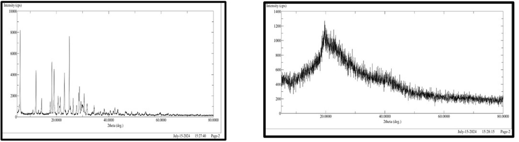
Figure 8 - Figure 8 - XRD study of pure drug and Nanofiber oral web
CONCLUSION –
The diameter, high surface-to-volume ratio, and high wettability such factors of nanofibers that make it a unique dosage form as compared to traditional oral film. Also, the XRD of furosemide-loaded nanofibers proves that the drug transitions from a crystalline nature to an amorphous nature. From the comparison between the nanofiber oral web and oral film, it is indicated that the nanofiber shows higher drug release in a short time.
REFERENCES

Smita More*, Pratiksha Londhe, Sanskruti Khedkar, Snehal Thorat, A Comparative Formulation Study of Furosemide Oral Film, Int. J. of Pharm. Sci., 2025, Vol 3, Issue 5, 2582-2589. https://doi.org/10.5281/zenodo.15429434
 10.5281/zenodo.15429434
