1,2,3Faculty Of Pharmacy, Sree Balaji Medical College and Hospital Campus.
4,5Sri Ramachandra College of Pharmacy.
Zebrafish (Danio rerio) have become a widely used model in neurotoxicity research due to their genetic similarity to humans, transparent embryos, and rapid development. This review deliberates the advantages of zebrafish for studying neurotoxic effects, methodologies used in zebrafish neurotoxicity assessments, and their applications in environmental and pharmaceutical toxicity studies. Additionally, challenges and future perspectives of zebrafish as a model for neurotoxicology are addressed. Neurodegenerative Disease Models in Zebrafish (Danio rerio) have become an increasingly popular model organism for studying neurodegenerative diseases, recognitions to their genetic similarity to humans, transparent embryos, rapid development, and ease of genetic manipulation. They offer numerous advantages over other models, including the ability to visualize the progression of neurodegeneration and the effects of potential therapeutic interventions in real time. This article will explore several key neurodegenerative disease models in zebrafish, focusing on Alzheimer's disease (AD), Parkinson's disease (PD), Huntington's disease (HD), and amyotrophic lateral sclerosis (ALS).
Neurotoxicity refers to the adverse effects of chemical substances on the nervous system, leading to functional or structural damage. With increasing concerns over environmental pollutants, pharmaceuticals, and industrial chemicals affecting neural health, animal models are essential for toxicity screening (Legradi et al., 2018). Zebrafish have emerged as an advantageous model due to their high genetic homology with humans, transparency during embryonic development, and high fecundity (Kalueff et al., 2014).
Advantages of Zebrafish in Neurotoxicity Research
Genetic Conservation
Zebrafish share approximately 70% of their genes with humans, including many genes associated with neurological disorders (Howe et al., 2013). This similarity makes them a reliable model for studying neurotoxic mechanisms relevant to human health.
Developmental Transparency
Zebrafish embryos develop externally and remain transparent, allowing real-time visualization of neurodevelopmental processes and toxicant effects (Kimmel et al., 1995).
High Reproductive Rate
A single zebrafish pair can produce hundreds of embryos weekly, facilitating large-scale screening studies (Parng et al., 2002).
Behavioral Relevance
Zebrafish exhibit complex neurobehavioral responses that can be assessed to determine functional deficits due to neurotoxic exposure (Stewart et al., 2014).

Fig.1: Behavioral Similarity of Zebrafish in Human studies
Similarities Between Zebrafish and Humans
Genetic Homology is about 70% of human genes have counterparts in zebrafish, making them effective for studying gene function and disease. Developmental Processes of Zebrafish embryos are transparent, allowing real-time observation of developmental stages, which aids in understanding human embryonic development and congenital disorders. Organ Systems of Zebrafish have similar organ systems (heart, brain, liver, kidneys) to humans, facilitating studies on organ development and disease. Nervous System is the structure and function of the zebrafish nervous system are comparable to those of humans, making them valuable for researching neurological diseases and drug effects. Zebrafish possess an adaptive immune system similar to mammals, enabling the study of immune responses and pharmacological effects on immunity. Behavioral Studies of Zebrafish display various behaviors that can be analyzed in response to drugs, including anxiety and social interactions, useful for neuropharmacological research. Drug Metabolism of their metabolic pathways are similar to those in humans, allowing for the investigation of drug absorption, distribution, metabolism, and excretion (ADME). Zebrafish can be genetically modified to model human diseases (e.g., cancer, cardiovascular diseases), aiding in the study of disease mechanisms and potential treatments.
Methodologies in Zebrafish Neurotoxicity Studies
Morphological Assessments
Fluorescent staining techniques, such as acridine orange and immunohistochemistry, are used to detect apoptotic cells and assess neuronal damage (Regnault et al., 2016).
Molecular and Genetic Approaches
Quantitative PCR, RNA sequencing, and CRISPR-Cas9 gene editing are employed to analyze gene expression changes and identify toxicant-induced molecular alterations (Sipes et al., 2011).
Behavioral Analysis
Locomotor activity, startle responses, and learning/memory tests are used to assess the functional impact of neurotoxicity (Tierney, 2011). Neuropharmacology experiments explore the effects of drugs or chemicals on the nervous system, helping researchers understand brain function, neurotransmitter systems, and neurological diseases. There are various types of neuropharmacological experiments, and they can range from basic research to clinical trials.
Neuropharmacological experiments
Receptor Binding Assays
To study how drugs, interact with specific receptors in the brain (e.g., dopamine, serotonin, or glutamate receptors). Radiolabeled ligands or fluorescent markers are used to track how drugs bind to receptors. This helps identify potential therapeutic drugs for conditions like depression, schizophrenia, and Parkinson's disease.
Behavioral Pharmacology
To evaluate how drugs, affect behavior in animals or humans. Drugs are administered to animals, and their behavior is observed in controlled settings. Common tests include the Morris water maze (for learning and memory), open field tests (for anxiety), or the elevated plus maze.
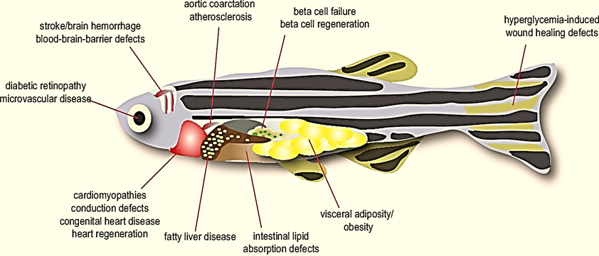
Fig.2: Parts Of Zebra Fish and its Pharmacology Activity
Electrophysiological Experiments
To measure the electrical activity of neurons in response to pharmacological interventions. Techniques like patch-clamping or multi-electrode arrays are used to measure changes in neuron firing, synaptic responses, or neurotransmitter release after administering drugs.
Neurochemical Analysis
To assess the impact of drugs on neurotransmitter levels in the brain. Drugs are given to animals, and their brain tissue is analyzed post-mortem to measure neurotransmitter concentrations using methods like high-performance liquid chromatography (HPLC) or mass spectrometry.
In Vivo Imaging Studies
To visualize brain activity and drug effects in living organisms. Positron Emission Tomography (PET), Functional Magnetic Resonance Imaging (fMRI), or optogenetics are used to study changes in brain activity or structure after pharmacological treatments.
Pharmacokinetics and Pharmacodynamics
To determine how drugs are absorbed, distributed, metabolized, and excreted in the body (pharmacokinetics), as well as the drug's effects and mechanisms of action (pharmacodynamics).
Blood samples, tissue biopsies, and brain imaging can track drug distribution, while behavioral or electrophysiological data can assess drug effects.
Neurodegenerative Disease Models
To investigate the potential of drugs to treat or prevent neurodegenerative diseases like Alzheimer's, Parkinson's, or Huntington's disease. Animal models with disease-like conditions (e.g., dopaminergic neurons in Parkinson's) are treated with drugs, and the effects on motor skills, cognition, and pathology are measured.

Fig.3: Behavioral Affinity of Zebrafish in Research
Gene-Drug Interaction Studies
To explore how genetic variations, affect the way individuals respond to pharmacological treatments. Genetic screening is performed to identify variations in genes encoding drug-metabolizing enzymes or drug targets, followed by treatment with various drugs to see how these genetic factors influence drug efficacy and side effects.
Drug Screening for Psychiatric Disorders
To identify compounds that may help treat psychiatric conditions like anxiety, depression, schizophrenia, or bipolar disorder. Researchers use behavioral tests and neurochemical assays to screen for drugs that modulate neurotransmitter systems implicated in these disorders (e.g., serotonin, dopamine, GABA).
Applications in Neurotoxicity Research
Environmental Toxins
Zebrafish are used to study neurotoxic effects of pesticides, heavy metals, and industrial chemicals. Studies have shown that exposure to mercury, lead, and bisphenol A impairs zebrafish neurodevelopment (Gonzalez et al., 2017).
Pharmaceutical Safety Testing
Zebrafish provide a cost-effective alternative for screening drugs for neurotoxic side effects before mammalian testing (McGrath & Li, 2008).
Neurodegenerative Disease Modeling
Zebrafish models of Parkinson’s, Alzheimer’s, and Huntington’s diseases help identify neurotoxic pathways and potential drug targets (Xi et al., 2011).
Advantages of Using Zebrafish in Neurodegenerative Research
Zebrafish have several advantages that make them an attractive model for neurodegenerative disease research: Genetic Similarity of Zebrafish share a high degree of genetic similarity with humans, particularly in the genes involved in neurodegeneration (Doyon et al., 2011). Transparency of the embryos of zebrafish are transparent, allowing researchers to directly observe neural development, neurodegeneration, and the effects of treatments using live imaging techniques (Richardson et al., 2017). Rapid Development of Zebrafish develop quickly, with embryos hatching within 24–48 hours, making them ideal for studying the early stages of neurodegeneration (Kalueff et al., 2014). Behavioral Assays of Zebrafish exhibit complex behaviors, including swimming, social interaction, and learning, which can be used to assess the effects of neurodegeneration on motor function and cognition (Guo et al., 2014).
Alzheimer's Disease Models in Zebrafish
Alzheimer's disease is characterized by the accumulation of amyloid-beta plaques and neurofibrillary tangles, leading to progressive cognitive decline. In zebrafish, transgenic models that express human amyloid precursor protein (APP) or amyloid-beta have been used to study AD pathology. Amyloid-beta Models using Zebrafish models expressing amyloid-beta develop plaques that resemble those found in human AD, providing a platform for studying amyloid deposition and testing potential therapies (Vandebroek et al., 2018). Fluorescent markers are often used to visualize amyloid plaques in vivo, allowing for the observation of disease progression in real-time. Tau Models using Tau protein, another key player in AD pathology, can also be expressed in zebrafish to study tauopathy. Transgenic zebrafish expressing human tau develop neurofibrillary tangles, making them suitable for investigating tau-related neurodegeneration (Batterham et al., 2018). Behavioral Impairments using Cognitive deficits in zebrafish AD models can be assessed using behavioral tests such as the Morris Water Maze and Novel Object Recognition test, which assess spatial memory and learning (Stachowicz et al., 2020).
Parkinson's Disease Models in Zebrafish
Parkinson's disease is characterized by the progressive loss of dopaminergic neurons in the substantia nigra, leading to motor deficits. Zebrafish models of PD can be created by expressing genes associated with the disease or by using neurotoxicants. Genetic Models of Transgenic zebrafish expressing alpha-synuclein, a protein implicated in PD, develop motor impairments and dopaminergic neuron loss. These fish models allow researchers to investigate the mechanisms of PD and screen for potential therapeutic compounds (Chung et al., 2016). Toxin-based Models of Zebrafish can be exposed to neurotoxins such as MPTP (1-methyl-4-phenyl-1,2,3,6-tetrahydropyridine), which induces dopaminergic neuron loss and PD-like symptoms (Abdullah et al., 2020). Motor Deficits is motor function of PD models can be assessed through swimming behavior, which is a sensitive indicator of motor dysfunction similar to human PD (Guo et al., 2014).
Huntington's Disease Models in Zebrafish
Huntington's disease is caused by a mutation in the HTT gene, which produces an abnormal polyglutamine repeat, resulting in neuronal toxicity. Zebrafish models expressing mutant huntingtin with expanded polyglutamine repeats recapitulate several features of HD.
Transgenic Models of Zebrafish expressing the mutant huntingtin protein exhibit motor deficits and cognitive impairments, making them suitable for studying the pathogenesis of HD (Di Caterina et al., 2017).
Behavioral Impairments of motor function in HD models can be assessed using swimming assays, while cognitive impairments can be measured using tests like the Novel Object Recognition and Conditioned Place Preference (Doyon et al., 2011).
Amyotrophic Lateral Sclerosis (ALS) Models in Zebrafish
ALS is characterized by the progressive degeneration of motor neurons, leading to muscle weakness and paralysis. Zebrafish models of ALS are typically created by expressing mutant SOD1 (superoxide dismutase 1), a gene associated with familial ALS. Transgenic Models of Zebrafish expressing mutant SOD1 develop motor neuron degeneration and motor deficits, providing a platform to study ALS pathophysiology and test potential therapies (Westerfield, 2007). Motor Function Testing: The progression of ALS in zebrafish can be monitored through swimming behavior, with a decrease in swimming speed or abnormal swimming patterns serving as indicators of motor dysfunction (Kalueff et al., 2014).
Techniques in Zebrafish Neurodegenerative Disease Models
Several cutting-edge techniques are used in zebrafish neurodegenerative disease research, including:
Fluorescence Microscopy is due to the transparency of zebrafish embryos; fluorescent markers are used to track disease progression in real time. This allows for detailed visualization of amyloid plaques, tau tangles, and neuronal degeneration (Vandebroek et al., 2018).
CRISPR/Cas9 is Gene-editing techniques like CRISPR/Cas9 are used to introduce specific mutations that mimic human neurodegenerative diseases, providing more accurate disease models (Chung et al., 2016).
Behavioral Assays of Zebrafish can be subjected to a variety of behavioral tests to assess the effects of neurodegeneration on motor and cognitive functions. These include swimming behavior, social interaction, and memory tasks (Guo et al., 2014).
Challenges and Future Directions
While zebrafish provide valuable insights into neurodegenerative diseases, several challenges remain:
Modeling Complex Human Pathology is the zebrafish brain is simpler than the human brain, and some aspects of human neurodegenerative diseases may not be fully replicated in zebrafish models (Di Caterina et al., 2017).
Translational Value is Although zebrafish models are useful for early-stage drug screening, translating findings to human treatments requires further validation in mammalian models (Vandebroek et al., 2018). Despite these challenges, zebrafish remain a powerful tool for understanding the mechanisms of neurodegeneration and developing novel therapeutic strategies. Despite their advantages, zebrafish have limitations, including differences in brain complexity compared to mammals and the need for standardized testing protocols (Scholz et al., 2008). Future research should focus on integrating zebrafish studies with advanced imaging and omics technologies for better neurotoxicity predictions (Van den Bulck et al., 2019).
CONCLUSION
Zebrafish have proven to be a powerful tool in neurotoxicity research, providing insights into the impact of toxic substances on neural development and function. Their continued use, combined with technological advancements, will enhance our understanding of neurotoxic mechanisms and aid in drug safety assessments. These similarities underscore the importance of zebrafish as a model organism in biomedical research, particularly in understanding human biology and developing new therapeutic strategies. Their use in research continues to expand, providing insights that can lead to advancements in medicine and pharmacology.
REFRENCES

Dr. Anamika P. K., Dr. Sandhiya, Dr. N. Deepa, Hemapriya, Jananee, Zebrafish in Neurotoxicity Research: A Comprehensive Review, Int. J. of Pharm. Sci., 2025, Vol 3, Issue 3, 462-469. https://doi.org/10.5281/zenodo.14992235
 10.5281/zenodo.14992235
