Department of Pharmaceutical Sciences, School of Healthcare and Allied Sciences, G.D Goenka University, Sohna-Gurgaon Road, Sohna, 122103, Haryana, India
Excessive melanin deposition causes hyperpigmentation and melasma, two common dermatological diseases that result in dark areas and uneven skin tone. Topical medications like hydroquinone, retinoids, and corticosteroids are examples of conventional treatments that frequently struggle with issues including poor skin penetration, irritability, and inconsistent effectiveness. Transferosomes, a new type of ultra-deformable vesicular carriers, have shown promise in recent years as a means of delivering medicinal substances to the skin's deeper layers. Phospholipids, surfactants, and edge activators provide transfersomes their remarkable deformability, which enables them to effectively pierce the stratum corneum. This characteristic reduces systemic absorption and adverse effects while increasing the bioavailability and effectiveness of encapsulated medications. Transfersomes have demonstrated better clinical results than traditional formulations when used to administer active ingredients such kojic acid, azelaic acid, arbutin, and botanical extracts for hyperpigmentation and melasma.The formulation, mode of action, and therapeutic potential of transfersomes in the treatment of melasma and hyperpigmentation are covered in this abstract. It draws attention to new developments, such as their capacity to alter melanogenesis pathways, lower oxidative stress, and offer targeted and persistent effects. Future research opportunities in improving these carriers for clinical usage are also discussed, along with issues including stability, large-scale production, and regulatory considerations. Transfersomes are a novel way to overcome the drawbacks of conventional therapies, providing a non-invasive, effective, and patient-friendly method of treating melasma and hyperpigmentation with little side effects.
Skin is the largest body organ which protects our body from many injuries and many microorganisms. There are many things which affect our skin layer and can cause many diseases like skin cancer, Hyperpigmentation, Melasma, Rashes etc. Many factors have different roles in skin issues. [1] When it comes to setting health priorities, skin conditions are sometimes viewed as minor players in the global league of illnesses when contrasted with illnesses that have a high death rate, such tuberculosis, HIV/AIDS, and community-acquired pneumonias. However, in tropical regions, skin conditions are typically among the most prevalent illnesses observed in basic care settings. [2] The Global Burden of Disease (GBD) 2013 Study examined the global disability and mortality rate from skin diseases. More than 1000 professionals from around the world have joined forces to build the GBD, which aims to develop an internally consistent, quantifiable, and systematic source of health information. [3] Furthermore, numerous drugs and dietary pollutants may trigger skin toxicity. [1] Early detection of the skin infection allows for the implementation of a recommended treatment plan that can cure the condition. A dermatologist's visual check and a comprehensive examination based on established clinical practice are typically used to make an appropriate diagnosis of the skin illness. Skin infections typically have a variety of reasons, and it is essential to recognize them as soon as possible. Inadequate care, poor hygiene habits, UV exposure, and bacterial or viral infections are among the common causes of skin infections. Since it is crucial to identify and treat skin infections as soon as possible, various diagnostic techniques have been investigated in the body of extant research. [4], [5].
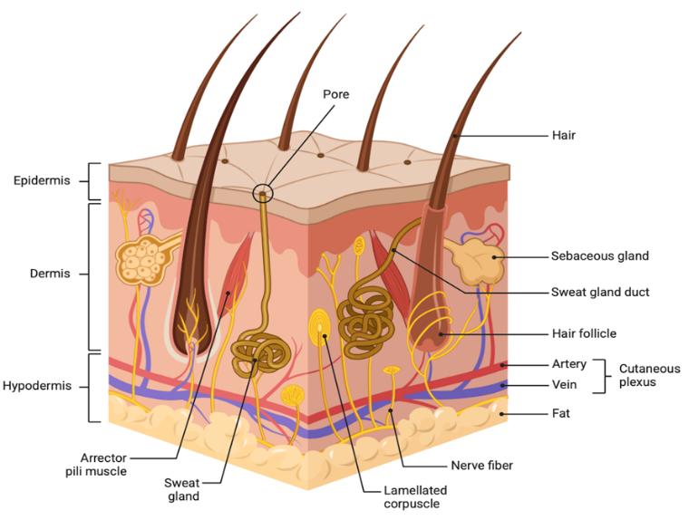
(Fig:1) Shows the skin anatomy and physiology and what are present beneath our skin or into our skin layer.
Numerous pathogens or microorganisms can cause various skin disorders; autoimmune disorders or drug toxicity may additionally be the cause; few examples of Skin Diseases are;
? Acne: it is caused by the bacteria or when skin pores get clogged with dirt and sweat. Acne vulgaris (AV) is a common chronic disorder that impacts over 85% of teenagers and young adults globally and contributes to between 15 and 20 percent of all skin disease cases. After dermatitis, it is quickly rising to the position of the second most prevalent dermatological condition. AV is an inflammatory condition that primarily affects the back, chest, and face.
? Psoriasis:Thick, red areas covered with silvery scales are the result of the rapid growth of skin cells in psoriasis, a chronic inflammatory condition. Although the occurrence might appear in many body parts, the lower back, elbows, scalp, and other skin areas are given special attention. [6]
? Eczema: The most prevalent chronic inflammatory skin condition, eczema (also known as atopic dermatitis, or AD) affects 15–30% of children and 2–10% of adults globally. [7]
Eczema typically shows up as evenly spread lesions around the body, which can display a variety of illness symptoms, including dryness and redness, with differing degrees of severity. Due to time restrictions, family practitioners still find it difficult to determine the severity of eczema. [8]
? Skin Cancer: Skin cancer is a global epidemic and a diverse group of cancers. There are two subtypes of cancer Melanoma skin cancer (MSC) and non-melanoma skin cancers (NMSCs). Approximately 90% of skin cancer are of this type. Skin cancer, of which NMSCs are the most prevalent, accounts for one out of every three malignancies diagnosed globally. [9] MSC is a malignant tumor that develops from defective and improperly proliferating melanocytes. Malignant melanoma or cutaneous melanoma are other names for it. Melanocytes, or pigment-producing cells, are derived from the neural crest. They create the brown pigment known as melanin, which gives skin its color and protects it from the sun's damaging rays. [10]
? Hyperpigmentation: Millions of people worldwide suffer from hyperpigmentation, a skin condition marked by uneven skin tone, mainly in the facial skin areas, which can have serious negative psychological and social effects. [11] Excessive melanin production by melanocytes and enhanced transfer of the melanosome, which stores melanin, from melanocytes to the keratinocytes of the epidermal melanin unit are the causes of skin hyperpigmentation. [12]
? Melasma: The most frequent cause of facial melanosis is melasma, which is characterized by the development of hyperpigmented macules on the face. When these macules are exposed to sunshine, they become more noticeable. [13] Melasma typically manifests as symmetrical, irregularly shaped macules and patches on the face. It primarily affects the face, though it can also occasionally affect the neck and forearms. [14]
2. Hyperpigmentation and Melasma:
Here we discuss one of the most common skin diseases Hyperpigmentation and Melasma. Although both are quite different from each other but yet have similarities. Millions of people worldwide suffer from hyperpigmentation, a skin condition marked by uneven skin tone, mainly in the facial skin areas, which can have serious negative psychological and social effects. Excessive melanin production by melanocytes and enhanced transfer of the melanosome, which stores melanin, from melanocytes to the keratinocytes of the epidermal melanin unit are the causes of skin hyperpigmentation. [11], [12]. This hyperactivity is affected by endocrine and autocrine mechanisms and has a strong correlation with excessive UV exposure. The distinctive hue of melanin is produced by combining two distinct pigments, pheomelanin and eumelanin. Tyrosinase (TYR), tyrosinase-related protein 1 (TRP-1), and tyrosinase-related protein 2 (TRP-2) are the three enzymes that synthesize these two colors [15]. For the reaction to take place, TYR needs to exist. It catalyzes the conversion of tyrosine to dopaquinone, the reaction's rate-limiting step, because of its monophenolase and diphenolase activity. The presence of MITF (Microftalmia Transcription Factor) initiates the manufacture of these three enzymes. A number of routes control the synthesis of enzymes necessary for the synthesis of melanin by activating or inhibiting MITF expression [16], [17].
Melasma is the type of hyperpigmentation, characterized by the development of hyperpigmented macules on the face. When these macules are exposed to sunshine, they become more noticeable. Melasma typically manifests as symmetrical, irregularly shaped macules and patches on the face. It primarily affects the face, though it can also occasionally affect the neck and forearms. Women and those with darker skin tones—particularly those with Fitzpatrick skin types IV–VI—are more likely to have this disorder. [13], [14]. According to several studies, between 1% and 50% of the general population is thought to have melasma. Furthermore, in 55% to 64% of cases, a positive family history has been documented. Several common risk factors have been found, including heredity, exposure to ultraviolet light, pregnancy, and the use of oral contraceptives or drugs like phenytoin, even if the precise etiology of melasma is still unknown [14], [18].
2.1 Pathology of Hyperpigmentation:
The overproduction of melanin or aberrant melanin deposition in the epidermis or dermis after inflammation causes post inflammatory hyperpigmentation, also known as hypermelanosis. Melanocyte hypertrophy and activity are triggered by inflammatory mediators, which raises the epidermis's production of melanin. Damage to basal keratinocytes causes them to emit a lot of melanin in deeper processes that reach the dermis. The skin becomes permanently discolored in a blue-gray hue as a result of the phagocytosed and deposited melanin. Compared to dermal hyperpigmentation, epidermal hyperpigmentation has a higher chance of clearing up. [19] [20] In other words, hyperpigmentation occur due to excess amount of melanin production into dermis layer of the skin, these melanin production can be enhanced due to UV ray (sun exposure), little tan is not hyperpigmentation as it occur on the outer layer of skin, aging, changes in hormones, and inflammation or damage to the skin. For example, exposure to the sun can result in solar lentigines, also referred to as sun spots or age spots, and hormonal factors can produce melasma, which is characterized by brown patches that are frequently observed in pregnant women or those on contraceptives. These are few common examples of how hyperpigmentation occurs.
2.2 Pathology of Melasma:
Melasma has a complex pathogenesis that is not fully understood. Melasma is more common in women as compare to the men because of the pregrancy, abortion or by using any kind of contraceptive pills, there appears to be a clear correlation between female hormonal activity and melasma. In fact, pregnancy is when half of melasma instances first appear. Furthermore, it seems that melasma lesions have an up-regulated expression of estrogen receptors. [21] [22]
There is ongoing research over the potential significance of hormone levels in the development of male melasma. Certain cosmetics, minor ovarian malfunction, and photosensitizing drugs are additional factors linked to the pathophysiology of melasma. Certain involved drugs are often the source of ochronosis, an exogenous form of melasma. Given that a substantial risk factor for the development of melasma is a family history of the condition, there is undoubtedly a role for genetic predisposition as well. Between 55 and 64 percent of melasma patients have a family history of the illness. [23] [24]. Sunlight exposure is the most significant element in the development of melasma. UV light causes the skin to produce reactive oxygen species, which in turn encourages melanogenesis. It is also known that ultraviolet (UV) radiation increases the production of [corticotropin, alpha-melanocyte-stimulating hormone, interleukin 1, and endothelin 1] , all of which help intraepidermal melanocytes produce more melanin. Melasma may also be caused by fibroblasts found in the skin's dermal layer; melasma lesions have been found to overexpress the tyrosine kinase receptor c-kit and certain stem cell factors, which are thought to promote melanogenesis. Without rigorous sun protection, melasma treatments that might work are doomed to fail. [25] [26]
2.3 Treatment for Hyperpigmentation and Melasma:
Some treatments are very costly or clinical and others are affordable for the treatment of hyperpigmentation and melasma. Although there are few ways which can prevent this from happening.
a) Topical; in this chemical or any drug applied over layer to let it absorb rapidly into the skin to show the action on target site.
b) Laser therapy; is a medical treatment that uses a highly focused, monochromatic beam of light to treat various skin and health conditions.
c) Sunscreen; this helps to prevent or reflect the UV ray from the skin so they can’t enter into the skin and harm it.
d) Microneedling; Using small needles to cause micro-injuries, microneedling improves product absorption and encourages the creation of collagen. Microneedling can help lighten pigmentation and enhance skin texture; it is frequently used in conjunction with tranexamic acid or vitamin C.
e) Oral Drugs; Tranexamic Acid Tablets: Melasma, especially hormonally driven instances, has been successfully treated with oral tranexamic acid. It is often administered under a doctor's supervision and for brief durations of time. Polypodium Leucotomos Extract: An oral antioxidant that might lessen hyperpigmentation and shield skin from UV rays.
2.3.1 Topical:
|
Drug |
Mechanism of Action |
Disadvantages |
References |
|
Hydroquinone and combinations |
Inhibit tyrosinase, reducing melanin production. |
Irritation, Ochronosis (if use it prolonged) |
[86] |
|
Retinoid |
Increases skin turnover, promoting exfoliation of pigmented cells. |
Burning, Dryness, Redness, Peeling, Irritation |
[87] |
|
Azelaic acid |
Inhibits tyrosinase and has anti-inflammatory properties |
Dryness, Burning, Irritation |
[88] |
|
Kojic acid |
Chelates Copper, Reducing tyrosinase activity |
Contact dermatitis, Irritation |
[89] |
|
Vitamin C |
Antioxidant that inhibits melanin synthesis and neutralised free radicals. |
Minimal Irritation |
[90] |
|
Cysteamine |
Antioxidant that inhibits melanin production |
Mild Irritation and odor. |
[91] |
2.3.2 Laser Therapy:
|
Non-ablative fractioning laser |
It works by creating microthermal zones that promotes controlled dermal remodeling and epidermal renewal. |
- Risk of post-inflammatory pigmentary changes in darker skin tones - Burns, erythema, pain - Requires optimization based on patient’s skin type |
[89] |
|
Low-fluence Q- switched laser |
High energy pulses that target melanin deposits energy through a photoacoustic effect. The laser is effective is treating deeper pigmentation while reducing the risk of epidermal injury. |
High rebound pigmentation if aggressive settings used.
|
[90] |
|
Picosecond laser |
It delivers ultra-short pulses that results in a photoacoustic effect, fragmenting pigment particles with minimal thermal damage to surrounding tissues. |
Its protocols are still being optimized and patients should be counseled about the possibilities of multiple sessions. |
[91] |
2.3.3 Chemical Peel:
|
Superficial Peel (Glycolic Acid and Lactic Acid) |
These are commonly used for epidermal melasma and mild-to-moderate hyperpigmentation. With little recovery time, they gently exfoliate the outer layers of the skin. |
Erythema, Redness, Dryness, Burning |
[92] |
|
Mild-depth Peel (Trichloroacetic Acid) |
More obstinate pigmentation can be addressed with TCA peels when applied at controlled concentrations (10–35%). To lessen the risk of PIH in patients with darker skin, however, lower doses and close observation are required. |
Redness, Burning, Dryness, Peeling, Irritation |
[92] |
3. Transferosomes:
The vesicular carrier systems that have drawn the most attention in recent decades as a transdermal medication delivery method are liposomes and niosomes. The characteristics of vesicle architectures have been understood by researchers in order to tag the vesicles for cell specificity and improve drug administration within their cavities. Vesicles are used in transdermal drug delivery because of their composition, which enhances penetration and acts as a drug carrier to transport entrapped drug molecules over the skin. A tailored transdermal medication delivery method uses transferosomes as its carrying body. These particular liposomes are made up of an edge activator and phosphatidylcholine. Phospholipid vesicles are also used by this technology as a transdermal medication delivery device. It enters the stratum corneum by the creation of an "osmotic gradient" either intracellularly or transcellularly. Transferosomes provide several benefits, including a broad spectrum of solubilities, improved penetration, and biocompatibility and biodegradability. Transferosome benefits include oxidative degradation, cost, etc [27] The conventional rotary evaporation sonication procedure was used to create the transfersomes. It has surfactant, phospholipids, and the medication that was made. The following are the transferosome evaluation parameters: Vesicle shape, entrapment efficiency, drug content, turbidity measurement, degree of deformability or permeability measurement, vesicle size distribution and zeta potential, Occlusion effect, penetration ability, charge density and surface charge, Physical stability, in vitro skin penetration studies, and in vitro drug release. Transferosomes can be used for transdermal vaccination, target delivery to peripheral subcutaneous tissues, controlled release, and the transportation of big molecules and weight substances. [27] The word Tranfersomes is made of two different words in Latin ‘tranferre’ refers to ‘to carry across’ and in Greek ‘soma’ refers to ‘the body’ which translates to ‘carrying body’. The active ingredient is contained within the core or between the bilayers, depending on its lipophilicity. When applied topically, transferosomes have a greater ability than liposomes to reach deeper, whole skin regions [28].
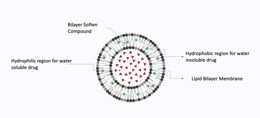
(Fig:3) Structure of Tranferosomes
The transfersome is a synthetic vesicle that is suitable for targeted and regulated drug administration since it resembles the characteristics of exocytotic cell vesicles. [29]
Extremely adaptable and resilient under stress, these are a complex aggregate. The vesicle's local composition and bilayer form independence allow it to be both self-regulating and self-optimizing. This makes it possible for transferosomes to effectively pass through a number of transport barriers before serving as a drug carrier for targeted, non-invasive medication delivery and prolonged release of therapeutic agents. [30]
Tween80, span80, sodium cholate, sodium deoxycholate, and other edge activators are frequently used in this kind of drug delivery system to promote flexibility, and soy phosphatidylcholine, egg lecithins, cholesterol, and other phospholipids are frequently utilized for vesicle formation. [31]
Few Examples of Transferosomes:
1. Egg Phosphatidyl CholineSoya Phosphatidyl choline, dipalamitoylphosphatidyl choline: it belongs to class phospolipids and used as vesicle forming component.
2. Ethanol, methanol, isopropyl alcohol, chloroform: it belongs to class solvents and used as solvent.
3. Sod. Cholate, Sod. Deoxycholate, Tween-80, Span 80, Tween 20: it lies under Edge Activators category (surfactant vesicles forming component)
4. Saline phosphate buffer (pH 6.4), Phosphate buffer pH 7.4: belongs to class buffer agent used as As a hydrating medium
5. Rhodamine-123 Rhodamine –DHPE Fluorescein –DHPE Nile-red: it belongs to class dye used a For CSLM study.
3.1 Advantages of Transferosomes: [27], [28], [29], [30], [31], [32], [33], [34],
1. The only drug delivery method that can transport therapeutic substances across a broad range of solubility is transferosomal carriers, which are composed of both hydrophilic and hydrophobic units.
2. Transferosomes are able to pass through narrow skin barrier constrictions, which are 5–10 times smaller than vesicle size, due to their ultra-deformability and elastic properties.
3. High vesicle deformability can be used for both topical and systemic therapy, allowing drugs to pass through the skin without significantly destroying vesicles.
4. These carriers are incredibly versatile and effective at holding a variety of agents, regardless of their size, shape, molecular weight, or polarity.
5. Natural phospholipids and edge activators are used in the preparation of these units. They are both biocompatible and biodegradable.
6. Just like other active substances, transferosomes are used to transport many proteins and peptides, insulins, cortico-steriods, anesthetics, NSAIDS, antitumor drugs and herbal treatment. Transferosomes are the best option for achieving both an expected and prolonged duration of activity and a sustained drug release.
7. These can improve the site sele 9 activity of bioactive agents and increase transdermal flow.
3.2 Limitations of Transferosomes:
1. The chemical instability of transferosome formulations is caused by their susceptibility to oxidative stress. The oxidation of transferosmes is significantly decreased when aqueous solutions are degassed and purged using inert gases like argon and nitrogen.
2. Getting pure natural phospholipids, which is hard to make, is another problem with employing transferosomes as drug delivery vehicles. Therefore, artificial phospholipids could be utilized as an alternative.
3.3 Composition and Mechanism of Action of Transferosomes:
A transferosome is a mixed lipid aggregate which is optimized and self-adapting.
As "edge activators," the surfactant molecules provide the transferosomes ultra-deformability, apparently allowing them to move through stratum corneum channels that are less than a tenth of the transferosome's width. The researchers who created them suggest that transferosomes up to 500 nm in size can naturally squeak across and penetrate the stratum corneum barrier in circumstances when liposomes are too big to fit through pores smaller than 50 nm. [35] Researchers hypothesize that the "transdermal gradient," resulting from the variations in water content between the fluid viable epidermis (almost 100%) and the almost dry epidermal surface (roughly 20% water), is what accelerates penetration into the skin. The proper proportion of surface-active agent is used to make transferosomes deformable. The formulation of transferosomes depends upon the concentration of the surface-active agent since these substances give vesicle membranes flexibility at sublytic concentrations and eliminate vesicles at larger concentrations. [44] When compelled against or forced into a small pore, the ultra-deformable transferosomes may modify their membrane composition locally and reversibly because to the consequent flexibility of the transferosomal membrane, which additionally minimizes the chance of complete vesicle rupture in the skin. This significantly reduces the energetic cost of membrane deformation and enables the very flexible particles that result to enter the pores and then swiftly and effectively flow through them. [46] At least one amphiphat (such as phosphatidylcholine) that self-resembles a lipid bilayer in an aqueous solvent and closes into a simple lipid vesicle makes up the carrier aggregation. [45] The flexibility and permeability of lipid bilayers are significantly enhanced by the inclusion of at least one bilayer softening component (such as an amphiphilic medicine or a biocompatible surfactant). Therefore, the transferosome vesicles can quickly and readily conform to their ambient shape by maximizing the ensuing flexibility and permeability. As a result, they can also modify the local concentration of each bilayer component in accordance with the bilayer's local stress. These vesicles' fundamental structure is largely comparable to that of liposomes. [47] The enhanced ability of transferosomes to bind and retain water is another advantageous effect of strong bilayer deformability. Avoiding dehydration is always the goal of an ultradeformable, highly hydrophilic vesicle; this may include a transport mechanism that is similar to but distinct from forward osmosis. In order to provide proper hydration, a transferosome vesicle put to an open biological surface, such as non-occluded skin, has a tendency to cross its barrier and move into the deeper strata that are rich in water. For the underlying hydration affinity and gradient to remain in place, barrier penetration requires reversible bilayer deformation, but neither the vesicle integrity nor the barrier characteristics must be unacceptable compromised. The transferosome must locate and enforce its own path through the organ because it is too big to diffuse through the skin. Therefore, the use of transferosomes in drug delivery depends on the carrier's capacity to enlarge and penetrate the hydrophilic pores in the skin or another barrier. The drug molecules can then spread and eventually bind to their target thanks to the drug carrier's following, progressive agent release. Unless the vesicle is actively taken up by the cell through a process known as endocytosis, drug transport to an intracellular action site may also include the fusing of the carrier's lipid bilayer with the cell membrane. [48] [49] [50]
Mechanism of Action of Transferosomes (Penetration):
Under the right circumstances, transferosomes can spread 0.1 mg of lipid every hour and square centimeter across healthy skin. Compared to the value usually influenced by the transdermal concentration gradients, this value is significantly higher. Naturally occurring "transdermal osmotic gradients," or another, much more pronounced gradient across the skin, are the cause of this high flux rate. Water loss via the skin is prevented by the osmotic gradient created by the skin penetration barrier, which also keeps the water activity difference between the virtually dry stratum corneum near the skin surface (15% water content) and the viable portion of the epidermis (75% water content). [51] [52] [53]
Even when the transdermal water loss is abnormally large, surrounding air acts as a perfect sink for the water molecule, which makes this gradient extremely stable. Every polar lipid contains some water. This results from the hydrophilic lipid residues' energetically advantageous interaction with their proximal water. Therefore, the majority of lipid bilayers naturally withstand an induced dehydration. As a result, every lipid vesicle derived from polar lipid vesicles migrates from a more arid area to one where the concentration of water is high enough. Lipid vesicles sense this "osmotic gradient" and attempt to avoid total drying by traveling along it when a lipid suspension (transferosome) is applied to the skin surface that has been partially dehydrated due to water evaporation loss. [54] Because surfactant-based transferosomes have better rheological and hydration characteristics than the vesicles that give them their greater deformability, they can only accomplish this if they are sufficiently deformable to fit through the skin's narrow pores. Standard liposomes and other less deformable vesicles are restricted to the skin's surface, where they completely dehydrate and fuse, giving them less penetration power than the transferosome. This allows transferosomes to be as flexible as possible, allowing them to fully use the transepidermal osmotic gradient (water concentration gradient). Transferosomes squeeze themselves along the intracellular sealing lipids of the stratum corneum to get past the barrier to skin penetration. [55] Researchers still not found the mechanism to enhance the delivery of the active substances across the skin. Two mechanisms of action have been proposed:
1. After penetrating the skin, transferosomes stay intact and function as drug vectors.
2. By upsetting the stratum corneum's highly ordered intercellular lipids, transferosomes serve as penetration enhancers, promoting the drug molecule's entry into and passage through the stratum corneum.
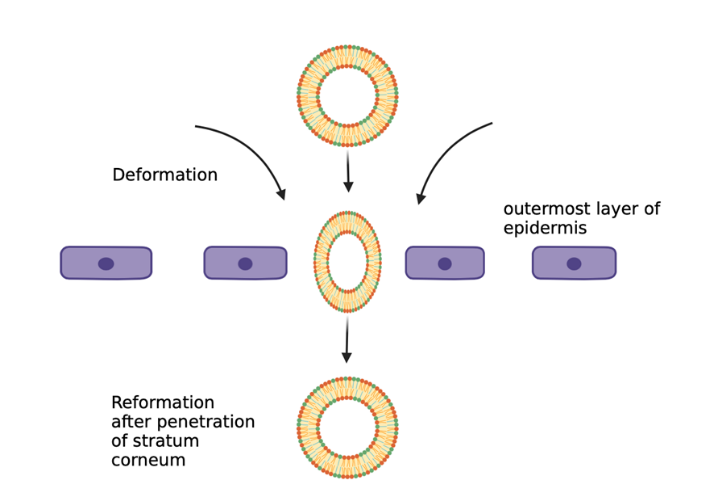
Mechanism of Transferosomes on Skin:
Transfersomes are very advanced phospholipid vesicles in transdermal drug delivery systems. Depending on the approach of administration or application, they can deliver the medicine to or through the skin in a reproducible and highly successful manner because of their self-optimized and ultra-flexible membrane properties. Vesicular transfersomes are appropriate for skin penetration because they are more porous than regular liposomes. Transfersomes squeeze along the intracellular locking lipid of the stratum corneum to get beyond the barrier of skin penetration. Because of the increased vesicle deformability that permits entry due to the surrounding mechanical stress, these are self-adapting characteristics of transfersomes. The combination of suitable surface-active chemicals with phospholipids regulates the transfersome membrane's flexibility. When used in a non-occlusive setting, the ensuing transfersome membrane flexibility allows transfersomes to follow the natural water gradient across the epidermis and reduces the risk of entire skin vesicle rupture. There are two internal lipid pathways via which transfersomes might spontaneously penetrate the intact stratum corneum, and the properties of their bilayers differ. According to confocal microscopy research, undamaged liposomes remain on the upper stratum corneum layer rather than penetrating the granular layer of the epidermis. The rate of drug release and deposition to the target site can be modified by altering the vesicular composition or surface properties. [56] [57] [58] [59] [60]
As you can see the Fig 4:
3.4 Preparation Methods of Transferosomes:
There are various methods by which you can prepare transfersomes:
1. Ethanol Injection Method
2. Thin Film Layer Method
3. Rotary Film Evaporation Method
4. Reverse Phase Evaporation Method
5. Freeze Thaw Method
3.4.1 Ethanol Injection Method:
A straightforward and popular approach for making transfersomes is the ethanol injection method. It involves infusing an ethanol-dissolved lipid solution into an aqueous phase, which causes vesicles to form on their own. [36] [37] [38] [39]
Material Required:
|
Phospholipid |
Phosphatidylcholine or lecithin |
|
Edge Activators (Surfactant) |
Sodium cholate, Tween-80, Span-80 |
|
Organic Phase |
Ethanol |
|
Aqueous Phase |
Distilled Water or PBS or Buffer Solution |
|
Active Ingredients |
Drug (Hydrophilic or Lipophilic) |
3.4.2 Rotary Film evaporation method:
One of the most typical techniques to produce transferosomes is the Rotary Film Evaporation Method. In order to produce multilamellar vesicles (MLVs), which can subsequently be converted into transferosomes, a thin lipid film must be formed and hydrated with an aqueous phase. [40] [41]
3.4.3 Reverse Phase Evaporation Method:
The versatile method to generate transferosomes is the Reverse Phase Evaporation Method (RPEM). It has ideal encapsulation efficiency and operates particularly well for encapsulating medicines that have properties that are both hydrophilic and lipophilic. This process creates vesicles by first producing a water-in-oil (w/o) emulsion and then removing the organic solvent. [42] [43]
3.5 Optimization of the formulation containing transferosomes:
The preparation and properties of the transfersomes may be influenced by several process variables. As a result, the training procedure was refined and verified. The formulation manufacturing method in consideration affects the process factors.
1. Lecithin: surfactant proportion
2. Effect of different solvents
3. Effect of different surfactants
4. Hydration medium
Optimization of the drug was accomplished via choosing the trap effectiveness of the drug. [61] [62] [63]
The phospholipid edge activator ratio should be precise since it significantly affects vesicle size, penetrating ability, and entrapment effectiveness. Generally speaking, it has been proposed that employing a surfactant with a higher concentration could lower EE. The arrangement of surfactant molecules inside the vesicular lipid bilayer structure may have increased the permeability of the vesicular membrane, creating pores that would improve fluidity and enable the rapid leakage of the entrapped drug. [72]
They use solvents such as methanol or ethanol. The choice of solvent is influenced by the compatibility and solubility of each formulation component in the solvent. To increase the stability with hydration all excipients and medication should properly dissolved into the solvent to form produce a clear and transparent solution. The use of solvents in formulation can potentially increase drug flux through membranes by acting as penetration enhancers. According to Williams and Barry, ethanol was used in numerous studies to enhance the transit of levenorgestrel, hydrocortisone, 5-fluorouracil, and estradiol through rat skin. [73] [74]
The medium is either water or a saline phosphate buffer (pH 6.5–7). The pH level of compositions must be appropriate in order to balance biological properties and biological uses, as well as the administration route. Unionized medications are last membrane-bound to the phospholipid bilayer and enter the cell through the intracellular pathway. The lipid bilayer of transferosomes is comparable to the phospholipid layer of a cell membrane. [75] [76]
3.6 Evaluation Parameter of Transferosomes:
Mostly Compare with Liposomes, Niosomes and Micelles.
In Dynamic light scattering technique (DLS) using Malvern Zetasizer's computerized examine system is used to determine vesicle size, size distribution, and zeta potential.
The percentage of trapping of the drug adds is a measure of its effectiveness. By initially separating the unentrapped drug, the mini-column centrifugation method was used to calculate the entrapment efficiency. Using 0.1 percent Triton X-100 or 50 percent n-propanol, the vesicles were interrupted after centrifugation. The effectiveness of encapsulation is expressed as: Efficiency of encapsulation = (complete quantity of encapsulated / amount of drug added) *100
Vesicle diameter is often determined using the DLS technique or spectroscopy of photon correlation. The produced sample in distilled water was filtered through a 0.2 mm membrane filter and diluted with filtered saline using photon correlation spectroscopy or DLS measurements. It is common practice to visualize transfersome vesicles using phase contrast microscopy and transmission electron microscopy (TEM).
By analyzing the vesicles' size and structure over time, one can ascertain their stability. Mean size and structural alterations were assessed by DLS and TEM, respectively.
This decision has significance for improving the method's structure and other elements. Transferosome formulations (without sonication) can be examined using optical microscopy with a hemocytometer after being diluted five times with 0.9% sodium chloride solution. Using the following formula, transfersomes are numbered and computed in 80 small squares: (Total transfersomes vesicle counted times x D.F x 4000) /total squares counted is the total number of transfersomes per cubic mm.
The drug content of a gel formulation needs to be processed by calculating the drug using the proper analytical procedures and converting equivalent weight to one-unit dose. Due to the viscosity of the gel, it is necessary to calculate at least six samples in order to ensure uniform drug distribution inside the gel. We can use advance modified technique high-performance fluid chromatography (HPLC) technique using a UV sensor, column oven, auto sample, pump, and computerized evaluation program.
The turbidity of the medication in an aqueous solution can be determined using a nephelometer.
One of the significant and unique factors for characterisation in the setting of transfersomes is the study of permeability. Pure water is used for the traditional deformability analysis. The process of making transfersomes involves passing them through a large number of known-sized pores (via a sandwich of several microporous filters, with pore diameters ranging from 50 nm to 400 nm, depending on the suspension starting transfersomes). Following dynamic light dispersion (DLS) studies, particle size distributions are seen.
Fluorescence microscopy can be used to measure the ability of transferosomes to penetrate.
For the calculation of surface charge and density charge of transferosomes are done via using zetasizer.
Skin occlusion is thought to be beneficial for medication penetration in conventional topical formulations.
However, the same shows detrimental effects on elastic vesicles.
The primary mechanism for vesicle penetration through the skin, from its relatively dry surface to deeper water-rich regions, is water hydrotaxis, or motion in the direction. Because occlusion stops bodily water from evaporating, it affects the hydration forces.
They were kept in sealed glass ampoules after the initial percentage of the medication trapped in the formulation was established. For a minimum of three months, the ampoules were kept at 4 ± 20C (refrigeration), 25 ± 20C (room temperature), and 37 ± 20C (body temperature). After 30 days, samples from each ampoule were examined to assess medication leakage.By keeping the initial drug trap at 100%, the percentage of drug loss was computed.
In order to determine the rate of penetration, in vitro drug release is studied. Prior to more costly in vivo investigations, the formulation is optimized using data from in-vitro research and the time needed to establish steady state permeation and permanent permeation flux.
By incubating the suspension at 32°C and extracting the free drug from samples taken at various intervals, minicolumn centrifugation is used to determine the penetration rate. Secondarily, the amount of drug released is calculated using the volume of drug entrapped at zero times.
A goat's skin is used in a phosphate buffer that has a pH of 7.4. Regular saline is used to moisturize the skin after the hair has been removed. Fat tissues should be removed with the cotton swab. A 50 mL column and an effective receiver compartment area of 2.50 cm2 are features of a modified Franz diffusion cell. With the stratum corneum facing the donor compartment and a stirring speed of 100 rpm, the skin can be maintained at a low temperature in IPA. Regularly, a 1 mL aliquot is taken and replaced with brand-new phosphate buffer.
3.7 Application of Transferosomes:
As drug delivery vehicles, transferosomes can strengthen labile medications and deliver the provided drug with regulated release.
Such large molecular weight medications applied topically via transferosomes are an efficient non-invasive treatment approach. Insulin is often delivered via a cumbersome subcutaneous route. All of these problems are resolved when insulin is encapsulated into transferosomes (transfersulin). The first indication of systemic hypoglycemia appears 90 to 180 minutes after transfersulin injection on undamaged skin, depending on the specific carrier structure. [76]
Corticosteroid delivery also uses transferosomes. By maximizing the medicine's epicutaneous dose, transferosomes improve the site specificity and overall drug safety of delivering corticosteroids to the skin. Transferosome-based corticosteroids have a dose that is several times lower than the formulation currently used to treat skin conditions and nevertheless have biological activity. [77]
Transfersomes were frequently employed as a means of transporting peptides and proteins. Large biogenic molecules like proteins and peptides are extremely difficult to transfer into the body and are totally broken down in the GI tract when taken orally. certain are the explanations for why certain proteins and peptides must still enter the body through injections. Numerous techniques have been developed to improve these conditions. Transferosome bioavailability is roughly equivalent to that of the identical protein suspension injected subcutaneously. The transferosomal preparations of this protein also produced a potent immune response following repeated epicutaneous application. [78] [79] [80]
Transdermal technology has been tried for transdermal administration of anti-cancer medications like methotrexate. The results were encouraging. This has provided a novel approach to the treatment of skin cancer in particular. [80]
In this regard, Xiao-Ying et al. produced transfersomes of capsaicin, which exhibit superior topical absorption when compared to pure capsaicin. Transfersomes can enter the stratum corneum and provide the nutrients locally to sustain its activities, resulting in skin maintenance. [81] [82]
4. Review work done on Transferosomes:
1. Report by Mbah CC et al. {2015} Using phopholipon 90 H and different concentrations of a variety of surfactants, including sorbitan monolaurate, SLS, and Tween 80, as well as increasing exicipients through solvent evaporation, the NIPRD-AF1 Transfersomal Veasicular Carrier System was developed. The British Pharmacopoeia standard and the fusion process were used to create the AF1 ointment formulation. It was found that Transferosomal AF1 had superior penetration when compared to the ointment AF1 formulation. [83]
2. Diclofenac sodium transferosomes were developed and assessed by Shabana Syeda et al. for transdermal medication delivery. These were created using a thin-film hydration method that involved lecithin in the organic phase and a ratio of surfactants (Span 20, 60, and 80). Because of its high entrapment efficiency and stability, span 60 was found to be the best surfactant for creating diclofenac sodium tranferosomes. [84]
3. In order to give the lipid vesicles the required range of particle size, PDI, Zeta potential, and ex vivo flux for crucial transdermal permeation and formulation stability, Mahmood S et al. reported the nano transferosomes vesicles of raloxifene HCL using phopholipon 90 G, sorbitan 80, and a rotary evaporation method. The new transfersomal formulation of sorbitan 80 and raloxifene HCL exhibited better results when applied transdermally. [85]
CONCLUSION
Excessive amounts of Melanin Production cause Hyperpigmentation and Melasma into the skin. It can be caused by many things, the most popular one is UV Rays. It can be by sunlight or Tan machine or laser. First Stage of darkern of the skin by melanin is Hyperpigmentation and if you do not treat it it slowly creates a dark permanent spot called Melasma, by the time of treatment it can fade but can not reduced properly. Transfersomes based formulation can work on Hyperpigmentation and Melasma nicely because it has the ability to go into deeper layers of the skin and penetrate drugs on the site of action. Transfersomes are also used in many other treatments like cancer. Niacinamide, also known as Nicotinamide (Vitamin B3), is mostly used in the cosmetic industry. Its ability to reduce spots and hyperpigmentation from the skin also promotes hair growth, useful for acne and inflammatory skin conditions, and can be very useful for anti-aging products. It can also be taken as multivitamin supplements for vitamin deficiency. Niacinamide based transfersomes work very well for the treatment of hyperpigmentation and Melasma as Tranferosomes helps it to go deeper into the layer and there it will work on the reduction of Melanin.
REFERENCES

Dakshita Dutta, Yashmi Jain, Manish Yadav*, Transferosomal Niacinamide: A Novel Approach for Hyperpigmentation and Melasma Treatment, Int. J. of Pharm. Sci., 2025, Vol 3, Issue 2, 1920-1936. https://doi.org/10.5281/zenodo.14913503
 10.5281/zenodo.14913503
