Department of Pharmaceutics, Centre for Pharmaceutical Sciences, University College of Engineering, Science and Technology, Jawaharlal Nehru Technological University, Hyderabad, Telangana, India 500085.
Transdermal drug delivery techniques may avoid gastrointestinal breakdown and hepatic first-pass metabolism, they have become a viable substitute for conventional methods of delivering peptides, vaccines, and other biologics. The potential of transdermal patches for macromolecule delivery has been greatly increased by recent developments in skin penetration enhancers, nanocarrier systems, and microneedle technologies. With an emphasis on vaccination applications, this study outlines the latest developments in transdermal patches for peptide and biologic administration. We look at many methods, such as physical, chemical, and biological ones, used to get beyond the skin's strong barrier qualities. Achieving uniform bioavailability, guaranteeing the stability of complicated biologics, and scaling up manufacturing are some of the hurdles that still exist despite promising preclinical and early clinical outcomes. In order to support the clinical translation of transdermal technology for biologic therapies, we wrap up by highlighting recent developments and identifying important areas for further study.
1.1 Transdermal Drug delivery (TDDS)
Although transdermal drug delivery has progressed the practice of medicine, it is not yet being fully realized as a viable replacement for hypodermic needles and oral availability [1]. In clinical examples, first generation transdermal delivery approaches are being explored via very small dosages of lipophilic, low molecular weight drugs [2]. Clinical products have been developed utilizing second generation systems such as iontophoresis, chemical enhancers and non-cavitational ultrasound with real time control of distribution rate [3]. In addition to possibly being a favourable substitute for oral drug delivery, transdermal dispersion will soon offer an alternative to hypodermic injections [4]. Transdermal drug delivery devices (TDDS) produce systemic effects by administering medicinal chemicals via the skin, hence removing the requirement for oral or parenteral administration. Longer drug release, simplicity of administration, improved patient compliance, and the avoidance of gastrointestinal degradation and hepatic first-pass metabolism are just a few advantages that these systems provide [5]. Since 1979, when the first scopolamine transdermal patch was authorized, TDDS has proven itself as a reliable technique for administering tiny compounds including nitroglycerin, fentanyl, and nicotine. A major obstacle to medication penetration is the epidermis, especially the outermost stratum corneum. Traditional TDDS has only employed small, lipophilic substances with molecular weights under 500 Da and adequate skin penetration [6]. Aiming to broaden this delivery method to encompass macromolecules including proteins, peptides, vaccines, and monoclonal antibodies, recent developments in transdermal technology. However, these biologics present serious issues due to their large molecular size, hydrophilia, and susceptibility to enzymatic degradation factors that often restrict passive diffusion through intact skin.
Importance & Benefits of Transdermal delivery of Macromolecules (Peptides, Biologics, Vaccines).
Peptide-based therapeutics comes from a fusion of disciplines. The emergence of new peptide-based drugs has been significantly increased through a combination of modern drug design and manufacturing processes, delivery systems, display library technology, bioengineering improvements, and artificial intelligence [7], effectively addressing their intrinsic problems of rapid clearance and breakdown that requires subcutaneous injection, thus causing some patient discomfort [8]. The transdermal delivery of macromolecules, including biologics, vaccines and peptides, may signify a paradigm shift in drug delivery (figure 1) [9]. Because of their great specificity and low off-target effects, these high-molecular-weight therapies have completely changed the available treatments for cancer, autoimmune diseases, infectious diseases, and chronic illnesses [10].
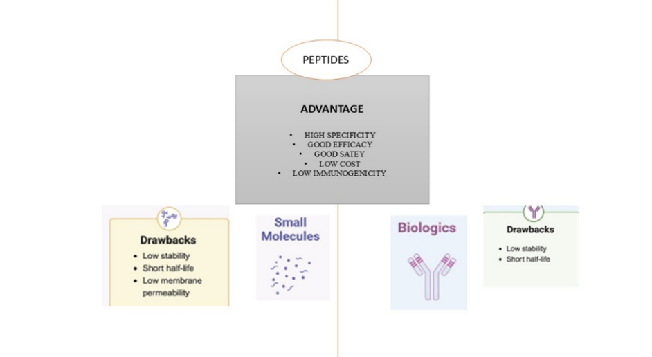
Figure 1. benefits and drawbacks of medications based on peptides. Three different types of therapeutic agents are represented by peptides, small molecules, and biologics. Comparing peptides to the other two classes reveals certain advantages. However, compared to peptides, small molecules have some drawbacks, while biologics have additional restrictions.
This review's main goal is to critically assess the state of transdermal patches for the administration of biologics, vaccines, and peptides [11]. Despite the increasing clinical importance of macromolecular therapies, formulation and delivery issues continue to hinder their efficient use. Consolidating recent developments, highlighting significant obstacles, and highlighting prospects for innovation in transdermal drug delivery systems (TDDS) that target high-molecular-weight molecules are the objectives of this study [12].
2. Skin Barrier and the challenge of Macromolecular Delivery
Gels are shear-thinning, semisolid formulations that are simple to apply and guarantee quick medication release as soon as they touch the skin. Effective facilitation of transport of hydrophilic macromolecules leads to improved skin penetration and decreased greasiness. However, because gels have a lower drug-loading capacity than other formulations, they might need to be administered more often [13]. A new method for the continual local delivery of anti-oncogenes. This method used a combination of the folate-poly (ester amine) (FA-PEA) polymer with DNA and a thermoresponsive PECE hydrogel. The results demonstrated the delivery of the biodegradable FA-PEA polymer targeted gene delivery. The composite may also prolong gene release when considering the PECE hydrogelation. This study demonstrated that it could be possible to perform targeted gene delivery using this method [14]. In an additional study developed two gelators, dextran-2-naphthylacetic acid conjugate (Dex-NAA) and hyaluronic acid-β-cyclodextrin conjugate (HA-CD). Because 2-NAA and β-CD, have a strong host-guest interaction, the gelators formed the supramolecular hydrogel. A variety of hydrogel characteristics can be changed by changing the HA-CD and Dex-NAA ratios. Altogether, the results suggest that the HA-Dex hydrogel may be used in tissue engineering as a cell scaffold [15]. On the contrary, according to the semisolid, oily association of ointments may exert a protective barrier on the skin, and reduce moisture loss while facilitating the infusion of medicinal components [16]. Lipophilic substances, such as fat-soluble vitamins (like A and D) [17] and certain hormones (like testosterone) [18], can be effectively transported by ointments. Even while ointments can provide extended medication release, some patients may not take them well and they may leave a sticky residue [19].
To get beyond the challenges posed by the skin barrier and enhance the topical delivery of macromolecules, mechanical-based techniques are essential. Iontophoresis, needle-free jet injection, laser-assisted delivery, microneedles, ultrasound-assisted delivery, and electroporation are some of these techniques also known as Strategies for Enhancing Transdermal Delivery of Macromolecules [20-23].
Iontophoresis improves transdermal medication delivery by electroosmosis and electrorepulsion [24]. Electroosmosis also affects the movement of ions such as sodium (Na?) and chloride (Cl?) in the skin by moving water from the anode to the cathode under the application of an electric current. When water moves in the form of bulk flow from anode to cathode, the negatively charged skin assists this bulk flow of positively charged water. Thus, the system facilitates the movement of uncharged water, which can facilitate the transport of other substances over the skin. [25]. Iontophoresis and chemical penetration enhancers can increase the delivery of drugs across the skin compared to independent drug delivery methods. The influence of iontophoresis and 5% terpenes (limonene, carvone, thymol, and cineole)/ethanol on the penetration of LHRH through the pig epidermis. When combined with ethanol, terpenes increased LHRH flow relative to the control; limonene shown the greatest benefit, increasing by 4.7 times [26].
Delivery with ultrasound assistance Lower frequencies, between 20 kHz to 3 MHz, are commonly used in ultrasound-assisted medication delivery to improve skin permeability and enable transdermal drug administration. These frequencies of ultrasound waves momentarily break down the skin's protective layer, improving medication absorption via the skin [27]. The degree of cavitation is dependent on the frequency of the ultrasonic waves. Low frequency, high-intensity ultrasonic waves (2-50 W/cm2) create very permeable localised transport zones (LTRs) in the stratum corneum. Sonophoresis relies on the bona fide cavitation as part of, and increases transdermal dispersion with heat effects and radiation pressures [28].
A potential noninvasive medication delivery technique with several advantages over transdermal drug administration is needle-free jet injection. It helps patients feel less pain and anxiety by doing away with the need for conventional needles, which is particularly beneficial for kids and those who are afraid of needles [29]. Better drug bioavailability and quicker drug delivery are made possible by this technique, which delivers drugs rapidly into the dermal or subcutaneous layer. Needlestick-free jet injection is helpful in many medical applications, including the delivery of insulin, the administration of local anaesthetics, and vaccination, despite a number of disadvantages, including skin irritation and injection site bruising [30].
These minuscule instruments create microchannels by puncturing the stratum corneum, which facilitates the delivery of medications, enhances transdermal absorption, and permits the injection of vaccinations [31]. Then, with the help of the aqueous pores, the drug is released from the patch into the deeper layers of the skin [32]. Arrays, which are collections of 150–650 microneedles/cm2, comprise the 200–750 microneedles [33].
yttrium-gallium garnet (Er: YAG, 2940 nm), yttriumscandium-gallium-garnet (YSGG, 2790 nm), and CO2 (10,600 nm) lasers for laser-assisted delivery leverages the ability of these lasers to create transport by altering the integrity of the skin barrier. There are three primary mechanisms of action associated with laser-assisted delivery: direct ablation, photothermal effect, and photomechanical wave (PW)-induced breakdown.
the laser travels at supersonic speed away from the skin's surface, breaking the target into small fragments. By lowering the epidermal barrier, this makes it easier for macromolecules to move effectively. A compressive, wideband, unipolar wave generated by lasers is known as a photomechanical wave [35]. The SC cannot be peeled, but the skin's surface and cell membrane are temporarily permeable. The creation of transient drug delivery holes is facilitated by the SC's lipid disruption and lacunar space expansion.
The formation of peptides, adjuvants, mechanical improvements, and nanosystems is evidence of the creative methods scientists are using to get over these obstacles. Particularly intriguing are adjuvants based on hyaluronic acid and chemical penetration enhancers, which alter the characteristics of the epidermal barrier to promote medication penetration. Conversely, peptides provide an advantage by improving penetration and facilitating distribution. By physically piercing the epidermal barrier, mechanical techniques like iontophoresis, ultrasound, and microneedles offer substitute tactics [36].
2.1 Structure and Function of the Skin
For the past 150 years, scientists have explored the structure and function of skin [39]. Primarily, the stratum corneum (SC), as the outer layer of skin and the boundary between skin and the environment, has more recently been concluded to serve as a barrier to limit excessive water loss and unwanted chemical loss from the body [37].
The main developments in our knowledge of this powerful membrane are outlined in this review. The SC's structure is described, along with methods for visualizing the barrier.
2.1.1 Structure of the Stratum corneum
The skin serves as the body's main line of defence and is composed of three main layers: the epidermis, dermis, and hypodermis. The stratum corneum (SC), the outermost layer of the epidermis, is the primary barrier to the dispersion of transdermal medications. The dead keratinocytes, or corneocytes, that make up the SC's structural core are densely packed and encased in a lipid matrix. Molecular mobility is restricted by this "brick-and-mortar" paradigm [38].
2.1.2 Visualization tools to study the SC
The primary visualization methods used to examine the SC thus far can be categorized as follows:
We have assessed advances in our understanding of the skin and the SC, which is its primary barrier component. Key visual aids that have contributed to our present understanding are emphasized, and the crucial function of lipids has also been taken into account. The SC's reactivity to experimental disruption and control over skin barrier formation emphasize its dynamic nature. The largest problem facing the business is extrapolating from animal data and various skin models utilized for human applications (SC membranes, cadaver skin,and skin equivalents). While the latter could be very different from human tissue, the former might have built-in defects or not have a repair response at all.
Transdermal drug delivery systems (TDDS) cannot effectively transport peptides and biologics due to their substantial physicochemical restrictions. Most significantly, these macromolecules are hydrophilic and have high molecular weights (typically over 1000 Da), It severely restricts their ability to penetrate the lipophilic stratum corneum [41]. According to the "500 Dalton rule," molecules larger than 500 Da have a very hard time entering undamaged skin, hence most therapeutic peptides and proteins are not suitable for passive transdermal diffusion [42]. Apart from their size and polarity, macromolecules like growth hormones, insulin, and monoclonal antibodies also have complicated structures and are sensitive to enzyme activity, pH, and temperature. These characteristics make formulation and storage more difficult since they might aggregate or denaturize during production or after skin application [43]. Moreover, proteins and peptides usually have brief half-lives and are vulnerable to enzymatic breakdown on both inside and on the surface of the epidermis. Their hydrophilic nature inhibits diffusion across the lipid-rich intercellular matrix, and their restricted lipid solubility decreases partitioning into the stratum corneum.
Diffusion and Permeability issues.
According to Fick's first rule, When the concentration gradient across SC and the drug's permeability coefficient controls the flow, passive diffusion through the skin takes place. Because of poor partitioning into the lipophilic SC matrix and steric hindrance, permeability is very low for macromolecules. Because of these restrictions, active augmentation techniques like iontophoresis, nano-formulations, and microneedles are required to break down the SC barrier or develop other delivery routes [44].
3. Applications in Peptides, Vaccines and Biologics
Non-invasiveness, potential for continuous release, and ability to prevent gastrointestinal degradation, transdermal drug delivery systems (TDDS) are being studied more and more for the administration of peptides, vaccines, and biologics. However, depending on the kind, size, and stability of the molecules, practical applications encounter particular difficulties.
Because of their strong pharmacological effects at low dosages, peptides like insulin, leuprolide, and calcitonin are appealing candidates for transdermal administration. However, passive penetration is limited by their hydrophilic nature and vulnerability to enzymatic breakdown in the skin.
3.1.1 Insulin: Insulin is one of the most studied peptide drugs for transdermal delivery because of its critical role in the treatment of diabetes mellitus, a condition that impacts over 400 million people globally. Insulin therapy, which is often administered by subcutaneous injection, can cause pain, injection-site reactions, and low patient compliance. These problems are intended to be addressed by transdermal drug delivery systems (TDDS). Insulin has been studied using nanocarriers and microneedles. In preclinical studies, MN patches that administer insulin have demonstrated effectiveness equivalent to subcutaneous injections [45]. One iconic peptide medication that has been thoroughly researched for transdermal administration is insulin. Passive skin penetration is minimal because to its hydrophilic nature and large molecular weight (~5800 Da). To improve its transdermal distribution, methods including iontophoresis, nanoparticle carriers, and microneedles have been studied. For instance, in diabetic mouse models, microneedle patches containing insulin in glucose-sensitive vesicles have shown quick glycemic control [46].
3.1.1.1 Challenges in Transdermal Insulin Delivery: Insulin is a big hydrophilic protein (about 5.8 kDa) that has many challenges while being delivered trans dermally. Molecules greater than 500 Da are significantly hindered by stratum corneum. On skin's surface, insulin is susceptible to enzymatic breakdown [47]. Without permeation-enhancing techniques, it is nearly difficult to achieve adequate bioavailability by passive diffusion.
3.1.1.2 Strategies for Enhancement: To overcome these constraints, several sophisticated distribution methods have been created. One of the most promising instruments is the microneedle patch [48]. Insulin may be directly delivered into the dermis thanks to the microchannels these minimally invasive procedures make in the skin. According to one noteworthy study, diabetic mice receiving microneedles filled with glucose-responsive vesicles experienced a rapid, closed-loop release of insulin that resembled pancreatic function. By using a modest electric field, iontophoresis has also demonstrated promise in improving transdermal insulin transfer; nevertheless, patient comfort and skin irritation remain issues.
3.1.1.3 Solid lipid nanoparticles and liposomes are examples of nanocarriers that have been developed to shield insulin and promote its penetration. But there are still problems with scalability, formulation complexity, and stability. Despite the fact that there is currently no insulin transdermal patch available for purchase, these technologies are still developing and hold promise for patient-friendly, painless diabetes care [49].
3.1.2 Leuprolide: used in malignancies linked to hormones, has demonstrated improved transdermal absorption when administered using iontophoresis and electroporation [61]. Because leuprolide is particularly big and hydrophilic, it has very little passive diffusion across the skin's stratum corneum. Molecule is also vulnerable to structural instability at the skin's surface and enzymatic breakdown [50].
3.1.2.1 Challenges in Transdermal Delivery: leuprolide is particularly big and hydrophilic, its passive diffusion through the stratum corneum of the skin is severely limited. The molecule is also vulnerable to structural instability at the skin's surface and enzymatic breakdown [51].
3.1.2.2 Enhancement Strategies: By using a small electrical current to push the peptide through the layers of the skin, iontophoresis has been utilized to enhance leuprolide distribution. Iontophoresis can reach therapeutic levels in systemic circulation and improve skin penetration rates, according to studies [64]. Additionally, electroporation has been studied, in which short high-voltage pulses produce temporary skin holes that allow macromolecules like leuprolide to enter without causing irreversible tissue damage.
3.1.3 Calcitonin: Although there is still little clinical translation, this medication, which is used to treat osteoporosis, has been encapsulated in flexible liposomes to enhance skin penetration. When formed into liposomes or flexible vesicles, calcitonin— It has shown improved skin absorption and is used to treat osteoporosis and Paget's disease. But there are still significant obstacles in preserving peptide stability and reaching therapeutic quantities through the skin [52]. A peptide hormone made up of 32 amino acids, calcitonin is mostly used to treat hypercalcemia, osteoporosis, and Paget's disease. Due to enzymatic breakdown in the gastrointestinal system, calcitonin, which is often delivered via nasal sprays or injectable formulations, has limited oral bioavailability. A non-invasive option that has gained popularity is transdermal drug delivery systems (TDDS), which may increase patient adherence, particularly for chronic illnesses that call for long-term treatment.
3.1.3.1 Challenges in Transdermal Delivery
Significant obstacles to efficient skin penetration are presented by calcitonin's hydrophilicity, relatively large molecular weight (~3.4 kDa), and instability at physiological pH. The polarity of the stratum corneum limits passive diffusion. On the skin's surface,it is prone to enzymatic breakdown. Controlled release and formulation stability are crucial yet challenging to accomplish [53].
3.1.3.2 Strategies for Enhancement
A number of strategies have been researched to get around these restrictions:
It has been demonstrated that ethosomes and flexible liposomes can improve calcitonin penetration. By breaking down the stratum corneum's lipid bilayers, these vesicular networks can penetrate the skin more deeply [54]. By avoiding the outermost barrier layers and delivering calcitonin into the viable epidermis and dermis, microneedle-based delivery devices have shown enhanced systemic bioavailability in animal models.
3.1.4 Clinical progress and Barriers.
Although preclinical research on the transdermal administration of peptides like insulin, leuprolide, and calcitonin has showed promise, a number of enduring obstacles have hindered clinical translation.
3. 2. Vaccines
As a painless, needle-free, and self-administrable substitute for conventional injections, transdermal delivery methods present a viable platform for vaccination administration. Because it can increase thermostability, decrease biohazard waste, and target immune-responsive skin layers—specifically, Langerhans cells in the epidermis—this strategy is particularly appealing for mass vaccination during pandemics or in environments with limited resources [58].
The virus's recurrent antigenic drift, influenza is a contagious respiratory disease that requires yearly immunization. Despite their effectiveness, traditional intramuscular (IM) injections have a number of drawbacks, such as needle-associated discomfort, biohazard waste, cold chain reliance, and limited compliance in some groups.
3.2.1.1 Transdermal Delivery of Influenza Vaccine
Particularly immunologically rich are the skin's epidermis and dermis, which include antigen-presenting cells including Langerhans cells and dermal dendritic cells. Using microneedle patches to deliver vaccinations directly to the skin has become a potent method of stimulating robust immune responses [59]. Dissolvable polymeric needles with recombinant hemagglutinin proteins or inactivated virus are commonly used in microneedle-based influenza vaccinations. These painless patches penetrate the outermost layer of skin and dissolve within minutes, releasing the antigen into immune-rich regions [60].
3.2.1.2 Clinical Evidence
A microneedle patch carrying an inactivated influenza vaccine showed similar seroprotection and seroconversion rates in large-scale Phase I clinical research to conventional intramuscular injection.
Additionally, subjects gave patch superior use and preference ratings, indicating that self-administration may be possible.
Benefits and Prospects Thermostability: Distribution in distant or low-resource environments is made easier by the capacity to keep microneedle patches without refrigeration. Administration without a needle lowers the risk of needlestick injuries and medical waste [61].
Increased compliance: Especially helpful for groups that are needle averse or during pandemics. Though further research is required for regulatory approval and commercial scalability, these features make transdermal administration of influenza vaccines a promising option for mass vaccination programs.
3.2.2 Hepatitis B Vaccine
The hepatitis B virus (HBV) causes hepatitis B, a deadly liver disease that affects hundreds of millions of people worldwide. Hepatitis B surface antigen (HBsAg) is administered intramuscularly three times as part of the current standard immunization regimen. However, issues including low compliance, needle fear, and cold chain regulations have spurred interest in other delivery systems.
Transdermal patches have been used to study the direct administration of HBsAg to the skin's immune-competent layers particularly microneedles, iontophoresis, and electroporation devices. These techniques seek to improve patient acceptance and thermostability while boosting the humoral and cellular immune response.
In animal experiments, microneedle patches have demonstrated the capacity to provide recombinant HBsAg, leading to improved T-cell responses and antibody titers that are on par with or better than intramuscular injections [62]. For instance, since microneedle vaccination targets dermal dendritic cells, it produces more immunogenicity in mice than traditional injection.
3.2.2.3 Iontophoresis and Electroporation
DNA-based hepatitis B vaccinations have been delivered using electroporation, a process that uses electrical pulses to temporarily rupture cell membranes, with encouraging preclinical outcomes. Despite the fact that this method is still in the experimental stage and might have problems like skin irritation or usability issues, it has also been demonstrated that iontophoretic administration of protein antigens increases the transdermal flux and systemic absorption of HBsAg.
3.2.2.4 Benefits and Prospects
Cold chain logistics are less necessary when vaccines are more stable at room temperature. Potential for self-administration might boost immunization rates, particularly in underprivileged communities [63]. To increase effectiveness even further, combination delivery methods like DNA plasmids or microneedles with adjuvants are being researched. The fact that no transdermal hepatitis B vaccinations have made it to late-phase clinical trials or been approved for sale despite these developments emphasizes the necessity of further thorough human research and formulation improvement.
The COVID-19 pandemic was brought on by the SARS-CoV-2 virus. Sparked an extraordinary worldwide effort to create and disseminate efficient vaccinations. Despite their widespread use, injectable mRNA and adenoviral vector-based vaccinations have a number of drawbacks, including needle aversion, cold chain dependence, injection site responses, and the requirement for skilled staff [64]. Interest in transdermal administration methods, especially microneedle patches, for COVID-19 immunization has increased as a result of these restrictions.
Microneedles provide a self-administered, minimally invasive delivery system that targets the immunocompetent layers of the skin, like Langerhans cells and cutaneous dendritic cells. Studies have demonstrated that COVID-19 vaccine antigens delivered by microneedle arrays elicit robust humoral and cellular immune responses in animal models [65].
3.2.3.2 Benefits
3.2.3.3 Transdermal Microneedle-Based COVID-19 Vaccines
The COVID-19 vaccine, micro needle (MN) patches have attracted a lot of interest among alternative platforms [66]. These patches directly transfer genetic material (DNA or mRNA) or components of viral proteins into the skin, which are widely distributed in antigen-presenting cells (APCs), including Langerhans cells and dermal dendritic cells.
Benefits
Distribution without needles lowers medical waste, increases vaccination acceptability, and does away with the requirement for trained staff. Mass vaccination in low-resource environments without ultra-cold storage is made possible by thermostatability. During pandemics, self-administration potential can reduce the strain on healthcare services [67].
The capacity of a vaccine to maintain its effectiveness at high temperatures without needing to be stored in a cold chain is known as thermostatability. Conventional vaccines, especially protein-based or mRNA formulations, are extremely susceptible to temperature changes and frequently need to be refrigerated between 2 and 8°C or even stored at extremely low temperatures (less than -70°C), as was the case with the first COVID-19 mRNA vaccines [68]. These specifications present major obstacles to worldwide dissemination, especially in environments with limited resources. Significant thermostability has been shown by microneedle vaccination patches; for example, formulations with SARS-CoV-2 and influenza antigens have maintained their effectiveness after weeks of storage at 25–40°C. These methods are particularly promising for mass vaccination campaigns since they enhance logistics, extend the shelf life of vaccines, and decrease waste [69].
The skin's epidermis and dermis are rich in Langerhans cells and dendritic cells, which are critical for antigen presentation and adaptive immunity. Microneedles can boost the immunogenicity of inactivated and subunit vaccines by delivering antigens directly to the stratum corneum [70]. Research indicates that transdermal patches that target Langerhans cells can trigger mucosal and systemic immune responses, which is advantageous over traditional intramuscular methods that circumvent the immunological architecture of the skin.
Examples in Development/Clinical Trials
There are a number of transdermal platform vaccination candidates in preclinical or early clinical stages:
4. Biologics
High-molecular-weight therapeutic proteins, including as growth hormones, cytokines, and monoclonal antibodies (mAbs), are employed to treat a range of conditions, such as hormone deficiencies, cancer, and autoimmune disorders [71]. These macromolecules are typically supplied by intravenous or subcutaneous injection, but the possibility of non-invasive, painless, and patient-friendly administration has increased interest in transdermal delivery systems (TDDS).
Large glycoproteins (~150 kDa) with intricate tertiary structures are known as monoclonal antibodies. Passive skin penetration is practically difficult due to their size, hydrophilicity, and structural fragility. Additionally, their sensitivity to pH, temperature, and enzymatic degradation makes transdermal administration formulation more difficult.
Despite the lack of an authorized TDDS for mAbs, research is being done on microneedle-based devices [72]. These seek to avoid the stratum corneum by delivering mAbs via microchannels. Although some preclinical research has shown that hollow or coated microneedles can deliver therapeutic antibodies locally or systemically, stability and repeatability are still major obstacles.
Small proteins (5–25 kDa) called cytokines are essential for regulating inflammation, hematopoiesis, and immunological responses. Common therapeutic cytokines include interleukin-2 (IL-2), interferons (IFNs), and tumour necrosis factor inhibitors (like etanercept). These biologics are used to treat viral infections, autoimmune disorders, and malignancies [73]. However, because of their high peak plasma levels, systemic administration is frequently linked to dose-limiting toxicities, repeated injections, and short half-lives.
Challenges for Transdermal Delivery
Cytokines still encounter major obstacles to transdermal distribution, even if their molecular size is comparatively less
Human growth hormone (hGH), a polypeptide hormone with a molecular weight of around 22 kDa, is secreted by the anterior pituitary gland. It is frequently used to address growth problems associated with chronic renal disease, Turner syndrome, and growth hormone deficiency (GHD) [74]. Daily subcutaneous injections are required for the traditional administration of human growth hormone (hGH), which may result in poor patient adherence, particularly in children and adolescents.
Challenges in Transdermal Delivery
Transdermal hGH delivery presents a number of technological difficulties:
4.4 Formulation and Delivery challenges.
The intrinsic properties of these big, fragile macromolecules, the formulation of biologics for transdermal drug delivery systems (TDDS) poses difficult problems. For biologics like growth hormones, cytokines, and monoclonal antibodies to remain stable, bioactive, and permeabilic across the skin, highly specific formulations are needed [76].
a) High Hydrophilicity and Molecular Weight: Biologics usually have molecular weights more than 10 kDa (often greater than 100 kDa for monoclonal antibodies), which severely restricts passive diffusion across the lipophilic stratum corneum. Without enhancing procedures, these compounds are not sufficiently lipophilic to pass through the skin.
b) Structural instability: When exposed to shear stresses during formulation, temperature fluctuations, and pH variations, biologics can denaturate, aggregate, and degrade. Bioactivity depends on their tertiary and quaternary structures remaining intact [77].
c) Skin Enzymatic Degradation: Proteases found in skin have the ability to break down proteins and peptides before they enter the bloodstream. Because of this, formulations with prolonged and regulated release are essential to guaranteeing therapeutic effectiveness.
D) Complexity of Formulation: Biologic-based TDDS frequently need to be incorporated into hydrogels, liposomes, Nano emulsions, or microneedles, which must maintain the drug's biological activity while improving penetration [78]. The viscosity, pH, osmolarity, and carrier compatibility of these systems need to be tuned.
e) Stability during Production and Storage: To avoid deterioration, many biologics must be stored in a cold chain. One major obstacle to scalability and worldwide accessibility is creating a stable product that maintains its efficacy at room temperature or above.
f) Low Bioavailability: Despite advancements in technology, transdermally administered biologics frequently have a lower systemic bioavailability than parenteral methods. This means that repeated administrations or formulations with penetration enhancers are required, which may irritate skin or trigger immunogenic reactions [79].
5. Regulatory & Manufacturing challenges.
A complicated regulatory and industrial environment surrounds the creation and marketing of biologics transdermal delivery systems (TDDS), including proteins, peptides, and vaccines. Because biologic-based TDDS is innovative, producers and regulatory bodies have particular challenges.
The European Medicines Agency (EMA) and the U.S. Food and Drug Administration (FDA) classify TDDS for biologics as combination products since they have both a drug/biologic and a device component [80]. Because these components are integrated, regulatory monitoring is increased.21 CFR Part 4, which specifies rules for combination products and mandates both medication and device quality requirements, must be followed, according to the FDA.
Similarly, the EMA mandates compliance with Directive 2001/83/EC for pharmaceuticals and Regulation (EU) 2017/745 for medical devices. To prove safety, effectiveness, and biocompatibility, both agencies require comprehensive clinical and non-clinical evidence.
Intrinsic instability, biologics can break down by oxidation, deamidation, aggregation, and hydrolysis. Biologic stability must be maintained by TDDS throughout:
To preserve shelf life and biological activity, excipients, packaging (such as moisture barriers), and processing techniques must be improved [81].
The medication and the device components, TDDS for biologics must follow Good Manufacturing Practices (GMP). Important elements of quality control include:
Cross-functional GMP inspections and documentation are typically necessary, complicating production and raising costs (ICH Q8/Q10 Guidelines).
Approval processes vary by location:
The Office of Combination Products (OCP) collaborates with CDER, CBER, and CDRH to evaluate the product in the United States. One marketing application (NDA, BLA, or PMA) may be used, depending on the predominant mode of action (PMOA) [82].
Under the MDR (Medical Device Regulation), combination goods in the EU must undergo a drug dossier and device conformance evaluation that involves recognized organizations.
Human factor testing, risk management evaluations, and bridge studies are frequently necessary for these approaches.
Determining the regulatory path requires that the combination product be properly classified:
Rejection or delays may result from incorrect categorization. It is crucial to have open lines of contact with regulators in the early stages of development [83].
6. Current limitations and challenges
Although transdermal drug delivery systems (TDDS) have advanced significantly, there are still many obstacles to overcome before these technologies may be used to macromolecules such as peptides, vaccines, and biologics. These difficulties are found in the fields of science, technology, and regulations.
6.1 Restriction of the Skin Barrier
The stratum corneum continues to be the largest obstacle to the transdermal transfer of large, hydrophilic compounds. Passive diffusion is severely limited by macromolecules such as proteins and antibodies, which have low permeability due to their huge molecular weight, charge, and hydrophilicity[101].
6.2 Limited Absorbance
Even with augmentation methods like iontophoresis, nano-carriers, or microneedles, the systemic bioavailability of macromolecules administered transdermally is sometimes much lower than that of parenteral delivery. It's still challenging to get steady therapeutic levels [84].
6.3 Degradation and Stability
Both during storage and after skin application, macromolecules are susceptible to enzymatic breakdown, denaturation, and aggregation, among other physical and chemical degradations. One of the main challenges is maintaining stability throughout the product lifespan, particularly in changeable conditions [85].
6.4 Device Scalability and Integration
Biologics formulation into stable, efficient, and producible patch systems that combine drug, excipient, and device components presents challenges for cost, user-friendliness, and GMP compliance. Microneedles are among the many delivery devices that are still in the early stages of testing and have issues with manufacturing scale-up.
6.5 Uncertainty in Regulation
Approval procedures are made more difficult by the need that combination items satisfy two regulatory requirements (drug + device) [86]. Many biologic-based TDDS have no regulatory precedent, which leads to uncertainty in terms of approval processes, clinical criteria, and categorization (FDA, 2023).
6.6 Acceptance by Patients and the Market
Transdermal biologics' novelty necessitates thorough training, usability testing, and proof of cost-effectiveness, particularly in mass vaccination or chronic therapy. Unless novel modalities demonstrably outperform well-established injection-based systems, patients and physicians may be reluctant to accept them [87].
7. Future Directions
Transdermal medication delivery for macromolecules might undergo a revolution thanks to interdisciplinary innovation. Emerging design methodologies and technological advancements aim to overcome current limitations and increase the therapeutic utility of biologic transdermal patches.
7.1 Smart and Compact Patches
The creation of incredibly thin, miniature patches with improved skin adherence, flexibility, and controlled release properties is made possible by developments in materials science and microfabrication [88]. Through discrete wearability, reduced skin irritation, and enhanced patient compliance, these patches are intended to provide therapeutic dosages of peptides, vaccines, and biologics.
The development of TDDS is progressively using machine learning (ML) and artificial intelligence (AI). AI is useful for:
A crucial advancement for widespread vaccination campaigns, particularly in low-resource environments, is the needleless, painless administration of vaccinations provided by microneedle and dissolving patch technology. These patches:
Get rid of the necessity for medical professionals with training.
Minimize the chance of cross-contamination and needlestick injuries provide the possibility of room-temperature storage, enhancing vaccination accessibility worldwide.
Specifically, microneedle patch-based COVID-19, influenza, and HPV vaccine candidates are making headway in clinical trials, indicating a change in public health vaccination approaches.
CONCLUSION
Transdermal drug delivery systems (TDDS), the main advancement, offer a possible non-invasive way to distribute macromolecules including peptides, biologics, and vaccines. These systems provide several benefits over traditional injection-based methods, including improved patient compliance, first-pass metabolism prevention, and self-administration potential. But the robust barrier of the stratum corneum, low bioavailability, and stability problems make it difficult to transfer big, hydrophilic macromolecules through the skin.
Microneedles, iontophoresis, electroporation, sonophoresis, and nanocarriers are examples of recent advancements in enhancement technologies that have shown promising results in enhancing skin permeability and therapeutic benefits. Transdermal patches can be effective delivery systems for insulin, leuprolide, calcitonin, several vaccinations, and monoclonal antibodies, according to clinical and preclinical research.
However, manufacturing scalability, regulatory complexity, and consistent efficacy across a range of patient groups continue to be major challenges. Combination goods are subject to strict regulations from organizations like the FDA and EMA, which emphasizes the necessity of strong quality control and adherence to changing standards.
The next generation of TDDS has significant possibilities because to the integration of artificial intelligence, tiny smart patches, and needle-free vaccination delivery methods. Transdermal patches might soon become commonplace instruments for administering intricate biological treatments in both individualized and public health settings with sustained multidisciplinary innovation and regulatory harmonization.
REFERENCES

Mekala Priyanka, M. Sunitha Reddy, K. Anie Vijetha, Transdermal Patches for Peptides, Vaccines and Biologics: Current Progress and Challenges, Int. J. of Pharm. Sci., 2025, Vol 3, Issue 8, 1373-1394. https://doi.org/10.5281/zenodo.16851658
 10.5281/zenodo.16851658
