We use cookies to make sure that our website works properly, as well as some ‘optional’ cookies to personalise content and advertising, provide social media features and analyse how people use our site. Further information can be found in our Cookies policy
Transdermal drug delivery systems (TDDS) offer a novel method for administering therapeutic agents through the skin to achieve systemic effects. These systems are engineered to deliver medications at controlled rates across the dermal barrier, providing an alternative to traditional oral or injectable methods. The primary benefits of TDDS include the elimination of first-pass metabolism, improved patient adherence due to their user-friendly nature, and the capacity to sustain stable drug levels in the bloodstream. This review highlights the essential elements of TDDS, such as drug formulation, penetration enhancers, and the significance of skin barriers. It also examines various transdermal devices, including patches, gels, and iontophoresis systems. Despite the many advantages, challenges persist regarding the skin penetration of larger or hydrophilic molecules. The future of TDDS appears promising with the emergence of advanced technologies like microneedles and nanocarriers, which improve drug delivery efficacy. The paper concludes by assessing the current clinical applications of TDDS, including pain management, hormone replacement therapy, and smoking cessation, while also considering future developments in this delivery method.
Keywords
Transdermal patch, Skin permeation, Rate controlling membrane, control release
Introduction
Transdermal Drug Delivery Systems (TDDS) represent an innovative approach to administering medication directly through the skin into the bloodstream. Unlike traditional methods such as oral tablets or injections, TDDS utilizes patches, gels, or similar products that adhere to the skin’s surface. The primary advantage of these systems is their ability to facilitate the gradual and continuous absorption of drugs, ensuring a consistent therapeutic effect over time TDDS is applicable in various therapeutic areas, including pain management (as seen with nicotine or hormone patches) and the treatment of chronic conditions. Common examples of TDDS include patches that deliver medication steadily through the skin and emerging technologies like microneedles, which create minuscule perforations in the skin to facilitate drug entry. A significant advantage of TDDS is its ability to bypass the gastrointestinal tract and liver, where many medications can be metabolized or altered by digestive processes. This characteristic enhances the efficacy of the drug, making it particularly beneficial for individuals who have difficulty swallowing pills or for whom injections are not suitable. In conclusion, transdermal drug delivery systems provide a practical and effective means of medication administration, enhancing patient comfort while ensuring a controlled and sustained release of therapeutic agents.
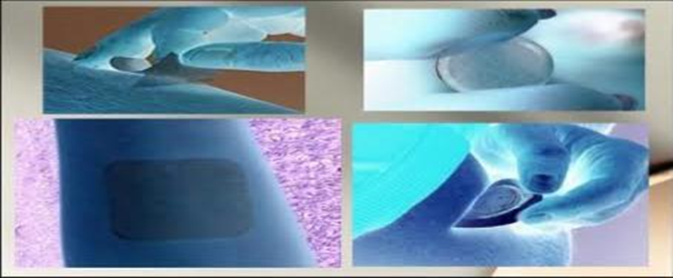
Fig 1 – Transdermal drug delivery systems
ADVANTAGES
Topical patches offer ease of use and are easy to remember.
Drug therapy can be quickly discontinued by simply removing the patch from the skin.
Self-administration is feasible, and the method is noninvasive, thus avoiding the complications associated with parenteral therapy.
In emergencies, transdermal systems can be quickly and easily recognized due to their physical characteristics, features, and identifying labels, making them useful for unresponsive, unconscious, or comatose patients.
Topical patches bypass gastrointestinal absorption issues related to pH, enzymatic activity, and interactions with food or other orally administered medications.
They generally have fewer side effects, making them a preferred choice for many individuals.
It serves as an alternative administration method for patients who are unable to tolerate oral medications, such as those experiencing vomiting.
The therapeutic efficacy of various drugs is enhanced by circumventing specific issues related to the medication, such as gastrointestinal irritation, poor absorption, and interactions with food, beverages, or other medications.
They are a cost-effective option for medication delivery.
It eliminates first-pass metabolism by bypassing the liver.
Transdermal medication provides a consistent release of the drug over an extended duration, thereby minimizing the risk of adverse side effects and therapeutic failures commonly linked to intermittent dosing.
DISADVANTAGES
Ionic drugs can pose complications.
Only highly potent medications are suitable for transdermal patches due to the skin’s natural barrier limiting drug absorption.
Drugs with extremely low or high partition coefficients may not effectively enter systemic circulation.
Certain patches, such as the scopolamine patch applied behind the ear, may cause discomfort.
Achieving elevated plasma drug concentrations can be difficult.
Drugs with a molecular weight greater than 500 Daltons are generally unsuitable for TDDS.
High concentrations of drugs may lead to skin irritation.
Adhesives may not adhere effectively to all skin types and can cause discomfort during wear.
Prolonged use may result in discomfort for patients.
Drugs with extremely low or high partition coefficients may not effectively enter systemic circulation.
Challenges in administering large doses, specifically those exceeding 10 mg/day.
LIMITATIONS
Drugs with large molecular sizes cannot be effectively delivered through TDDS.
Transdermal drug delivery systems (TDDS) are unable to deliver ionic drugs.
TDDS can result in elevated drug levels in blood or plasma.
Development is not feasible if the drug or formulation irritates the skin.
COMPONENTS
Active Pharmaceutical Ingredient (API)
Penetration Enhancers
Polymer Matrix (Backing Layer)
Adhesive Layer
Release Liner
Reservoir or Drug Matrix
Other Components
Transdermal drug delivery systems (TDDS) are engineered to transport medications through the skin into the bloodstream, offering benefits such as avoiding the gastrointestinal tract and enabling a controlled release of the active pharmaceutical ingredient (API). The elements that comprise TDDS are essential for ensuring effective delivery, stability, and regulated drug release. Below is a comprehensive overview of the components of TDDS, incorporating insights from the latest developments in 2024.
Active Pharmaceutical Ingredient (API) [ 10]
The API is the core substance responsible for the therapeutic effect. For effective transdermal delivery, the API must exhibit specific characteristics, including sufficient lipophilicity for skin absorption, a low molecular weight, and efficacy at minimal doses. Typically, TDDS are utilized for medications that require lower dosages, such as nicotine, fentanyl, and estradiol.
Factors for API
Molecular Weight: The ideal drug should have a molecular weight below 500 Da.
Partition Coefficient: Suitable drugs for TDDS should maintain a balance between lipophilicity and hydrophilicity, facilitating penetration through the stratum corneum, the outermost skin layer.
Penetration Enhancers [11]
These agents are incorporated to aid the drug’s movement through the skin, effectively overcoming the barrier presented by the stratum corneum. Penetration enhancers can modify the skin’s structure or fluidize the lipid bilayers to facilitate drug absorption.
Types of Penetration Enhancers:
Chemical Enhancers: Examples include ethanol, dimethyl sulfoxide (DMSO), terpenes, fatty acids, and surfactants.
Physical Techniques: Methods such as iontophoresis (utilizing electrical currents) and microneedling.
Recent Developments: Researchers are exploring the use of nanocarriers, including liposomes and nanostructured lipid carriers (NLCs), to improve drug penetration. These carriers can encapsulate medications and facilitate deeper skin absorption, especially for larger molecules.
Polymer Matrix (Backing Layer) [12]
backing layer represents the outermost component of the transdermal drug delivery system (TDDS) and fulfills several essential functions:
It serves as a barrier to prevent premature drug loss prior to application.
It provides mechanical integrity to the system.
It regulates the drug release process.
Materials: Commonly used polymers include ethyl cellulose, polyvinyl alcohol, hydroxypropyl methylcellulose, and silicone-based polymers. The selection of materials significantly influences the patch’s stability and the kinetics of drug release.
Adhesive Layer [13]
This layer is crucial for ensuring that the TDDS remains securely attached to the skin while enabling controlled drug release. Adhesives must be biocompatible, non-irritating, and capable of forming a robust bond with the skin.
Types of Adhesives
Acrylic-based adhesives: These are commonly utilized due to their ease of application and non-irritating characteristics.
Silicone-based adhesives: Renowned for their high tolerance, these are frequently employed in areas of sensitive skin.
Innovations in Adhesives: Recent advancements have introduced pressure-sensitive adhesives that allow for the patch to be removed without causing skin damage, while still ensuring effective adhesion during use.
5. Release Liner [ 14]
The release liner serves as a protective barrier that inhibits premature interaction between the adhesive and the drug with the external environment prior to the application of the patch on the skin. This component is generally constructed from non-permeable materials such as polyethylene or polyester.
Function: Its primary role is to maintain the integrity of the patch and to prevent any unintended release of the drug until the moment of application.
6. Reservoir or Drug Matrix [ 15]
The reservoir or matrix is responsible for containing the drug and regulating its release over a specified duration. There are two main categories:
Reservoir-based systems: In this configuration, the drug is housed within a distinct compartment, and a membrane that controls the release rate is employed.
Matrix-based systems: Here, the drug is integrated within a polymer matrix, with the release rate being influenced by the drug’s diffusion through the polymer.
Recent Developments: Innovative matrix formulation, including hydrogel-based systems and microparticle-loaded matrices, have been investigated to enhance the consistency of drug release.
7. Other Components
Plasticizers: Enhance the flexibility of the patch.
Antioxidants and Stabilizers: Maintain the stability of the drug throughout the product’s shelf life.
Colorants: Incorporated for aesthetic reasons to distinguish the patches or to enhance patient adherence.
ANATOMY
The skin’s anatomy is intricate and diverse, comprising multiple layers that each serve crucial functions in safeguarding the body and facilitating essential physiological activities. As the largest organ, the skin plays a pivotal role in shielding underlying tissues from various environmental threats, including pathogens, chemicals, and harmful UV rays. Additionally, it aids in temperature regulation, delivers sensory feedback, and participates in vital metabolic processes such as the synthesis of vitamin D.
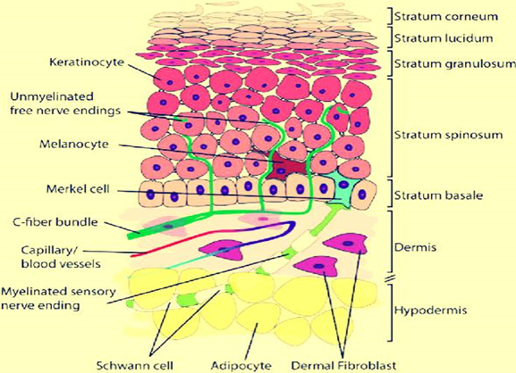
Fig 2 - Skin anotomy
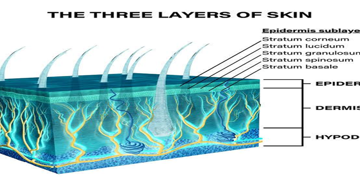
Skin Structure Overview
The skin is primarily divided into three key layers:
Epidermis (the outer layer)
Dermis (the middle layer)
Hypodermis (the deepest layer)
Each layer possesses unique functions and structures that enhance the skin’s overall contribution to bodily health.
Fig 3 – skin structure overview
Epidermis: [16]
The epidermis serves as the skin’s outermost layer, acting as a waterproof barrier and the first line of defense against external threats. It is mainly made up of keratinocytes, which generate keratin, a protein that fortifies the skin. This layer is avascular, meaning it does not contain blood vessels and instead depends on nutrient diffusion from the dermis below.
Key Layers of the Epidermis:
Stratum Corneum: The outermost layer, consisting of dead, flattened keratinocytes. These cells, filled with keratin, create a robust protective barrier that minimizes water loss and guards against environmental harm.
Stratum Lucidum: Present only in areas of thick skin (such as the palms and soles), this thin, translucent layer of dead keratinocytes offers extra protection.
Stratum Granulosum: This layer contains cells that are in the process of dying and losing their nuclei. These cells produce keratin and lipids that contribute to the skin’s waterproof barrier.
Stratum Spinosum: Known as the “prickle cell layer,” it features keratinocytes linked by desmosomes, providing structural integrity. This layer also includes Langerhans cells, which play a role in immune responses.Stratum Basale
(Stratum Germinativum): This is the innermost layer of the epidermis, made up of basal keratinocytes that are in a constant state of division to generate new skin cells. Additionally, this layer contains melanocytes, which are responsible for producing melanin, the pigment that gives skin its color, as well as Merkel cells, which are involved in sensory perception.
Functions of the Epidermis:
Shields against UV radiation
Aids in preventing water loss
Acts as a barrier to pathogens and toxins
Contributes to immune defense
Dermis: [17]
The dermis is situated beneath the epidermis and is significantly thicker. It is essential for the skin’s strength and elasticity contains various structures, including blood vessels, nerves, hair follicles, and glands.
Key Components of the Dermis:
Papillary Dermis: The uppermost layer of the dermis, characterized by loose connective tissue, small blood vessels, and sensory neurons. The papillary dermis interlocks with the epidermis, forming dermal papillae that increase the surface area for nutrient exchange and contribute to the formation of fingerprints.
Reticular Dermis: The deeper and thicker section of the dermis, made up of dense, irregular connective tissue rich in collagen and elastin fibers. This layer provides the skin with tensile strength and elasticity, and it contains larger blood vessels, hair follicles, sweat glands, and sebaceous glands.
Specialized Structures in the Dermis:
Hair Follicles: Found within the dermis, hair follicles are responsible for hair production. They are accompanied by sebaceous glands that produce sebum (oil) to keep the hair and skin moisturized.
Sweat Glands: These are divided into eccrine glands, which are distributed throughout the body and assist in temperature regulation, and apocrine glands, located in areas such as the armpits and groin, which release a protein-rich fluid, particularly during stress or sexual excitement.
Nerve Endings: The dermis is home to various sensory receptors, including Meissner’s corpuscles (responsive to light touch) and Pacinian corpuscles (responsive to pressure and vibration), enhancing the skin’s sensory capabilities.
Function
Provides structural integrity to the skin
Contains blood vessels that deliver nutrients and help regulate temperature
Facilitates sensory perception and supports the immune response
Houses sweat glands for thermoregulation
Hypodermis (Subcutaneous Tissue): [18]
The hypodermis, or subcutaneous layer, is situated beneath the dermis and primarily serves as insulation and an energy reserve. It is predominantly composed of adipose tissue (fat cells), collagen fibers, and blood vessels.
Function-
Thermal insulation: The adipose tissue in the hypodermis aids in maintaining body temperature by functioning as a thermal barrier.
Energy storage: The fat cells store energy as triglycerides, which can be utilized during times of energy shortage.
Cushioning: The hypodermis acts as a protective layer, absorbing shocks and safeguarding underlying muscles and organs from injury.
Structure of the Hypodermis:
Adipocytes (Fat Cells): These cells are responsible for storing triglycerides, contributing to both energy reserves and insulation.
Blood Vessels and Nerves: The hypodermis contains large blood vessels and nerves that supply the dermis and epidermis.
EVALUATION PARAMETERS:
Physicochemical properties of the drug and patch
Skin Permeation and Penetration
Performance Under Different Conditions
Safety and Toxicity Studies
Release Mechanism
Bioavailability
Release kinetics
Skin Irritation and Sensitization
Physical Characteristics of the Patch
METHOD:
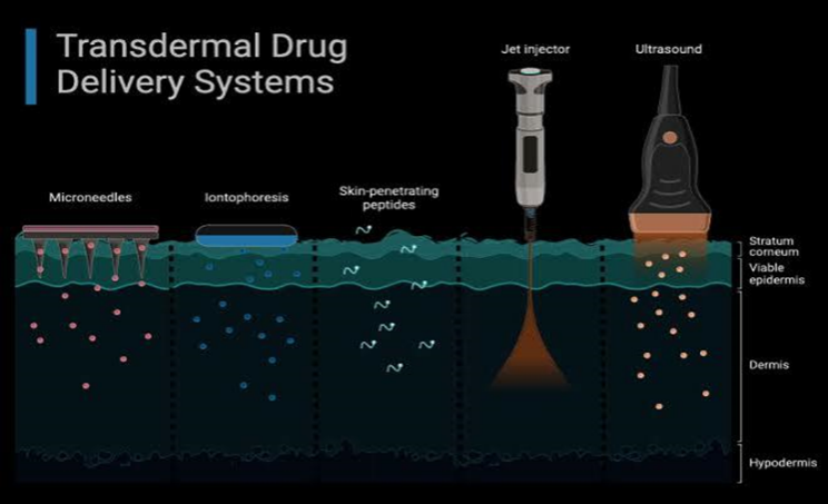
Systems Based on Passive Diffusion: [ 24]
These devices don’t require any additional energy or augmentation techniques; instead, they rely on the natural diffusion of medicines via the skin. To regulate the release and guarantee continuous distribution, they usually employ a drug matrix, adhesive, and backing layer. “Matrix patches” are medications that are embedded in a polymeric matrix (such as polyvinyl alcohol or ethylcellulose) that releases the medication gradually as it permeates the skin.
“Reservoir Patches”: This design uses a rate-controlling membrane to regulate a liquid or gel reservoir that holds the medicine.
Iontophoresis: [ 25]
Iontophoresis increases the penetration of charged (ionic) medications through the skin by applying a little electrical current. By rejecting the charged drug molecules, the administered current makes it easier for them to penetrate the layers of the skin and helps them get past the skin barrier.
Benefits: Non-invasive, targeted medicine delivery, and no requirement for chemical boosters.
Uses: utilized to transport proteins, peptides, and other macromolecules.
Microneedling: [ 26]
Tiny Needling To enhance drug absorption, tiny, needle-like structures called microneedles form microscopic channels in the skin. Drug coating or increasing the skin’s permeability to topical formulations are two possible uses for the microneedles.
Microneedle Types:
Solid microneedles: These are used to make temporary holes that make it easier for medications to enter.
Drug-coated microneedles: These needles are inserted straight into the deeper layers of the skin.
Benefits: include being almost painless, minimally invasive, and enabling the delivery of a variety of medications.
Formulations Based on Lipids: [ 27]
By interacting with the lipid bilayer of the skin, lipid-based systems, including liposomes, ethosomes, and transferosomes, are intended to increase skin permeability. Additionally, both hydrophilic and lipophilic medications can be encapsulated in these systems.
Ethosomes: They are now lipid vesicles with increased ethanol concentrations that improve skin penetration.
Transferosomes: pliable liposomes with the ability to bend and fit through small skin openings.
Sonophoresis: [ 28, 29]
Sonophoresis is the process of temporarily altering the structure of the skin with ultrasonic waves to increase permeability. Drug penetration is made possible by the lipid bilayers in the skin being loosened by the mechanical vibrations of the ultrasonic waves. The non-invasive method improves the penetration of both big and tiny molecules.
Benefit: It works well with both hydrophilic and lipophilic medications.
Fig 4 -Transdermal drug delivery systems method
FORMULATION APPROACHES:
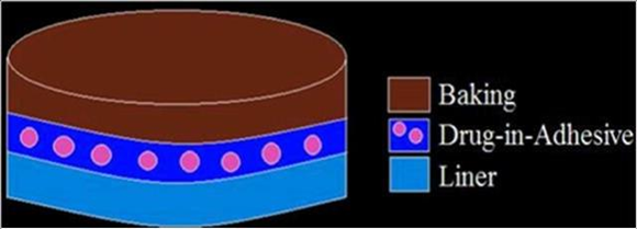
Single-layer medication in adhesive:
This system's sticky layer contains the medication. The adhesive layer in this patch not only holds the layers together and the system to the skin, but it also releases the medicine. Drug release from this mechanism depends on its dispersion across the skin. The sticky layer is surrounded by two layers: temporary linear and backing.
Fig 5: Single layer medication in adhesive
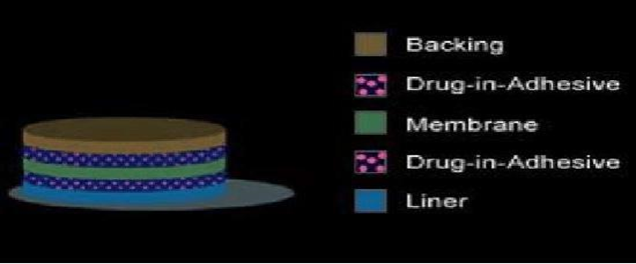
The multi-layer drug:
In adhesive patch works similarly to the single-layer device, releasing the drug via both layers of adhesive. One layer is for immediate medication release, while the other is for controlled release from the reservoir. The multi-layer patch also includes a temporary linear layer and a permanent backing.
Fig 6: Multi-layer drug adhesive
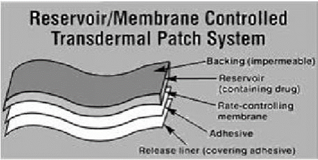
Reservoir system:
The reservoir transdermal system differs from adhesive systems in that it has a distinct drug layer. The drug layer is a liquid compartment that contains a drug solution or suspension and is separated by an adhesive layer. This type of system has a zero-order release rate.
Fig 7: Reservoir system
Vapour Patch:
The adhesive layer not only holds the layers together, but also releases vapour. The vapour patches are new to the market, and they may release essential oils for up to 6 hours. The vapour patches release essential oils and are mostly used for decongestion. Other vapor patches on the market are controlled and can improve sleep quality. There are vapor patches on the market that can lessen the amount of cigarette smoke in the mouth.
Fig 8: Vapour patch
CONCLUSION:
To sum up, transdermal drug delivery systems (TDDS) are a viable substitute for conventional drug delivery techniques, offering advantages like regulated release, enhanced patient adherence, and prevention of first-pass metabolism. These systems use the skin to administer drugs, and new technologies make it possible for both big and small molecules to penetrate the skin more effectively. There are still issues, though, like the possibility of skin irritation, variations in skin conditions, and resistance to the skin barrier. These restrictions are being addressed by ongoing research and development, which will make TDDS a competitive and growing pharmacotherapy option. A variety of therapeutic disciplines stand to gain from the potential for more efficient and focused drug administration as technology develops further.
REFERENCES
Prausnitz, M. R., & Langer, R. (2008). Transdermal drug delivery. Nature Biotechnology, 26(11), 1261–1268.
Tiwari, G., Tiwari, R., & Rathi, V. (2012). Transdermal drug delivery: An overview. International Journal of Drug Development and Research, 4(4), 1–10
Singh, J., & Kim, M. S. (2014). Advances in transdermal drug delivery systems: A review. Pharmaceutical Development and Technology, 19(5), 435-448.
Chen, Y., & Wang, Y. (2016). Recent advances in transdermal drug delivery systems: Technologies and challenges. BioMed Research International, 2016, 8.
Shaila L, Pandey S and Udupa N. Design and evaluation of matrix Type membrane controlled Transdermal Drug Delivery System of Nicotin by suitable for use in smoking cessation. Indian journ. Pharm. Sci. 2006;68: 179-184
Raza R, Mittal A, Kumar P, Alam S, Prakash S, Chauhan N. Approaches and evaluation of transdermal drug delivery System. Int J Drug Dev Res 2015; 7:222-33.
Jhawat VC, Saini V, Kamboj S, Maggon N. Transdermal Drug delivery systems: Approaches and advancements in Drug absorption through skin. Int J Pharm Sci Rev Res
Patel et al. (2024) on formulation techniques for TDDS emphasizes that the choice of API is vital for influencing the system’s permeability and stability. (Source: Patel, A., Patel, A., & Patel, H. (2024). Advances in Transdermal Drug Delivery Systems: A Review, International Journal of Pharmaceutics).
Zhang, L., et al. (2024). “The Role of Penetration Enhancers in Transdermal Drug Delivery Systems.” Journal of Controlled Release, 355, 123-134.
Gupta, R., & Gupta, K. (2024). “Recent Developments in Backing Materials for Transdermal Patches.” Materials Science and Engineering B, 289, 1-12.
Liu, S., et al. (2024). “Next-Generation Adhesives in Transdermal Drug Delivery: An Overview.” Journal of Adhesion Science and Technology.
Patel, A., et al. (2024). “Evaluation and Design of Release Liners for Transdermal Drug Delivery.” Journal of Drug Delivery Science and Technology, 78, 1029-1038.
Sharma, A., & Sinha, V. (2024). “Matrix and Reservoir Systems in Transdermal Drug Delivery.” European Journal of Pharmaceutical Sciences, 162, 78-85.
Dermal-Epidermal Junction: Structure and Function by H. K. S. Peinado et al. (2023), Journal of Dermatological Science.
The Role of Dermal Fibroblasts in Wound Healing by C. S. Chung et al. (2023), Wound Repair and Regeneration.
Structure and Functions of the Hypodermis” by L. M. Smith et al. (2023), Cell and Tissue Research.
Sharma, A., & Gupta, P. (2017). Evaluation of transdermal drug delivery systems: A review. International Journal of Pharmaceutical Sciences and Research, 8(5), 2087-2097.
Khurana, S., & Jain, R. (2018). Transdermal drug delivery system: A review of evaluation parameters. Journal of Pharmaceutical Innovation, 13(3), 198-213.
Kwon, G. S., et al. (2024). “Recent Advances in Passive Transdermal Drug Delivery Systems.” Journal of Controlled Release, 345, 10-25.
Gao, J., et al. (2024). “Iontophoretic Transdermal Drug Delivery Systems: Recent Innovations and Clinical Applications.” Drug Development and Industrial Pharmacy, 50(2), 135-144.
Wang, L., et al. (2024). “Microneedle -Based Systems for Transdermal Drug Delivery: A Review of Recent Progress.” Journal of Drug Targeting, 32(5), 491-505.
Iqbal, Z., et al. (2024). “Lipid-Based Formulations for Transdermal Drug Delivery: A Comprehensive Review.” Pharmaceutical Research, 41(4), 145-157.
Zhang, L., et al. (2024). “Sonophoresis in Transdermal Drug Delivery: Mechanisms and Applications.” International Journal of Pharmaceutics, 588, 119777.
Singh MC, Naik AS, Sawant SD. Transdermal drug delivery system with major emphasis on transdermal patches: a review. J Pharm Res. 2010;3(10):2537-2543.
Joshi K, Selvaduary G. Transdermal drug delivery system and their use of polymers. MatE 175- Biomaterials. 1st Ed. 2008:1-28
Zhang, Y., et al. (2024). “Recent advancements in transdermal drug delivery systems: A comprehensive review.” Journal of Drug Delivery Science and Technology, 63, 102674.
Patel, K., et al. (2023). “Microneedle-based transdermal drug delivery systems: Current trends and future directions.” Pharmaceutical Research, 40(5), 757-769.
Reference
Prausnitz, M. R., & Langer, R. (2008). Transdermal drug delivery. Nature Biotechnology, 26(11), 1261–1268.
Tiwari, G., Tiwari, R., & Rathi, V. (2012). Transdermal drug delivery: An overview. International Journal of Drug Development and Research, 4(4), 1–10
Singh, J., & Kim, M. S. (2014). Advances in transdermal drug delivery systems: A review. Pharmaceutical Development and Technology, 19(5), 435-448.
Chen, Y., & Wang, Y. (2016). Recent advances in transdermal drug delivery systems: Technologies and challenges. BioMed Research International, 2016, 8.
Shaila L, Pandey S and Udupa N. Design and evaluation of matrix Type membrane controlled Transdermal Drug Delivery System of Nicotin by suitable for use in smoking cessation. Indian journ. Pharm. Sci. 2006;68: 179-184
Raza R, Mittal A, Kumar P, Alam S, Prakash S, Chauhan N. Approaches and evaluation of transdermal drug delivery System. Int J Drug Dev Res 2015; 7:222-33.
Jhawat VC, Saini V, Kamboj S, Maggon N. Transdermal Drug delivery systems: Approaches and advancements in Drug absorption through skin. Int J Pharm Sci Rev Res
Patel et al. (2024) on formulation techniques for TDDS emphasizes that the choice of API is vital for influencing the system’s permeability and stability. (Source: Patel, A., Patel, A., & Patel, H. (2024). Advances in Transdermal Drug Delivery Systems: A Review, International Journal of Pharmaceutics).
Zhang, L., et al. (2024). “The Role of Penetration Enhancers in Transdermal Drug Delivery Systems.” Journal of Controlled Release, 355, 123-134.
Gupta, R., & Gupta, K. (2024). “Recent Developments in Backing Materials for Transdermal Patches.” Materials Science and Engineering B, 289, 1-12.
Liu, S., et al. (2024). “Next-Generation Adhesives in Transdermal Drug Delivery: An Overview.” Journal of Adhesion Science and Technology.
Patel, A., et al. (2024). “Evaluation and Design of Release Liners for Transdermal Drug Delivery.” Journal of Drug Delivery Science and Technology, 78, 1029-1038.
Sharma, A., & Sinha, V. (2024). “Matrix and Reservoir Systems in Transdermal Drug Delivery.” European Journal of Pharmaceutical Sciences, 162, 78-85.
Dermal-Epidermal Junction: Structure and Function by H. K. S. Peinado et al. (2023), Journal of Dermatological Science.
The Role of Dermal Fibroblasts in Wound Healing by C. S. Chung et al. (2023), Wound Repair and Regeneration.
Structure and Functions of the Hypodermis” by L. M. Smith et al. (2023), Cell and Tissue Research.
Sharma, A., & Gupta, P. (2017). Evaluation of transdermal drug delivery systems: A review. International Journal of Pharmaceutical Sciences and Research, 8(5), 2087-2097.
Khurana, S., & Jain, R. (2018). Transdermal drug delivery system: A review of evaluation parameters. Journal of Pharmaceutical Innovation, 13(3), 198-213.
Kwon, G. S., et al. (2024). “Recent Advances in Passive Transdermal Drug Delivery Systems.” Journal of Controlled Release, 345, 10-25.
Gao, J., et al. (2024). “Iontophoretic Transdermal Drug Delivery Systems: Recent Innovations and Clinical Applications.” Drug Development and Industrial Pharmacy, 50(2), 135-144.
Wang, L., et al. (2024). “Microneedle -Based Systems for Transdermal Drug Delivery: A Review of Recent Progress.” Journal of Drug Targeting, 32(5), 491-505.
Iqbal, Z., et al. (2024). “Lipid-Based Formulations for Transdermal Drug Delivery: A Comprehensive Review.” Pharmaceutical Research, 41(4), 145-157.
Zhang, L., et al. (2024). “Sonophoresis in Transdermal Drug Delivery: Mechanisms and Applications.” International Journal of Pharmaceutics, 588, 119777.
Singh MC, Naik AS, Sawant SD. Transdermal drug delivery system with major emphasis on transdermal patches: a review. J Pharm Res. 2010;3(10):2537-2543.
Joshi K, Selvaduary G. Transdermal drug delivery system and their use of polymers. MatE 175- Biomaterials. 1st Ed. 2008:1-28
Zhang, Y., et al. (2024). “Recent advancements in transdermal drug delivery systems: A comprehensive review.” Journal of Drug Delivery Science and Technology, 63, 102674.
Patel, K., et al. (2023). “Microneedle-based transdermal drug delivery systems: Current trends and future directions.” Pharmaceutical Research, 40(5), 757-769.
Sakshi Kulkarni
Corresponding author
S. V. S. institute of Pharmacy, Mungase, Malegaon.
S. V. S. institute of Pharmacy, Mungase, Malegaon.
Prof .Zahid Anwer Ansari Shahid Ahmed
Co-author
S. V. S. institute of Pharmacy, Mungase, Malegaon.
Prof Tufail Dana
Co-author
S. V. S. institute of Pharmacy, Mungase, Malegaon.
Prof. Manohar Nikam
Co-author
S. V. S. institute of Pharmacy, Mungase, Malegaon.
Sakshi Kulkarni*, Prof. Zahid Anwer Ansari Shahid Ahmed, Prof. Madhuri Shirsath, Prof. Tufail Dana, Prof. Manohar Nikam, Review on Recent Advances in Transdermal Drug Delivery Systems, Int. J. of Pharm. Sci., 2025, Vol 3, Issue 2, 2048-2058. https://doi.org/10.5281/zenodo.14936820


 10.5281/zenodo.14936820
