United College of Pharmacy, Periyanaickenpalayam, Coimbatore – 641020, Affiliated to the Tamilnadu Dr MGR Medical University, Chennai.
This study aimed to formulate and evaluate diclofenac sodium copper oxide (CuO) nanoparticles for improved drug delivery. Diclofenac sodium CuO nanoparticles were synthesized via a chemical precipitation method using copper chloride dihydrate as a cross-linking agent and sodium borohydride as a reducing agent. Three formulations (F1, F2, and F3) with varying diclofenac sodium concentrations (0.25M, 0.37M, and 0.5M, respectively) were prepared and characterized. The nanoparticles were characterized using FTIR spectroscopy to confirm the presence of diclofenac sodium and CuO. Key characterization parameters included drug content, particle size, polydispersity index (PDI), zeta potential, and entrapment efficiency. In vitro dissolution studies were conducted to evaluate the drug release behavior. Results indicated that formulation F3 (0.5M diclofenac sodium) exhibited the highest entrapment efficiency, optimal particle size, and a favorable zeta potential compared to F1 and F2. Specifically, F3 demonstrated [Insert specific values for entrapment efficiency, particle size, and zeta potential here, e.g., “An entrapment efficiency of X%, a particle size of Y nm and a zeta potential of Z mV”]. These findings suggest that increasing the diclofenac sodium concentration within the nanoparticle formulation can enhance key properties relevant to drug delivery. F3 shows promise as an intermediate for further dosage form development. Future studies will focus on stability analysis, in vivo evaluation, and incorporation of the nanoparticles into suitable dosage forms.
1. TARGETED DRUG DELIVERY SYSTEM
Drug delivery (DD) involves methods, formulations, technologies, and processes for transporting pharmaceuticals within the body to achieve therapeutic effects. It includes approaches for administering medicinal compounds to humans and animals. Recent advancements focus on smart DD, optimizing drug administration in terms of timing, dosage, and location for maximum safety and efficacy. Novel DDSs (NDDSs) enhance therapeutic effectiveness with targeted, managed, and sustained delivery, meeting appropriate drug demand. DD is a rapidly growing field with five generations of DDSs, with targeted delivery in the fourth generation.
2. PRINCIPLES AND APPLICATIONS
The basic principle of drug targeting is to deliver a high concentration of the drug to the target site while minimizing its concentration in non-targeted areas. This optimizes therapeutic effects and reduces side effects, multi-target interactions, and non-target concentrations. Drug targeting also prevents unwanted interactions with bioenvironmental factors that may affect drug access to target sites.
Drug targeting involves coordinated drug behavior, the target site, and a pharmaceutical carrier. The target can be a specific organ, cell, or group of cells in need of treatment. The carrier is a specially engineered molecule or system that transports the drug to preselected sites. Ideally, a drug-targeting complex should be atoxic, nonimmunogenic, biochemically inert, biodegradable, biocompatible, and stable in vivo and in vitro. It should have predictable and controllable drug release patterns, simple and reproducible preparation, easy elimination from the body, and minimal drug leakage during transit.
3. TYPES OF DRUG DELIVERY SYSTEM
II. VEHICLE/CARRIER SYSTEMS FOR TARGETED DRUG DELIVERY
1. COMMONLY USED CARRIERS FOR TARGETED DRUG DELIVERY
2. POLYMERIC CARRIERS FOR TDDSs
Polymeric carriers are essential in pharmaceutical drug delivery systems (DDS) due to their unique properties. Advances in polymers have improved medical treatment efficiency, effectiveness, and safety, enabling controlled and targeted drug release through micro- and nanospheres. Polymeric nanocarriers like poly(D,L-lactide-co-glycolide) enhance drug solubility, reduce toxicity, prolong circulation time, and improve drug utilization. Amphiphilic polymers, which contain both hydrophilic and hydrophobic blocks, are widely studied as carriers. They form nanostructures like micelles and vesicles, delivering both hydrophilic and hydrophobic drugs. Polymeric micelles have a hydrophobic core and hydrophilic shell, while polymeric nanovesicles have a bilayer structure isolating hydrophilic drugs within and integrating hydrophobic molecules in the membrane.
Polymeric micelles entrap drug molecules, improving transport and preventing early release. This enhances the therapeutic window of lipophilic drugs, reduces embolism risk, and improves drug accumulation in tumor cells, making them promising for advanced diagnostic and therapeutic applications.
3. MONOCLONAL ANTIBODIES AND FRAGMENTS
Monoclonal antibodies (mAbs) are gaining traction as therapeutic agents for treating chronic conditions like cancer and infectious diseases. They can be combined with chemotherapeutic drugs, radioisotopes, bacterial toxins, cytokines, and enzymes to enhance their tumor-targeting effects. Currently, human mAbs are being developed as antitumor agents, with adalimumab being the first human mAb approved for clinical use.
4. NANOPARTICLES
Nanoparticles, materials smaller than 100 nm, are vital in modern medicine for uses like contrast agents in medical imaging and gene delivery carriers. Their unique properties, including chemical reactivity and biological mobility, set them apart from bulk materials. Known as "zero-dimensional" nanomaterials, all their dimensions are nanoscale, unlike one-dimensional (nanowires) or two-dimensional (self-assembled monolayer films) nanomaterials.
Nanoparticles enable analyses and therapies that other materials cannot, but they also present environmental and societal challenges, particularly related to toxicity. This overview highlights their medical contributions and discusses challenges, focusing on technologies in clinical use or in vivo experimentation, particularly in medical imaging and drug/gene delivery.
5. TARGETED NANOPARTICLES
Nanotechnology is widely applied in energy production, industrial processes, and biomedical research. One key use is nanoparticles (NPs) in biology and medicine, with unique functionalities for imaging cellular processes and detecting low concentrations of analytes. Targeting NPs in biological research is essential. In proteomics, they focus on specific proteins, while in genetics, gene targeting helps determine functions. Drug discovery and therapies, like cancer treatments, benefit from small molecules and monoclonal antibodies. NPs offer many advantages, such as imaging, sensing, and drug delivery. They provide insights into cell functions, apoptosis, cell division, and stem cell fate. NP devices aid in understanding and manipulating biological interactions.
NP techniques enhance understanding of biochemical pathways in disease and injury. Targeted NP systems bind or react to specific molecules with high affinity and selectivity, improving detection and molecular imaging. They address limitations of conventional delivery methods, such as insolubility and instability of hydrophobic compounds and nonspecific targeting.
6. CuO NANOPARTICLES
Metal oxide nanoparticles, such as copper oxide (CuO), are recognized for their antimicrobial and biocide properties, making them valuable in biomedical applications. CuO, a semiconductor metal with unique optical, electrical, and magnetic properties, is used in supercapacitors, near-infrared filters, magnetic storage media, sensors, catalysis, and semiconductors.
Key synthesis parameters involve controlling particle size, morphology, and crystallinity, achieved through various methods like sonochemical, sol-gel, laser ablation, electrochemical, chemical precipitation, and surfactant-based techniques.
Despite their biomedical potential, CuO nanoparticles pose toxicity risks, potentially harming mammalian cells, vertebrates, and invertebrates by increasing reactive oxygen species production, leading to oxidative stress, DNA damage, and mitochondrial harm.
7. DICLOFENAC SODIUM
Diclofenac (DCF), a non-steroidal anti-inflammatory drug (NSAID), is used for pain relief and as an antipyretic. It has antitumoral properties but can cause gastric toxicity. To reduce side effects and improve therapeutic efficiency, modified release systems with site-specific targeting have been developed, particularly for cancer treatment. These systems enhance drug stability, permeability, and bioavailability. Encapsulating DCF in biodegradable polymeric nanoparticles (NPs) increases its therapeutic activity and half-life at the target site, reducing systemic exposure and adverse effects. In this study, DCF was incorporated into CuO nanoparticles for improved delivery and reduced toxicity.
III. METHODOLOGY
1. CHARACTERIZATION OF ACTIVE INGREDIENT
Characterization of Active Pharmaceutical Ingredient (API) is one of the essential steps involved in any formulation development. Drug characterization step involves identification and confirmation of the drug by infra-red spectroscopy.
a) FT-IR Spectroscopy
Fourier Transform Infrared (FT-IR) spectroscopy plays a crucial role in the characterization of pharmaceutical ingredients. By analyzing the IR spectrum of a drug sample and comparing it to the standard spectrum of the pure drug, scientists can identify any significant shifts in functional peaks and verify the non-involvement of certain functional groups. This process ensures the integrity and purity of the pharmaceutical substances.
2. ANALYTICAL METHOD FOR DICLOFENAC SODIUM
100mg of standard was taken in 100 ml buffer solution.(A) 1ml was taken from solution A and diluted to 10ml with buffer (100microgram/ml) solution(B). 5ml from solution B was taken and diluted to 50ml with buffer solution (10microgram/ml).Solution (C) Take 1ml, 2ml, 3ml, 4ml, 5ml, 6ml from solution C and diluted with buffer solution to obtain concentration 10µg/ml, 20µg/ml, 30µg/ml, 40µg/ml, 50µg/ml, 60µg/ml respectively. Absorbance of above prepared solution was measured by using UV visible spectroscopy.
3. PREPARATION OF DICLOFENAC COPPER NANOPARTICLES
Diclofenac copper nanoparticles were prepared by dissolving 9g of copper chloride dihydrate and 5.4g of sodium boro hydride pellet in ethanol. The copper chloride and sodium hydroxide was dissolved separately in ethanol. Then sodium hydroxide is added dropwise to copper chloride with constant stirring at room temperature. The colour change is seen from green to bluish green then finally turns black in colour. 200mg of diclofenac sodium dissolved in 200ml of deionized water. The solution was added drop by drop into copper nanoparticle solution. Then 5% poly vinyl pyrrolidine was added to the copper nanoparticle solution. The solution is probe sonicated at 60-70% for 15minutes.
Table No: 1 Preparation of Diclofenac Copper Nanoparticles
|
S. No |
FORMULATION CODE |
CONCENTRATION OF CuCl2.2H2O |
CONCENTRATION OF DRUG (Diclofenac Sodium) |
|
1. |
F1 |
0.25M |
100mg |
|
2. |
F2 |
0.37M |
100mg |
|
3. |
F3 |
0.5M |
100mg |
IV. CHARACTERIZATION OF DICLOFENAC COPPER NANOPARTICLES
A) Drug Content (%)
The drug content was determined using an indirect method. Firstly, 10mg of prepared Diclofenac was accurately weighed and transferred into a 100ml volumetric flask, then dissolved and the volume was made up to 100ml with buffer solution. Subsequently, 10ml of this solution was transferred into another 100ml volumetric flask and diluted to 100ml with buffer solution. Then, 1ml of this solution was pipetted into a 10ml volumetric flask and diluted to 10ml with buffer solution. The absorbance of this solution was measured at 275 nm using UV visible spectroscopy. For the centrifuged sample (Diclofenac Copper Nanoparticles), the supernatant was taken, diluted to the standard concentration, and measured at 275 nm using UV spectroscopy.
Drug content = (concentration of sample/concentration of standard) x 100
B) Entrapment Efficiency
Twenty-five milligrams of drug-crushed copper nanoparticles were dispersed in 100 ml of water and sonicated for 20 minutes. The dispersion was filtered using a Whatman filter paper. The filtered solution was then analyzed by UV spectrophotometry at 275 nm. The drug entrapment efficiency was determined using the following equation.
Drug entrapment efficiency= (Total drug conc-supernatant drug conc)/ total drug conc x 100
C) Particle Size, PDI and Zeta Potential
Diclofenac copper nanoparticles are characterized based on particle size, PDI, and zeta potential using a Nano size analyzer at 25°C. To remove unentrapped drug, the formulations undergo dialysis. The shape and form of the optimized formulation are confirmed using transmission electron microscopy.
D) In vitro dissolution study
In vitro dissolution was conducted using the US Pharmacopoeia dissolution type II apparatus at 37 ± 0.5°C with a rotation speed of 50 rpm/min, utilizing 900 ml of dissolution medium per vessel. The nanoparticles were immersed in a phosphate buffer (pH 6.8) for 1 hour. Diclofenac Sodium in the sample solutions was analyzed by measuring UV absorbance at 275.8 nm using a UV spectrophotometer. Each sub-sample was identified with a specific vessel and position. Every 15 minutes, a 5 ml sample was withdrawn and replaced with an equal volume of dissolution medium. The withdrawn samples were filtered, diluted, and analyzed at 275.8 nm for Diclofenac Sodium using a UV spectrophotometer. The drug amounts in the samples were calculated from a calibration curve of standard Diclofenac Sodium.
V. RESULTS AND DISCUSSIONS
1. CHARACTERIZATION OF ACTIVE INGREDIENT BY UV SPECTROSCOPY
Table No: 2 Analytical Method for Diclofenac Sodium
|
Concentration (µ/ml) |
Absorbance at λmax 275nm |
|
10 |
0.142 |
|
20 |
0.276 |
|
30 |
0.432 |
|
40 |
0.552 |
|
50 |
0.695 |
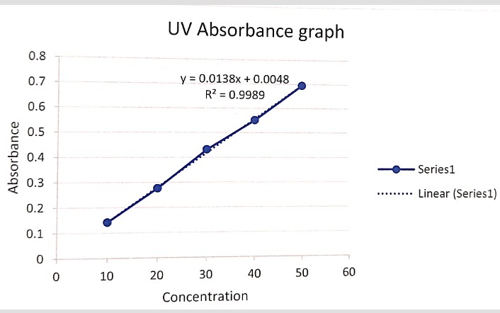
Figure No: 1 UV Spectroscopy
2. FOURIER- TRANSFORM INFRARED SPECTROSCOPY:
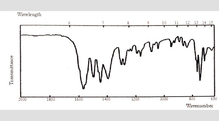
Figure No: 2 FTIR Spectrum of Diclofenac Sodium

Figure No: 3 FTIR Spectrum of Copper Oxide
3. CHARACTERIZATION OF DICLOFENAC COPPER NANOPARTICLES
For F1:
Drug content = (7.097/10)×100=70.97.
For F2:
Drug content = (7.042/10) ×100= 70.42.
For F3:
Drug content = (7.456/10)×100=74.56.
Table No: 2 Drug Content (%)
|
FORMULATION CODE |
% DRUG CONTENT |
|
F1 |
70.97 |
|
F2 |
70.42 |
|
F3 |
74.56 |
Table No: 3 Entrapment Efficiency
|
FORMULATION CODE |
ENTRAPMENT EFFICIENCY |
|
F1 |
79.32 |
|
F2 |
80.10 |
|
F3 |
83.29 |
Table No: 4 Particle Size
|
FORMULATION CODE |
PARTICLE SIZE |
|
F1 |
174.5nm |
|
F2 |
161.5nm |
|
F3 |
159.8nm |
Table No: 5 PDI
|
FORMULATION CODE |
PDI |
|
F1 |
0.371 |
|
F2 |
0.366 |
|
F3 |
0.381 |
Table No: 6 Zeta Potential
|
FORMULATION CODE |
ZETA POTENTIAL |
|
F1 |
-25.1mV |
|
F2 |
-21.4mV |
|
F3 |
-23.5mV |
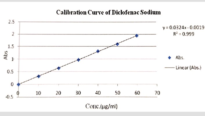
Figure No: 4 In vitro dissolution study (Calibration Curve of Diclofenac Sodium)
VI. SUMMARY AND CONCLUSION
1. SUMMARY:
2. CONCLUSION:
REFERENCES


Dr. Christopher Vimalson D.*, Dr. Alagarraja M., Dinesh P., Latha Mary T., Aravindh Rao S., Gowrishankar T., Monica K., Siva B., Yuvarajan K, Research On Preparation and Evaluation of Diclofenac Sodium Copper Oxide Nanoparticles, Int. J. of Pharm. Sci., 2025, Vol 3, Issue 3, 2199-2206. https://doi.org/10.5281/zenodo.15079444
 10.5281/zenodo.15079444
