Ideal Institute of Pharmacy, Posheri, Wada Palghar 421303. Dept-B pharmacy.
Researchers often encounter a significant challenge when it comes to making sure that drugs reach their intended target without impacting other areas of the body. Fortunately, this issue can be tackled by creating innovative colloidal carriers known as nanosponges. These cutting-edge nanocarriers enable the precise and controlled delivery of both topical and systemic medications. Nanosponges are tiny, sponge-like particles, measuring less than 1 µm in diameter—roughly the size of a virus. Their small and highly porous structure allows them to encapsulate drugs that typically struggle with solubility, thereby boosting their bioavailability. Once they’re in the body, nanosponges circulate and specifically interact with target tissues, releasing the drug in a steady and controlled way. They can be produced using various methods, including sonication, ultrasound-assisted preparation, solvent diffusion, melt method, and solvent evaporation. All in all, nanosponges offer an effective, site-specific drug delivery system that minimizes side effects and improves solubility compared to traditional nanocarriers.
Nanosponges are being crafted as cutting-edge drug delivery systems made from porous polymers, known for their unique, highly porous, spherical shapes. These tiny particles come with a host of benefits, such as lower drug dosages, better retention on the skin, and reduced systemic absorption, as they naturally guide cosmetic and pharmaceutical ingredients to specific areas of the skin.[3] Their design allows for a slow and steady release, maintaining contact with the skin, which helps to lower toxicity, ensures consistent absorption, and boosts patient adherence by lengthening the time between doses.[4] Solid nanosponges can be made into various forms, including topical, oral, injectable, or inhalable dosages, and can be mixed with excipients, diluents, lubricants, or anticaking agents that are ideal for tablet or capsule creation.[5] Some common types of nanosponges include those based on titanium, silicon, hyper-crosslinked polystyrene, and cyclodextrin. Among the metallic nanoparticles, silver nanoparticles (AgNPs) are particularly popular due to their impressive antibacterial properties and beneficial physical characteristics, making them useful in water purification, medical devices, and household disinfectants.[6] However, the widespread production and use of AgNPs raise concerns about their potential release into the environment, which could pose health and ecological risks. Interestingly, silver nanoparticles aren't a recent development; citrate-stabilized silver colloids were first documented by Lea back in 1889, and silver colloids, like “Collargol,” have been utilized in medicine since 1897. The first commercial biocidal silver product, “Algaedyn,” was registered in the U.S. in 1954 and is still in use today.[7]
Objective
To critically review and summarize recent advancements in nanosponges, emphasizing their novel applications, underlying mechanisms, and future prospects in diverse fields—particularly as a cutting-edge approach for drug delivery, biomedical applications, and environmental solutions—while highlighting their ability to improve solubility and enhance drug loading [2].
Experimental Methods
We used a transmission electron microscope (TEM, Hitachi 7100) to take a closer look at carbon-coated filters that had silver nanoparticles suspended in a 0.5% carboxymethylcellulose (CMC) solution. The samples were set on a 200-mesh Veco grid (Eerbeek, Holland) for imaging. We measured the particle diameters at a magnification of 50,000×, analyzing 400 randomly selected particles. To dive deeper into the elemental makeup of the silver nanoparticles, we also utilized an energy-dispersive X-ray analyzer (EDX-200, Horiba, Japan) at an accelerating voltage of 75 kV [8].
Silver Nanoparticles
Spherical silver nanoparticles in three different sizes—20 nm, 80 nm, and 110 nm—were sourced from NanoComposix in San Diego, CA, USA, and were then dispersed in a 2 mM phosphate buffer with a pH of 7.4. The purified nanosilver suspensions displayed no signs of aggregation or sedimentation, and their physicochemical properties were thoroughly characterized [9].
Histopathological Evaluation
Tissue samples were taken from various organs, including the stomach, small intestine, liver, spleen, kidneys, testicles, and ovaries, and then preserved in a 4% neutral buffered formaldehyde solution. After embedding in paraffin and applying Giemsa staining, these tissues were examined under a Leica DM 6000B microscope (Wetzlar, Germany) to assess any histopathological changes [10].
Preparation of Lansoprazole Nanosponges
To create Lansoprazole nanosponges, we used the emulsion solvent diffusion method, mixing different amounts of ethyl cellulose, polyvinyl alcohol (PVA), and Pluronic F68. First, PVA was dissolved in 100 mL of an aqueous continuous phase. Then, we gradually added the disperse phase, which included 100 mg of lansoprazole and the necessary amount of ethyl cellulose (as detailed in Table 1) dissolved in 30 mL of dichloromethane. This mixture was stirred at 1000 rpm for 2 hours with a magnetic stirrer. Finally, the nanosponges were collected through vacuum filtration and dried in an oven at 40°C for 24 hours [11].
Characterization Of Nanosponges
• You can measure the particle size of nanosponges using dynamic light scattering (DLS) with a 90Plus particle size analyzer that has MAS OPTION software. This technique gives you insights into the average diameter and the polydispersity index of the particles. All measurements were taken at a consistent scattering angle of 90°, with samples properly diluted using Milli-Q water [12].
• Typically, nanosponges are smaller than 1 µm and feature porous cavities that can be adjusted for polarity. By tweaking the crosslinker-to-polymer ratio, you can create nanosponges with specific sizes and polarities [13].
• These nanocarriers are not only porous and biocompatible but also non-toxic, showing stability at temperatures as high as 300?°C. Plus, they’re mostly insoluble in common organic solvents [14].
• When they’re made into dosage forms, nanosponges keep their thermal stability up to 130?°C and remain stable across a broad pH range of 1–11 [15].
• In summary, nanosponges are stable, porous, and non-toxic, capable of withstanding high temperatures (up to 300?°C) and resisting dissolution in most organic solvents [16].
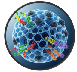
Fig [1] Nanosponges
APPLICATIONS OF NANOPONGES
Nanosponges for Various Drug Types
Nanosponges are pretty amazing when it comes to delivering both lipophilic and hydrophilic drugs, even those tricky poorly water-soluble compounds and BCS class II drugs. They really boost the delivery and bioavailability of these medications [17].
Antiviral Applications
These little wonders have a lot of potential in delivering drugs through the lungs, nose, and eyes. They could play a key role in treating viral infections like HBV, HSV, and HIV, with researchers currently looking into using drugs such as zidovudine, saquinavir, interferon-α, and acyclovir in nanosponge-based delivery systems [18].
Absorbent Properties
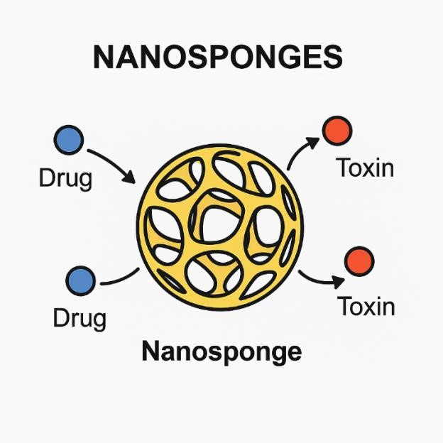
Thanks to their incredible absorbent qualities, nanosponges can help cleanse the bloodstream by soaking up toxins, acting as a detoxifying agent [19].
Other Applications
You’ll find nanosponges being used in a wide range of areas, from biomedical applications and antifungal treatments to various analytical uses [20]
Anticancer therapy
Researchers at Vanderbilt University have come up with some fascinating nanosponges that can deliver anticancer drugs right to the tumor sites. This innovative approach not only improves targeting but also helps in reducing side effects [21].
Oilngonucleotides nanosponges
When it comes to oligonucleotides, these nanosponges play a crucial role in protecting them from degradation. Antisense oligonucleotides, which are used to inhibit gene expression, are often employed in treating cancer and viral infections. Thanks to nanosponges, their stability and effectiveness are significantly enhanced [22].
DNA enzyme nanosponges
In the realm of innovation, we now have bioinspired, self-catabolic DNAzyme nanosponges that offer customizable drug delivery and gene silencing features. These advanced nanosystems enable targeted and controlled drug release, showing great promise for gene therapy and personalized biomedical applications [23].
For covid 19
As for COVID-19, research indicates that SARS-CoV-2 can harm various organs, either directly or through immune responses [24]. To tackle this issue, scientists are developing cellular nanosponges that act as therapeutic barriers. These nanosponges are made from membranes of human cells that the virus targets and include the same receptors the virus uses to enter cells, effectively neutralizing viral particles [25].
Cyaclodextrins based NS
Lastly, cyclodextrin-based nanosponges are proving to be quite effective in water treatment. They have strong sorption capabilities for heavy metal ions, making them excellent for removing both organic and inorganic pollutants. This offers a sustainable solution for water purification [26].
Table 1: Applications Of NS
|
Application |
Description |
|
General Application |
Both lipophilic and hydrophilic drug molecules, as well as BCS class II and poorly water-soluble drugs, can be effectively utilized by nanosponges. [17] |
|
Antiviral Use |
Advantageous in pulmonary, nasal, and ocular delivery routes. Used in HBV, HSV, and HIV treatments with drugs like zidovudine, saquinavir, interferon-α, and acyclovir. (18) |
|
As an Absorbent |
Can remove harmful substances from the bloodstream by absorbing toxins. (19) |
|
Other Applications |
Biomedical applications, anti-mycotic therapy, and analytical applications. [20] |
|
Cancer Therapy |
Developed to deliver anticancer drugs directly to tumors (e.g., Vanderbilt University research). [21] |
|
Oligonucleotides NS |
Prevent degradation of oligonucleotides; antisense oligonucleotides used in cancer and viral infection treatment. [22] |
|
DNAzyme Nanosponges |
Bioinspired, self-catabolic nanosponges with customizable drug delivery and gene silencing for intelligent nanosystems. [23] |
|
COVID-19 Therapy |
Cellular nanosponges created as a therapeutic barrier against SARS-CoV-2 by incorporating virus-targeted receptors. [25] |
|
Water Treatment |
Cyclodextrin-based nanosponges remove heavy metal ions and contaminants from water solutions. [26] |
β-Cyclodextrin Nanosponges (β-CD-NSs)
Five different types of nanosponges were created by mixing various molar ratios of β-cyclodextrin (β-CD) with diphenyl carbonate (DPC): 1:2, 1:4, 1:6, 1:8, and 1:10. The polymer and crosslinker reacted together for five hours at a temperature of 90?°C. Once the crosslinking was done, the mixture was allowed to cool to room temperature, and double-distilled water was added to wash away any unreacted β-CD. The resulting product was then purified using a Soxhlet extractor with acetone for up to four hours to get rid of impurities and byproducts. Finally, the nanosponges were dried under vacuum [27].
Gliclazide Nanosponges (GLZ-NS)
GLZ nanosponges were made using the emulsion solvent diffusion method, which combines both an organic and an aqueous phase. The organic phase included Eudragit S100 and Gliclazide, dissolved in a suitable organic solvent, while the aqueous phase was made up of polyvinyl alcohol (PVA). The organic phase was slowly added to the aqueous phase and stirred for two hours or more to form the nanosponges. The final product was then filtered, purified, and dried either at room temperature or in a vacuum oven at 40?°C for 24 hours [28].
Gabapentin Nanosponges for Dry Suspension
A solution of Gabapentin (GBP) in dichloromethane was combined with the necessary amount of nanosponges in an evaporating dish and stirred continuously until the solvent completely evaporated. The resulting solid was dried overnight in a hot air oven (Itherm AI 7981, Biomedica) set at 60?°C [29].
Etoricoxib Nanosponges (ETX-NS)
Etoricoxib (ETX), a selective COX-2 inhibitor and nonsteroidal anti-inflammatory drug (NSAID), comes in film-coated tablets ranging from 30 to 120 mg. To create nanosponges, we used the emulsion solvent diffusion technique. We started by dissolving ETX and ethyl cellulose (EC) in 20 mL of either acetone or dichloromethane, which served as our internal (organic) phase. This mixture was then gradually introduced into an external aqueous phase that contained polyvinyl alcohol (PVA) dissolved in water, using a syringe pump for precision. The two phases were stirred together for two hours at different speeds with a magnetic stirrer. Afterward, we collected the resulting nanoparticles through filtration and allowed them to dry at 40?°C for a full 24 hours [30]. Etoricoxib (ETX), a selective COX-2 inhibitor NSAID available in 30–120 mg film-coated tablets, was used to prepare nanosponges via the emulsion solvent diffusion technique. ETX and ethyl cellulose (EC) were dissolved in 20 mL of acetone or dichloromethane as the internal (organic) phase. This phase was slowly added to an external aqueous phase containing PVA dissolved in water using a syringe pump. The two phases were stirred for two hours at varying speeds with a magnetic stirrer. The formed nanoparticles were collected by filtration and dried at 40?°C for 24 hours [30].
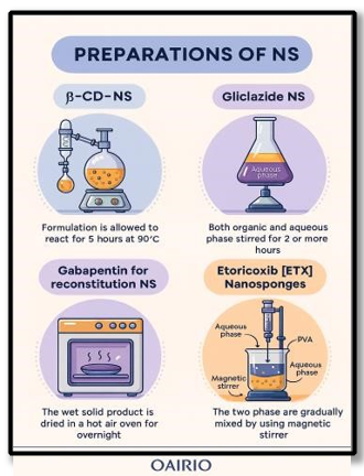
Preparations of fig.2
Table 2 preparation of NS
|
Formulation |
Method Used |
Key Materials |
Processing Steps |
Drying Conditions |
|
β-CD-NSs |
Solventbased crosslinking |
β-Cyclodextrin, Diphenyl Carbonate (DPC), Acetone |
React at 90 °C for 5 hrs → Cool → Add water → Soxhlet extraction (4 hrs) |
Vacuum drying |
|
Gliclazide NS |
Emulsion solvent diffusion |
Eudragit S100, Gliclazide, Polyvinyl Alcohol, Organic solvent |
Dissolve in organic phase → Add to aqueous phase → Stir (2+ hrs) → Filter & purify |
Air dry or vacuum oven at 40 °C (24 hrs) |
|
Gabapentin NS |
Solvent evaporation |
Gabapentin, Dichloromethane, Nanosponges |
Stir until solvent evaporates in dish |
Hot air oven at 60 °C overnight |
|
Etoricoxib NS |
Emulsion solvent diffusion |
Etoricoxib, Ethyl Cellulose, PVA, Acetone or Dichloromethane |
Dissolve in organic phase → Inject into aqueous phase via syringe pump → Stir (2 hrs) → Filter |
Dry at 40 °C for 24 hrs |
Nanosponges are created using a mix of different polymers and cross-linking agents.
1. Polymers: Some of the most commonly used polymers are cyclodextrins, hypercross-linked polystyrene, and their derivatives, which include hydroxypropyl β-cyclodextrins, methyl β-cyclodextrins, and alkyloxycarbonyl cyclodextrins. Additionally, copolymers like poly(valerolactone), poly(valerolactone-allyllactone oxepanedione), ethyl cellulose, and polyvinyl alcohol are also utilized [31].
2. Cross-linking Agents: The cross-linkers that are often used include diisocyanates, diphenyl carbonate, carboxylic acid dianhydrides, dichloromethane, carbonyl diimidazoles, glutaraldehyde, pyromellitic anhydride, 2,2-bis(acrylamido)acetic acid, and epichlorohydrin [31].
3. Artificial Polymers: In the realm of synthetic polymers, options like polycaprolactone, polycarbonate, polyacrylonitrile, as well as polyethylene, polyether, polydimethylsiloxane, polydioxane, polydiol, polylactic acid, and polydimethylsiloxane are also incorporated into nanosponge formulations [32].
4. Natural Polymers: On the other hand, natural polymers, which come from biological sources, tend to be water-compatible. Examples of these include proteins, cellulose, DNA, silk, and wool [33].

Fig.3 various polymers
MATERIALS AND METHODS
1. Interfacial Condensation Method
In this method, we use an organic crosslinker like methylene chloride, butanone, or chloroform, while cyclodextrin (CD) is fully dissolved in an alkaline aqueous solution with a pH above 10 [34].
2. Emulsion Solvent Diffusion Method
This technique involves two phases that don’t mix—an internal and an external phase—created through emulsification. The internal phase forms when a crosslinker is added drop by drop to a solution containing CD and the analyte in a polar aprotic solvent, usually DMF, while continuously stirring. At room temperature, this internal phase is slowly introduced into the external aqueous phase with vigorous stirring. After lyophilization, we collect and dry the resulting cyclodextrin nanoparticles (CDNSs) [35].
3. Ultrasound-Assisted Synthesis
By using ultrasonic vibrations, we can promote CD crosslinking with the right linker in a specific molar ratio, all without solvents, making this an eco-friendly option. This method yields uniform, spherical nanoparticles [36].
4. Chain-Growth Polycondensation Method
In the classic step-growth polycondensation, monomers react with each other and then with new monomers, creating functional groups that have similar reactivity. This process gradually leads to the formation of crosslinked polymeric structures [37].
5. Hot Melting Procedure
This straightforward, reproducible, and solvent-free method involves melting CD together with a carbonyl-based linker, most commonly diphenyl carbonate (DPC). The mixture is blended at temperatures between 90–130?°C for at least 5 hours to ensure complete crosslinking, with additional heating needed for further crosslinking. The end result is a fine, uniform powder [39, 40].
6. Microwave-Assisted Synthesis
Microwave irradiation offers a consistent and controlled way to heat materials, speeding up reactions by as much as four times compared to traditional melting methods. This technique is not only reliable but also easily scalable. When cyclodextrin (CD) interacts with a suitable crosslinker, usually DPC, in polar aprotic solvents like DMF, it forms highly crystalline cyclodextrin nanospheres (CDNSs) with a tight size distribution [41, 42, 43]. Additionally, microwave-assisted synthesis can enhance solvent condensation reactions. For instance, Vasconcelos and colleagues utilized a tin octanoate catalyst to drive the reaction between βCD and HDI in DMF, applying microwave irradiation at 80?°C for 30 minutes [44, 45].
ADVANTAGES
This strategy effectively reduces negative effects while skillfully trapping active ingredients. It also enhances stability, visual appeal, and formulation flexibility, allowing for a steady drug release over a span of 12 to 24 hours [46]. Nanosponges can alleviate drug-induced irritation without compromising therapeutic effectiveness. The materials used in their creation act as a protective shield, preventing the drug from being eliminated too soon from the body [47]. Other benefits include improved stability, refined formulation characteristics, and adaptability. Generally, nanosponges are non-toxic, non-irritating, non-allergenic, and non-mutagenic, and their biodegradable components help to prevent the early breakdown of the encapsulated medication [48]. These systems can be easily scaled up for commercial production. By tailoring drug release profiles for immediate, intermediate, or delayed release, dosing can be optimized, which helps to minimize the chances of under- or over-dosing [49]. Additionally, tweaking the cross-linker-to-polymer ratio allows for control over particle size, ensuring fewer interactions with healthy tissues and thus reducing unwanted side effects [50].
DISADVANTAGES
Nanosponges have the unique ability to encapsulate only small-molecule drugs. They can take on either crystalline or paracrystalline forms. The capacity for drug loading in nanosponges is mainly influenced by how crystalline they are. Interestingly, paracrystalline nanosponges can show different loading capacities based on their structural characteristics.
Factors Influencing Nanosponges
Factors Affecting Drug–Nanosponges Complexation
Evaluation Of Nanosponges
The particle size of nanosponges plays a critical role in their optimization. It can be measured using laser light diffractometry or a Zeta sizer.
Zeta potential assesses the surface charge of nanosponges and can be determined using a Zeta sizer [64].
Scanning Electron Microscopy (SEM) and Transmission Electron Microscopy (TEM) are employed to analyze the morphology and microscopic features of nanosponges [9, 30]. SEM is particularly useful for examining particle surface characteristics.
Production yield (PY) is calculated by comparing the initial weight of raw materials to the final weight of the prepared nanosponges.
Thermo-analytical techniques help determine whether the drug undergoes any changes prior to the thermal degradation of the nanosponge [65].
Powder X-ray diffractometry is used to confirm inclusion complex formation in solid-state nanosponges. For liquid drugs lacking distinct diffraction patterns, the diffraction profile of the formed complex differs significantly from that of the uncomplexed nanosponge.
The phase solubility method, described by Higuchi and Connors, is commonly applied to study drug solubility enhancement due to nanosponge complexation. Phase solubility diagrams provide insight into the extent of complex formation [66].
IR spectroscopy is employed to examine interactions between drug molecules and nanosponges in the solid state. Minor shifts in nanosponge bands may indicate complex formation; however, if less than 25% of the drug is encapsulated, these bands may be masked by the nanosponge spectrum.
In TLC studies, a reduction in the Rf value of the drug molecule indicates the formation of a drug–nanosponge complex [67].
DISCUSSION
The size of the nanosponges was significantly influenced by the ratio of drug to ethyl cellulose (EC). When drug concentrations were higher, there was less polymer available for each nanosponge, which resulted in thinner walls and smaller sponge structures [68]. In formulation LS4, increasing the EC fourfold led to a notable increase in particle size (p < 0.001). This is likely because a denser polymer network boosts emulsion viscosity, which in turn prevents smaller droplets from forming, resulting in larger nanosponges [69]. On the flip side, lower levels of EC allowed dichloromethane to move more effectively from the internal organic phase to the external aqueous phase, speeding up droplet formation and leading to smaller particles [70]. Additionally, a higher concentration of polyvinyl alcohol (PVA) also contributed to larger particle sizes, probably due to frothing that encourages the particles to clump together [71]. Scanning electron microscopy (SEM) images showed that the nanosponges had a consistent spherical shape, porous interiors, and smooth surfaces. The tiny surface pores likely formed from the diffusion of dichloromethane during the preparation process [72]. In vitro release studies indicated that LX in solution released slowly even after 6 hours, likely due to its limited solubility [73]. However, the nanosponge formulations significantly improved LX release, driven by an initial disruption at the surface followed by a steady diffusion over the 6-hour period [74]. Interestingly, increasing the PVA content reduced LX release, probably because the thicker polymer walls lengthened the diffusion path [75–77]. Among the formulations we tested, LS1 stood out with the highest release after 6 hours, measuring at a particle size of 545.5 ± 1.19 nm. Its larger surface area made it the best candidate for further studies [78]. We also found that release kinetics were affected by factors like pore size, porosity, and the distribution of particle sizes [79]. Our kinetic analysis pointed to an unusual non-Fickian diffusion pattern, suggesting that the release of LX was influenced by both diffusion and the relaxation of the polymer. The way the solvent penetrated ethylcellulose—a non-swelling, water-insoluble polymer—played a key role in controlling the diffusion rate through its microporous structure [80]. A physicochemical evaluation of the LX-loaded nanosponge hydrogels showed that they had acceptable drug content and pH levels, making them suitable for topical use. Achieving a uniform consistency is crucial for dermal applications, and the hydrogels' non-Newtonian, shear-thinning properties helped reduce resistance under shear stress, ensuring an even distribution [81, 82]. Adding propylene glycol boosted skin penetration and improved drug release. Among the different formulations, G1 released more LX compared to G2 and G3, likely due to a higher concentration of CMCNa and increased viscosity [83].The release followed a Fickian transport model based on the Korsmeyer–Peppas equation, except for G0, which showed non-Fickian behavior. Draize tests confirmed that the formulations were non-irritating, ensuring they are safe for topical application. The anti-inflammatory effect was assessed using Carrageenan-induced paw edema. The nanogel reached its peak edema inhibition at 5 hours, showcasing its prolonged effectiveness. After 6 hours, the control gel (plain LX) demonstrated significantly less inhibition compared to the nanogel. Formulation G1 aligned with the release profiles, showing a 50.78% release of LX and a 66.63% reduction in edema at the 3-hour mark, while G0 exhibited a 32.82% release and a 42.11% inhibition. The highest inhibition for G1 was 92.91% at 5 hours, which corresponded to a 69.25% release of LX, indicating effective controlled release and therapeutic performance. Overall, these results confirm that nanosponges enhance transdermal drug permeation and significantly boost anti-inflammatory outcomes.
RESULT
CONCLUSION
Research shows that nanosponge-based systems, known for their high porosity, easy functionalization, unique structural design, environmental friendliness, and cost-effectiveness, are paving the way for targeted drug delivery. These innovative systems have demonstrated potential across various fields, including pharmaceuticals, cancer treatment, and even COVID-19 therapies. However, more research is needed to fully grasp their properties, enhance their performance, and ensure their safety. Beyond their biomedical uses, nanosponges also play a crucial role in environmental cleanup, effectively trapping pollutants and contaminants. They find applications in industries as well, helping recover valuable materials from production waste. The structural characteristics of polysaccharide-derived nanosponges significantly affect the combustion behavior and flammability of certain polymers. This is mainly because they help protect phosphorus-based additives when exposed to heat. Furthermore, studies on combustion and flammability indicate a synergistic relationship between nanosponges and phosphorus compounds, boosting the fire-retardant qualities of these materials.
REFERENCES


Payal Mane, Shiv Prasad Dhage*, Dr. Sonali Uppalwar, Nanosponges: A Novel Approach of Drug Delivery System, Int. J. of Pharm. Sci., 2025, Vol 3, Issue 10, 958-974 https://doi.org/10.5281/zenodo.17320264
 10.5281/zenodo.17320264
