Dept of Pharmaceutics, St. Joseph’s college of pharmacy, Cherthala, Kerala, India 688524.
Nanogels are a class of nanoscale, crosslinked hydrogel particles that have become a versatile substrate for biomedical applications, especially targeted drug and gene delivery. They are ideal for controlled therapeutic release due to their unique blend of high-water content, customizable size, biocompatibility, and responsiveness to physiological stimuli (such as pH, temperature, and redox conditions). Over the past decade, there has been considerable advancement in the design and manufacturing of nanogels using both natural and synthetic polymers, enabling precise control over drug loading, release kinetics, and target selectivity. The purpose of this review article is to briefly portray a comprehensive overview of the current state of nanogel research, with a focus on fabrication methods, physicochemical properties, characterization techniques, and therapeutic applications. Important advancements in multifunctional and stimuli-responsive nanogels are highlighted, with a focus on their applications in regenerative medicine, gene delivery, cancer treatment, and vaccine platforms. Furthermore, the review critically looks into the advantages and limitations of nanogels, including challenges in large-scale production, stability, and regulatory approval. Notwithstanding their great promise, the clinical application of nanogels remains constrained. Ongoing studies in bio responsive materials, smart nanogel systems, and hybrid nanostructures, however, are opening the door for next-generation personalized treatment strategies. Nanogels may become even more clinically relevant when combined with cutting-edge technology like biosensors, nanorobotics, and artificial intelligence. All things considered, nanogels are a vibrant and quickly developing field with a lot of room for precision treatments innovation.
Nanotechnology has revolutionized the field of medicine by enabling the development of nanoscale materials with unique physicochemical properties tailored for biomedical applications. It is the science and engineering focused on the design, synthesis, characterization, and use of materials and devices with a lowest functional size of at least one nanometer. Over the past decade, there has been active research and growth in utilizing nanoparticles as reservoirs for drug and gene delivery. Various nanotechnological techniques like protein based nanoparticles, lipid based nanoparticles, nanoemulsions, nanocrystals, nanosuspensions and nanogels have been introduced as an advanced drug delivery system in which nanogels have been introduced in the market due to its maximum advantages over other drug delivery system techniques. Nanogels are nanoparticles composed of a hydrogel with cross-linked hydrophilic polymer with 100–200 nm particle (Garg et al. 2012b). Physically and chemically cross-linked synthetic polymers (Bencherif et al. 2009) or biopolymers (Kabanov and Vinogradov 2009) constitute nanogels. The controlled and prolonged release of medications is made possible by nanogels. Nanogels have swellable and degradation properties with high drug loading capacity, high stability, sustained and targetable manner, large surface area. Because of their three-dimensional architecture, they easily entrap medicines, polymers, and liquid phases in suspension using nanogels. Since they possess inherent soft hydrogel structure and nanoscale size characteristics, together with tunable performances, nanogel has emerged as an extremely versatile class of nanocarriers in the biomedical field. Nanogels in general may be developed from heterogeneous polymerization of monomers or synthesized from polymer precursors. Like hydrogels, nanogels possess the capacity of retaining a great quantity of water or biological fluids within its structure while preserving its arrangement, which is devoted to the existence of hydrophilic groups like –OH,–CONH–, –CONH2–, and–SO3H in the polymer. Yet, nanogels display swelling feature instead ofbeing liquefied due to the presence of cross-links in the nanogels. Such exceptional feature renders nanogels a favorable aspirant for a variety of uses. Engineering of nanogels can achieve improved control over the release kinetics because of their small size and higher surface area, and they can be internalized by cells, surmounting the selective control of the cell membrane and releasing the drug in response to intracellular cues. Various studies have promoted nanogels as a model drug delivery vehicle because of its outstanding drug loading capability, great stability, biologic consistence (i.e. density, firmness or viscosity) and response to a diversity of environmental stimuli, which is unique for common pharmaceutical nanocarriers. One of the main advantages is the potential of nanogels to be responsive to external (for example, temperature, light, magnetic field) or internal (for example, pH, redox potential , enzymes) stimuli. They also exhibit excellent colloidal stability and can be administered through various routes including intravenous, oral, ocular, transungual and transdermal pathways.
There are different methods to synthesise nanogels. These include;
The physical self-assembled polymer approach is used to synthesize nanogels by using amphiphilic polymers, in which the drug and solvent interact by hydrogen bonding, van der Waals interaction, electrostatic interactions etc. During self-assembly, micro and macro molecules are entrapped within the nanogel. The right blend of polymer content, amphiphilic nature, functional groups, pH, ionic strength and temperature influence the size of nanogels. For example, polysaccharide-based nanogels can be prepared by this way. Polysaccharides are used as hydrophilic polymers which are modified by hydrophobic groups. In the case of such modified polymers, the hydrophobic moieties interact with each other and thus the formation of nanoparticles suitable for the transport of active substances is significantly increased (Oh et al. 2009). Another example of hydrophobic interaction based nanogel formation are systems based on cholesterol modified pullulans. These systems can be used for transporting various bioactive molecules, for example insulin (Akiyoshi et al. 1998). Hydrophobic interaction can occur even between two types of polymers. For example, lauryl-modified dextrans with β-cyclodextrins containing polymers were used for successful nanogel formation (Daoud-Mahammed et al. 2007).
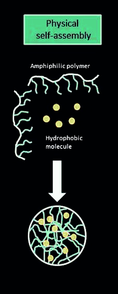
Figure 2.1 Schematic representation of the physical self-assembly method
2.2 Chemical crosslinking
This method is used for the production of nanogel with a large particle size. It is possible to use cross-linked cationic nanogel for polynucleotide delivery. In the method of cross-linking polymerization, nanogel can be prepared by solvent evaporation of an oil in water emulsion whereas PEG can be in conjugation to a branched polyethylene amine in an aqueous system. The cross-linking method has been successfully used for the synthesis of various functionalized nanogels for the transport of drugs. The chemical cross-linking process used to create those nanogels gives them a variety of morphologies, including spheres, rods, and toroids. The size, shape, surface properties, and composition of the particles are all controlled by this cross-linking process. For example, this procedure was used to synthesize the first cross-linked cationic nanogel for the transport of polynucleotides (Vinogradov et al. 1999). In this case, double-activated PEG was conjugated to branched PEI in an oil/water emulsion.
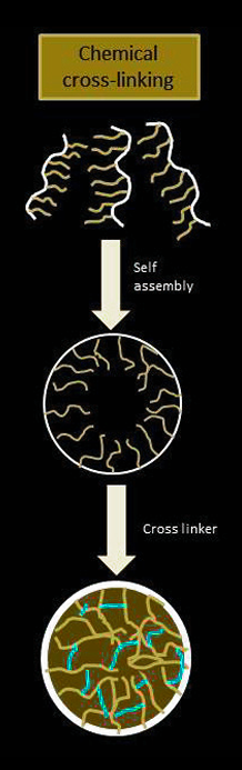
Figure 2.2 Schematic representation of chemical cross-linking
2.3 Polymerization
Polymerization of monomers in a uniform phase or in a microscale or nanoscale diverse setting. In this approach, a polymer colloidal suspension is prepared through the uniform nucleation of water-soluble monomers that are utilized to create stabilized nanogels. Particle size is specifically controlled with this technique. An ionic surfactant can be used to create nanogels with lower particle sizes while also improving the formulations' colloidal stability. As the surfactant concentration decreases, particle size increases in the nanogel formulation (Nayak and Lyon 2005). Polymerization appropriate for nanogel creation can be categorized into two: emulsion and inverse emulsion polymerisation. In the latter, a medium for the polymerization of monomers is an inverted water in oil nano-emulsion. In fact, certain co-monomers that act as bifunctional cross-linkers are added to create stable nanogels. The Khmelnitsky group (Khmelnitsky et al. 1992) described covalently immobilized enzymes in polymer nanogels based on the copolymerization of acrylamide with N, N-methylene-bis acrylamides. It is also possible to perform polymerization that results in nanogels in an aqueous suspension or in an oil-in-water nano-emulsion.
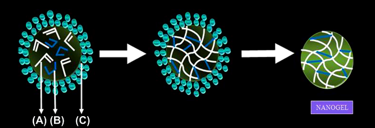
Figure 2.3.1Synthesis of nanogels by copolymerization in colloidal environments. Copolymerization of monomers [A] and bifunctional cross-linkers [B] in w/o microemulsions stabilized by surfactants [C] produces nanogels which can be then transferred into aqueous media after removal of surfactants and organic solvent
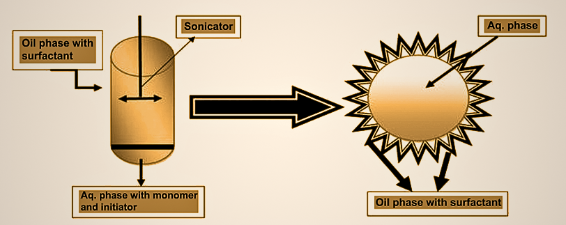
Figure 2.3.2 Inverse micro emulsion process
In certain instances, the polymerization can commence in a uniform aqueous solution, that transforms into a cloudy appearance during polymerization suspension that includes the expanding nanogel. The final product is then separated from suspension by freeze drying.
DeSimone developed (Gratton et al. 2007) this new imprinting method, also known as particle replication in non-wettable templates (PRINT), which is suitable for the production of nanogels. This technique can be used to create polymeric nanoparticles that are tens of nanometers to several micrometers in size. Particle size, composition, shape, surface functionality, and good loading control of pharmaceuticals and biomacromolecules.
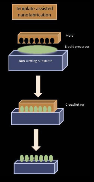
Figure 2.4 schematic representation of template assisted nanofabrication
For instance, this technique has been used to create PEG-based monodispersed swelling particles by UV-induced copolymerization of several monomers, including p-hydroxystyrene, O-(methacryl)-O'methylpolyethylene glycol, and trimethylolpropane ethoxylate triacrylate. Lithographic methods are used to make the master template. Liquid fluoropolymer is applied to the surface of this template. As this fluoropolymer saturates the template, It is cross-linked through photochemical processes. Due to the low surface energy and high gas permeability, the organic liquid precursor could fill the cavities through capillary action. Nanoparticles prepared using this method have the same precise shape of the master template they were derived from (Rolland et al. 2005, Gratton et al. 2007, Napier and DeSimone 2007).
Depending on the specific applications and characteristic properties, Nanogels can be composed of Natural or Synthetic polymers. Natural polymers include chitosan, alginate, hyaluronic acid and polysaccharides like in natural gums.
Advantages
Disadvantages
Advantages
Disadvantages
4.1 Biocompatibility and Degradability
They have excellent biocompatibility and biodegradability
Nanogels have rapid swelling or de-swelling characteristics.
Nanogels typically range in size of 20–200 nm in diameter and hence are effective in avoiding the rapid renal exclusion but are small enough to avoid the uptake by the reticuloendothelial system (Labhasetwar et al., 2007).
Nanogels are able to solubilize hydrophobic drugs and diagnostic agents in their core or networks of gel.
Nanogels could be prepared without employing energy or harsh conditions such as sonication or homogenization, which is critical for encapsulating biomacromolecules.
This type of drug delivery system usually does not produce any immunological responses.
Over the surfactant micelles nanogels or polymeric micellar nanogel systems have a higher stability exhibit slower rate of dissociation, lower critical micelle concentrations, and longer retention of loaded drugs.
In the biomedical field and biodistribution the softness of nanogel is crucial parameter and it can be adjusted by variation on the structure of nanogel.
4.9 Higher Drug Loading Capacity
The properties of higher drug loading capacity of nanogels depend on the functional group present in the polymeric unit. These functional groups have a tremendous effect on drug carrying and drug-releasing properties, and some functional groups have the potential to conjugate with drugs/antibodies for targeting applications. to conjugate with drugs/antibodies for targeting applications. These pendent functional groups of polymeric chains contribute toward establishing hydrogen bonding or Vander Waals forces of interactions within the gel network and thus facilitate the drug-carrying efficiency. Moreover, the presence of functional groups at interface with drug/protein molecules is also responsible for higher loading.
4.10 Stimuli-Responsive behaviour
Stimuli-responsive systems nanogels has been widely used on their stability, high drug-loading capacity, simple synthesis and talent to be modified in multiple ways. Importantly, As compared to other drug delivery system (DDSs), nanogels are more highly aware of mutative environments through their unique 3D network structures, which may be easily adjust in several environments to regulate drug release.
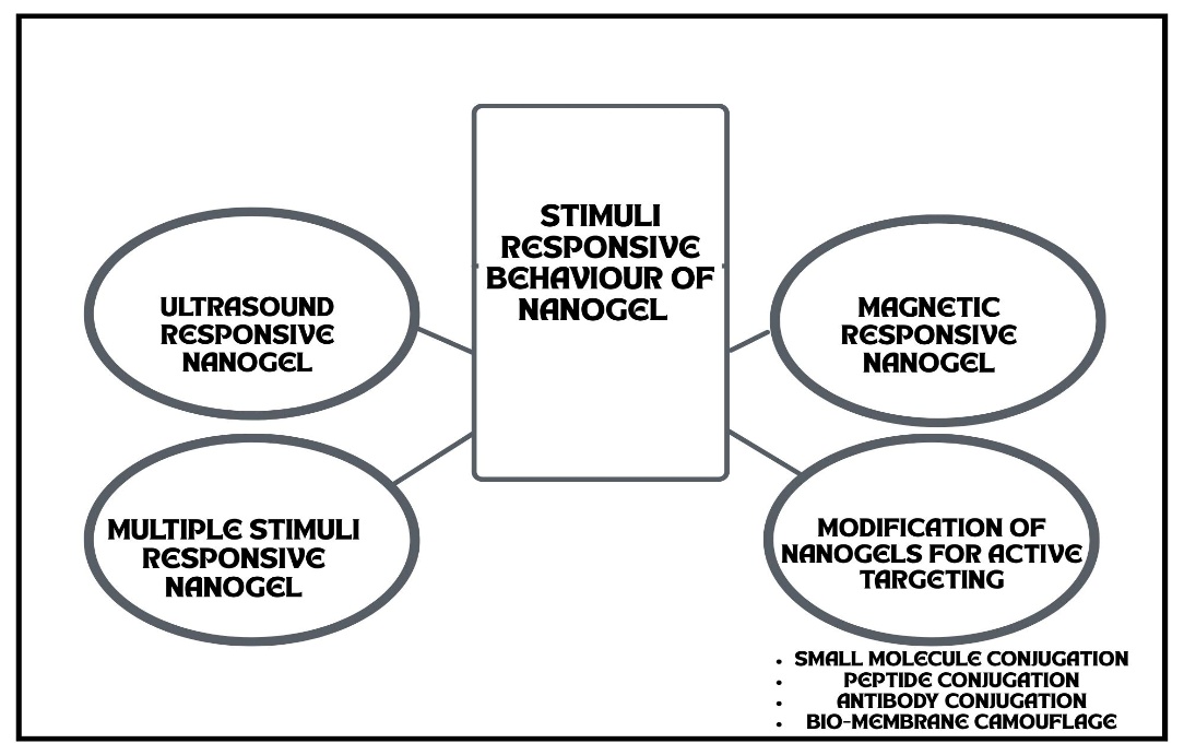
Figure 4.10 mechanism of nanogel
4.11 Others
Both type of drugs (hydrophillic and hydrophobic drugs and charged solutes) can be given through nanogel. Such properties of nanogel are significantly influenced by temperature, presence of hydrophilic/ hydrophobic groups in the polymeric networks, the cross-linking density of the gels, surfactant concentration, and type of cross-links present in the polymer networks.
Dynamic light scattering (DLS) is also known as Quasi- Elastic Light Scattering (QELS), or Photon Correlation spectroscopy (PCS). This technique is generally used to determine the size and distribution of the nano molecules Jayakumar et al. determined the particles size with the help of DLS in a doxorubicin–chitin nanogel (Jayakumar et al. 2012). Shen et al. prepared biocompatible and thermo-sensitive core– shell nanogel that was synthesized by RAFT aqueous dispersion polymerization. They prepared two types of nanogels: one is a linear poly (ethylene glycol) s (l-PEGs) and another is a nonlinear analogue as polymers derived from oligo (ethylene glycol) (meth) acrylates (g-PEGs); they are characterized by DLS for their particles structure and size distribution in the absence of surfactant. DLS was performed by a Malvern Zetasizer 3000 HSA at 25°C (Shen et al. 2011). Daoud Mahammed et al. synthesized nanogel by the spontaneous addition of b-cyclodextrin and modified dextran (modified with alkyl chain) with hydrophobic Benzophenone (guest molecules). In this kind of nanogel, DLS is used to determine the mean diameter and size distribution using a Coulter Nanosizer (Model N4MD, Coultronic, France) (Daoud- Mahammed et al. 2009). Inomoto et al. determined the structure of hydrogel nanoparticles, formed by self-aggregation of cholesterol-bearing pullulan (CHP); this was studied by dynamic light scattering (DLS). DLS measurements were performed by a static/Dynamic Compact Goniometer (DLS/ SLS-5000), which determined that a system has unimodel distribution (Inomoto et al. 2009).
Drug entrapment in nanogel is determined by UV-Vis spectroscopy method. UV-Vis spectroscopy method refers to absorption spectroscopy or reflectance spectroscopy in the Ultraviolet-Visible spectral region (Goyal et al. 2013). Daoud-Mahammed et al. (2009) determined the entrapment efficiency by UV spectroscopy of two hydrophobic molecules (benzophenone and tamoxifen). Wang et al. (2008) also determined the entrapment efficiency by UV spectroscopy method in gelatinized thermo-sensitive nanogels.
Scanning electron microscopy (SEM) is a type of electron microscopical technique that produces an image of a sample by scanning it with a focused beam of electrons. SEM is able to detect the surface topography of nanogels, composition, three-dimensional structure, chemical composition, crystalline structure, and orientation of the formulation. SEM can achieve a resolution better than 1 nanometer (Kataria et al. 2014). Jayakumar et al. (2012) studied doxorubicin-loaded pH-responsive chitin nanogel for drug delivery; they determine the particle size and shape by the SEM method. Kettle et al. (2011) also used SEM to determine particle size range of the nanogel.
Transmission Electron Microscopy (TEM) is a method in where a stream of electrons is passed through an ultra thin specimen, interacting with the given sample. A black and white high-resolution image is formed by the TEM when the sample and electrons are interacting. TEMs are able to give information of surface features, shape, size, and structure of the nanogel. Flaws, fractures, and damages occurring in the nanogel formulation can also be determined by TEM. Peng et al. also used TEM to analyse the morphology of dual-responsive cisplatin nanogel (Peng et al. 2013). Gref et al. (2006) determined the structural formation of self-assembled nanogel formed by b-cyclodextrin polymers (pb-CD) and Dextran bearing hydrophobic lauryl side chains.
FTIR methods are employed to validate the structure of main functional group in the nanogel formulation. The FTIR method was used to assess the absorption, emission, and photoconductivity of the nanogel. An FTIR spectrometer gathers spectral information across a broad range of the specified sample. FTIR spectrophotometer (Nicolet 6700) was used to obtain spectra data of copolymeric nanohydrogels. Guerrero-Ramírez et al. used FTIR to characterize copolymeric nanohydrogel of p-nitro phenol acrylate (NPA) and N-isopropylacrylamide (NIPA) by inverse microemulsion polymerization method (Guerrero-Ramírez et al. 2008). The nano hydrogels spectrums of FTIR were collected using Attenuated Total Reflectance Smart Orbit accessory. Kettel et al. used FTIR to confirm polymerization in the nanogel of doxorubicin. They also used FTIR for cyclodextrin-based aqueous nanogels. Cyclodextrin content in nanogels has been measured by quantitative analysis of the band at 1032cm-1

. The integrals of this band measured for the nanogel samples were compared with a calibration curve obtained by mixing different amounts of b-CD with KBr (Kettel et al. 2011).
Nanogels have demonstrated considerable potential in preclinical research for drug delivery, cancer treatment, vaccine creation, and regenerative medicine. Nonetheless, their clinical application is still in the initial phases when compared to other nanocarrier systems such as liposomes and polymeric nanoparticles. At present, the majority of nanogel-based formulations are in preclinical assessments or early-stage clinical trials, concentrating on enhancing therapeutic effectiveness, reducing systemic toxicity, and facilitating targeted delivery. Numerous nanogel formulations have progressed to Phase I/II clinical trials for use in oncology, ocular drug delivery, and immunotherapy. For instance, carriers based on nanogels have been studied for transporting anticancer drugs like paclitaxel and doxorubicin, demonstrating enhanced pharmacokinetics and minimized off-target effects. In ophthalmology, nanogel eye drops have been investigated for prolonged drug delivery to manage ongoing issues such as glaucoma and dry eye syndrome. Moreover, nanogel systems are being assessed as vaccine adjuvants to improve immune reactions to infectious diseases and cancer antigens. Although there have been encouraging outcomes, no nanogel-based drug delivery system has obtained complete FDA or EMA approval for extensive clinical application. Main obstacles consist of mass production, guaranteeing reproducibility and stability, surpassing biological limitations, and tackling regulatory challenges concerning polymer safety and biodegradability. Ongoing research, encompassing the development of stimuli-responsive and multifunctional nanogels, along with hybrid systems that integrate nanogels with inorganic nanoparticles, is anticipated to hasten clinical translation. Advancements in personalized therapeutics and precision medicine position nanogels as key elements of future drug delivery systems.
Upcoming progress in nanogel research is anticipated to concentrate on development of "smart" nanogels that can respond dynamically and accurately to particular physiological stimuli. These systems can intelligently adjust drug release based on environmental signals like pH changes, enzyme function, temperature variations, or oxidative stress levels. New designs also incorporate various responsiveness systems, allowing for immediate and location-specific therapeutic intervention. These intelligent nanogels offer potential for enhancing treatment effectiveness while reducing overall toxicity.
Nanogels hold considerable promise in the age of personalized medicine because of their adjustable physicochemical characteristics and capacity to encapsulate a wide array of therapeutics, such as biologics and nucleic acids. Personalized nanogel formulations can be designed according to a person's genetic profile, disease condition, and treatment efficacy, facilitating precise drug delivery with enhanced dosing regimens. Employing biomarkers sourced from patients for nanogel functionalization will further improve targeting precision and treatment outcomes.
Combination therapy is becoming prominent as an approach to tackle complex diseases like cancer and multi drug resistant infections. Nanogels can be designed to concurrently deliver various therapeutic agents, such as small molecules, proteins, and nucleic acids, within one platform. This strategy enables combined drug effects, minimizes the emergence of resistance, and enhances the likelihood of successful treatment outcomes. Future studies will probably concentrate on designing nanogels that enable spatial and temporal control over the release of multiple drug payloads.
The integration of nanogels with artificial intelligence (AI) and nanorobotics is expected to transform biomedical applications. Modelling and predictive analytics powered by AI can speed up nanogel design, enhance drug loading efficiency, and personalize therapeutic regimen. Furthermore, incorporating nanogels into nanorobotic systems might allow active movement through intricate biological settings, improving tissue penetration and targeted drug delivery. These intelligent, autonomous systems signify a groundbreaking shift for future precision therapeutics.
Nanogels represent a rapidly evolving and highly promising platform in the field of advanced drug delivery systems, offering unique advantages such as high biocompatibility, tunable size, exceptional drug-loading capacity, and responsiveness to physiological stimuli. Their versatility has enabled significant progress in applications ranging from targeted drug delivery and cancer therapy to vaccine development and regenerative medicine. Despite these advances, widespread clinical translation remains limited due to challenges in large-scale manufacturing, stability, reproducibility, and regulatory approval. Ongoing research is driving the development of smart, stimuli-responsive, and multifunctional nanogels that can deliver therapeutics with enhanced precision and reduced systemic toxicity. Furthermore, the integration of nanogels with cutting-edge technologies such as artificial intelligence, biosensors, and nanorobotics holds immense potential for personalized and site-specific treatment strategies. With continued interdisciplinary research and optimization, nanogels are poised to play a pivotal role in shaping the future of precision medicine and next-generation therapeutics.
REFERENCES


Jayasankar K. R.*, Boby Johns G., Arya Ashok, Nanogel; A Comprehensive Review, Int. J. of Pharm. Sci., 2025, Vol 3, Issue 9, 870-883 https://doi.org/10.5281/zenodo.17075720
 10.5281/zenodo.17075720
