Caritas College of Pharmacy, Caritas College Road, 101 Junction, Ettumanoor, Kottayam- 686631
Microneedles are medical devices used for micro needling, mainly for collagen induction therapy, medication diagnosis, and illness diagnostics. Known for their minimally invasive and precise nature, MNs consist of arrays of micro-sized needles ranging from 25?m to 2000?m. [1][2] Microneedle systems are a rapidly growing and promising technology for delivery of drugs, such as vaccines, small molecules, or biologics and for aesthetic skin treatment in local clinics; however, they remain relatively new from a regulatory perspective. There are strong demands for established procedures, test requirements for approval, and recent trends that industries/researchers related to microneedle systems can refer to. Some microneedle systems are commercially available, many are currently undergoing clinical trials, and some are pending approval for commercialization. [3][4][5]This review focuses on microneedle systems that are either on the market or in clinical trials, their applicability and characteristics, and the critical evaluation and test methods necessary for their development and approval.
The microneedling process, or percutaneous collagen induction (PCI), with microneedling devices like Dermaroller and Dermapen, and manufactured microneedle patches (MNs), including solid, hollow, dissolved, coated, and hydrogel MNs, have all found widespread usage in the cosmetic industry. Microneedling is noted for its minimal invasiveness, painlessness, and self-administration, and this can lead to satisfactory patient compliance, along with diminished hazardous waste sharps [6]. These benefits indicate that there is a wide market for microneedling uses in the cosmetics industry.
Microneedling or PCI was first proposed in 1990 for treating scars, and later its application for drug delivery became more general after 1990. Microneedling has in recent times been used to improve the delivery of active ingredients that are applied in cosmetics, including ascorbic acid (AA), retinoids, melanin, proteins, and peptides [6][7]. With the application of microneedling devices and manufactured MNs, micro-needles can penetrate through the SC, generating micro-channels of aqueous transport routes that facilitate the transport of molecules across the skin at therapeutically significant doses. This will ultimately boost the cosmetic agent absorption against what is delivered topically. It is interesting to note that microneedling disruption of the SC molecular structure is reversible and causes less pain or bleeding than hypodermic needles, especially for short microneedles of less than 600 μm in length. Pain and bleeding have been found to have a dramatic correlation with the length of the microneedle. For example, microneedles of 500–1500 μm length would cause a sevenfold escalation in human pain score. This is due to the fact that pain receptors innervate epidermis and dermis; hence, penetration of longer microneedles would stimulate more receptors. Moreover, microneedles ranging from 1000–1500 μm in length have been noted to puncture minute blood capillaries and leave spots of blood on the skin surface. It should further be indicated that the thickness of the skin greatly differs in the human body, something that must be factored in when using microneedles on particular parts of the body [6,7,8].
DEVICES UESD IN MICRONEEDLING
Microneedling with microneedling devices like Dermaroller and Dermapen in cosmetic treatment is becoming more popular, especially for rejuvenating the appearance of the skin and blemishes and scar treatment. The devices have progressively developed since they were first discovered during the early twentieth century. A few years ago, dermatologists added microneedling to anti-aging and rejuvenation therapy to obtain silky and youthful-looking skin. These devices prick the skin with micron-sized needles without pathogenicity, stimulating the underlying cells to increase the production of collagen and elastin, the essential dermal elements. Besides, these devices have been employed in order to increase the delivery of cosmetic agents [7,8].
Dermaroller® is a basic handheld microneedling device with very fine needles of 200–3000 µm in length and an effective diameter of 0.1 mm. The Dermaroller® needles are made from silicon or stainless steel by reactive ion etching. The silicon and stainless-steel needles are resistant and durable. Dermarollers® are applied commonly in treating acne scars, skin care, burn scars, pigmentary disorders, and PCI. They can also be applied in facial rejuvenation, stretch marks, and hair loss. It is, however, due to the needles' length that this treatment is not ideal since it might lead to skin bleeding [7].
There have been proposed two mechanisms for microneedling : One mechanism is that microneedling results in the release of growth factors that induce the synthesis of collagen and elastin in the papillary dermis. To put it briefly, the needles puncture the SC and produce tiny holes (micropunctures) without destroying the epidermis. These micro-injuries cause minimal superficial bleeding, triggering the wound-healing cascade and stimulating platelets and neutrophils to release growth factors (transforming growth factor (TGF)-alpha, TGF-beta, and platelet-derived growth factor) and stimulate collagen and elastin synthesis in the papillary layer of the dermis [6,7]. This finally results in fibroblasts depositing collagen. In addition, micropores formed by microneedling improve permeation of skincare products, thereby increasing their effectiveness. The second mechanism postulates that microneedling produces a demarcation current and not physical injury when microneedles enter the skin. This current stimulates a cascade of growth factors that promote the healing process. This process relies on the principle of bioelectricity, whereby epidermal damage changes the electric potential in cells to a negative electric potential of −70 mV, relative to the epidermis, which has a positive potential. It is also expected that the shift in the potential induces the migration and proliferation of fibroblasts to the injured area, resulting in the deposition of collagen at the site of injury.
Another type of microneedling tool commonly used is Dermapen®. Dermapen® is a handheld pen-like device with disposable needles used to treat acne, burn scars, and photo-aging . The needle’s length can be adjusted to drive the needle up and down the treatment site. It has a battery that can be recharged. Nine to twelve needles are stacked in rows on the needle tip.
Moreover, Dermapen® has two operating modes: high speed (700 cycles per minute) and low speed (412 cycles per minute) . Dermapen® has benefits over Dermaroller®, as it provides disposable needles, uniform application of pressure on the skin, and minimum risk of tips breaking in the skin . In addition, it can treat delicate regions around the eyes, lips, and nose without damaging the skin [6,7,8].
THE SAFETY PROFILE OF MICRONEEDLE
Microneedling with Dermaroller, Dermapen, Dermastamp, and engineered MNs has safety complications related to transdermal delivery, including skin irritation and sensation of pain. The FDA reported that risk related to microneedling, in terms of skin irritation, mild bleeding, bruising, redness, itching, rashes, and peeling, can remain for short (few days) or long (few weeks) durations. Furthermore, utilization of the gadget can sometimes be followed by skin infection, irritant and allergic contact dermatitis, hyperpigmentation, abnormal scarring, and irritant and allergic granulomas. The FDA explained safe utilization of these gadgets, including cleaning and disinfection of the reusable elements in between patients, whereby re-use of needle cartridges can trigger or transmit infection. Moreover, specific care may be required after microneedling, as skin may be more responsive to sunlight and skin products containing retinol, glycolic acid, or alcohol. Due to the fact that microneedling being relatively akin to traditional hypodermic injection, they come as sterile products. Additionally, fabrication of gadgets from metals commonly utilized in a dermatological facility renders their sterilization and re-sterilization an uncomplicated process. To reduce microneedling's side effects, patients who have experienced recent sunlight can be advised to postpone the process of microneedling until suntan traces have disappeared to prevent post-microneedling dyspigmentation. Moreover, patients bearing oral herpes labialis may be under high risk of reactivation of virus after microneedling. Additionally, microneedling over inflammatory or active lesions of acne can cause bacterial micro-abscesses or granulomas. In addition, pre-procedure skin cleansing and hygiene prior to microneedling are essential, in which adequate cleansing of the skin exfoliates makeup and debris from the skin surface and minimizes introducing bacteria to deeper skin layers, reducing superficial skin infections [7,8].
For polymeric MNs, due to the small size of MN arrays, adequate mechanical strength, in terms of strength, geometry of tips, aspect ratio of height-to-base diameter, and sharpness of needles, must be there to penetrate the needle successfully in the skin without breakage. It is noteworthy to mention here that once MNs are inserted in the skin, there is no means to remove the needles from the body. The degree of MNs' safety variation depends upon types of MNs and materials utilized in fabrication. The microbiological characterization of hydrogel-forming MNs and potential for microbes to migrate into the skin after MNs' penetration were explained by Donnelly et al. There were not any evidences of microbial penetration through swollen MNs. Study on human volunteers showed that whereas MNs were utilized for transdermal drug delivery, skin or systemic infection was relatively scarce. In summary, chemical and biological safety of materials utilized in MNs' fabrication, generally accepted as safe (GRAS), must be taken into account in order to have successful and safe administration. MNs must have a fracture force above insertion force utilized for successful microporation, wherein higher the fracture force, better and safer would-be MN insertion. Microporation through MNs exhibited minimum invasiveness, diminished pain, and less tissue injury in human volunteers, and very low average pain score in 0–100 mm visual analog pain scale, in comparison to hypodermic needle. The sensation of pain is in an inverse ratio to MNs' number and length. In addition, pain intensity also depends upon needle tip angle and needle geometries. Bleeding can be caused by MNs, and this also depends upon MN array length and their penetration in the skin [8]
STRUCTURE OF SKIN
The skin serves as the largest organ in human anatomy because it creates a defensive barrier which protects internal body systems. The skin contains three primary layers which consist of the epidermis as the top layer and the dermis as the second layer and the subcutaneous tissue as the third and final layer. The various layers operate together as a system to defend body structure and protect against environmental dangers and regulate body temperature and detect sensory information. The skin shows different thickness levels throughout the body because the palms and soles contain more skin than the eyelids.[9)].
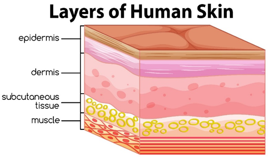
Figure 1: Layers of human skin
MICRONEEDLE DRUG DELIVERY SYSTEM
Microneedles consist of an array of micron-sized needle tips attached to a base. The fibers measure between 100 and 3000 μm in length while their diameters fall within 50 to 250 μm range. The standard-length selection between 250 and 1500 μm depends on the intended use of the fibers. There are mainly five types of microneedles are as follows[10].
Hollow Microneedles
The medical device known as Hollow microneedles (HMNs) operates through hollow needles which function as delivery channels for injecting fluid and liquid formulations. The drug delivery system of HMN functions through the method which scientists call poke and flow. The HMN system enables steady molecule delivery because it lets drug solutions move through MN bores by diffusion and pressure and electrical flow methods [10].
Solid Microneedle
The solid microneedle system for transdermal drug delivery requires two distinct phases which start by placing MN arrays onto the skin surface before their removal to produce microchannels followed by the application of standard drug formulations or transdermal patches. The procedure requires physicians to create skin openings through which they place transdermal drug patches. The method functions through the creation of skin openings which enables medication delivery through patches [10].
Dissolving Microneedle
Scientists created dissolving microneedles to achieve total skin absorption of the needles after insertion which prevents any hazardous medical waste from remaining after treatment. The method of drug delivery operates by inserting the drug into the skin through a poke and dissolve system. The dissolving drug delivery system provides three main benefits which include drug loss prevention during encapsulation and absorption and exact dosing capability because microneedles dissolve in skin and a patch- or pump-free delivery method [11][12][13][14].
Hydrogel Forming Microneedle
Hydrogel-forming microneedles consist of swelling materials which typically include aqueous polymer gels. The hydrogel forming microneedle system allows drug and biotherapeutic placement in two different locations between the microneedle matrix and the reservoir backing. The skin absorbs interstitial fluid when microneedles penetrate its layers which causes the microneedles to expand. The MN cross-linked structure develops pores which enable straightforward drug release. The system operates through a two-step process which starts with poking followed by gel application [15][17].
Coated Microneedles
Solid microneedles function as both skin-piercing tools and drug delivery systems which transport medication molecules into body tissues. The Solid microneedle requires a drug formulation that meets coating standards to be applied on its surface for drug delivery into the skin after insertion. The method functions through two separate actions which involve coating followed by skin penetration Painless administration [16][18].
ADVANTAGES OF MICRONEEDLES
DISADVANTAGES OF MICRONEEDLES
APPLICATION OF MICRONEEDLE
2. BIOSENSING AND DIAGNOSTICS
The Microneedle patches operate as devices that deliver instantaneous health indicator monitoring.
3. COSMETIC AND DERMATOLOGICAL APPLICATIONS
The skin receives active ingredients through the process of micro needling.
4. GENE AND NUCLEIC ACID DELIVERY
Skin cells receive genetic material through the delivery system which uses microneedles.
5. WOUND HEALING AND REGENERATIVE MEDICINE
The delivery system allows for the transfer of growth factors together with stem cells.
THE ROLE OF MICRONEEDLES IN COSMETIC DERMATOLOGY
Microneedles have become the latest technology in cosmetic dermatology, offering a relatively painless treatment to enhance skin health and appearance. The micro-device between 50 and 900 micrometres in size pierce the stratum corneum to allow transdermal delivery of active substances without causing significant pain or damage to tissues [26].
Rejuvenation of the skin is possibly the best application of microneedles in cosmeceuticals. By producing controlled micro-injuries, microneedling triggers the skin's natural wound-healing cascade, leading to increased production of collagen and elastin. The treatment relaxes the texture of the skin, reduces fine lines, and increases firmness overall. Clinical studies have confirmed microneedling to significantly enhance photoaged and scarring skin due to acne [26,27].
Microneedles are also engaged in drug transdermal delivery, particularly low-permeability drug molecules, actively. Active chemicals such as hyaluronic acid, retinoids, and peptides are better delivered using microneedle-assisted systems with better hydration, control of pigmentation, and anti-aging properties. Soluble microneedle patches that release actives at insertion are well-liked by their ease of use and localized benefits.
Apart from facial treatment, microneedling has also proved effective against baldness. With the enhancement of topical drug delivery like minoxidil and platelet-rich plasma, microneedles stimulate follicular growth and hair development in people suffering from androgenetic alopecia.
Although they are beneficial, microneedles should be applied properly. Poor technique or sterilization has been noted to create adverse reactions such as erythema, infection, or post-inflammatory hyperpigmentation. Consequently, professional advice and compliance with safety guidelines are important for optimal gains [26,27].
COMPARISON OF MICRONEEDLES AND CONVENTIONAL COSMETICS
Conventional cosmetic formulations such as creams, serums, and lotions rely primarily on passive diffusion through the stratum corneum, the skin’s outermost barrier. This barrier function significantly limits the penetration of many active ingredients, particularly those with high molecular weight or poor lipophilicity, such as peptides, hyaluronic acid, and certain vitamins [26]. As a result, conventional cosmetics often deliver only superficial benefits, with limited efficacy in targeting deeper dermal structures [27].
In contrast, microneedle-based cosmetic systems overcome this limitation by creating transient microchannels in the skin, enabling direct delivery of active compounds into the viable epidermis and dermis [28]. This approach enhances bioavailability and therapeutic outcomes, allowing for more effective anti-aging, brightening, and hydrating effects compared to traditional topical applications [26,29]. For example, dissolving microneedle patches loaded with hyaluronic acid have demonstrated superior wrinkle reduction and skin hydration compared to topical creams alone [30].
Another distinction lies in the mechanism of action. Conventional cosmetics primarily act through surface hydration, occlusion, or antioxidant activity, whereas microneedling not only facilitates ingredient delivery but also induces controlled dermal injury. This stimulates collagen and elastin synthesis, leading to long-term improvements in skin texture and firmness [27,28]. Thus, microneedles provide both a delivery platform and a rejuvenation stimulus, offering dual benefits not achievable with conventional products.
However, microneedle systems are associated with higher costs and require careful application to avoid adverse effects such as erythema, infection, or post-inflammatory hyperpigmentation [27]. Conventional cosmetics, while less effective in deep delivery, remain more accessible, affordable, and user-friendly for daily use. Therefore, microneedles represent a complementary advancement rather than a complete replacement for traditional cosmetic formulations.
COMPARISON OF MICRONEEDLES AND CONVENTIONAL COSMETICS
|
Feature |
Conventional Cosmetics |
Microneedle-Based Cosmetics |
|
Skin Penetration |
Limited to the stratum corneum; poor delivery of large or hydrophilic molecules [26,27] |
Create microchannels that bypass the stratum corneum, enabling deeper dermal delivery [26,28] |
|
Active Ingredient Efficacy |
Reduced bioavailability; many actives remain on the surface [27] |
Enhanced absorption of peptides, hyaluronic acid, retinoids, and growth factors [26,29] |
|
Mechanism of Action |
Hydration, occlusion, antioxidant activity, and surface-level effects[27] |
Dual action: (a) physical stimulation of collagen/elastin synthesis, (b) transdermal delivery of actives [22,23] |
|
Clinical Outcomes |
Gradual, often superficial improvements in hydration and tone [27] |
Significant improvements in wrinkles, scars, pigmentation, and firmness [26,28,30] |
|
Safety Profile |
Generally safe, minimal irritation; low infection risk [27] |
Safe when applied correctly, but risks include erythema, infection, or post-inflammatory hyperpigmentation [27,30] |
|
Cost & Accessibility |
Widely available, affordable, easy for daily use[27] |
Higher cost; often requires professional application or specialized patches [21,30] |
CONCLUSION
Evading first-pass effects decreases the possibility of toxicity to normal tissues, while MN-assisted topical drug delivery can improve local drug use at target lesions through the skin. Hence, depending on the condition that is being treated, the size and geometry of MNs may be customized, and a simpler method for preparing MNs with extra creative structures and lowered preparation costs has been under consideration. Even further, MN-assisted therapy has not been related to inflammation in skin diseases, as the latest studies state. MNs, therefore, lie promisingly as medical devices that might improve patients' quality of life by drawing on the benefits from many biomedical, nanomaterial, nanomedical, and photonic technologies, aided, of course, by the current development of new intelligent treatment platforms and approaches. Being one of the most versatile platforms available, MNs will surely become the major focus of the emerging nanotech and therapy spheres that value painlessness, shallowness, accuracy, precision, and portability. One of the constraints of clinical application of synthesized MN patches is that they only cover small regions of the skin. But efforts have been made to expand the size of MN patches to cover more areas of the skin surface by preparing MN patches in a larger size. Moreover, the constraints of the time are the complexity of the application in utilizing a combinational treatment that includes microneedling and topical preparation. Secondly, synthetic MNs have a limited capacity to load. In order to overcome the drawbacks of clinical applications of synthetic MN patches, other materials with favorable MN properties like enhanced mechanical properties (strength and flexibility) and biocompatibility need to be identified. Besides, new synthetic methods for MNs that can enhance the delivery of different types of MNs for different therapeutic applications need to be studied. New materials, new processes, and commercialization will be discussed in detail in further studies to achieve medical effectiveness, economic efficiency, and mass production of synthesized MNs. Further, new MN applications, such as disease detection, management, monitoring, diagnosis, and personalized medicine, will be investigated. Furthermore, commercially marketed microneedling devices, especially those sold for home use, require stringent regulation of production. For instance, one would maximize the surveillance of the devices regarding materials employed in production, mechanical characteristics, insertion force, dosing and release precision, and needle depth.
REFERENCES


Teresa George Poozhikanadakel, Aiswarya P Sajua, Chandichan Thomasa, Meenakshi Ma, Anjaly Krishnaa, Microneedles: An Emerging Trend for Cosmetics and Pharmaceuticals, Int. J. of Pharm. Sci., 2025, Vol 3, Issue 12, 3591-3600. https://doi.org/10.5281/zenodo.18047735
 10.5281/zenodo.18047735
