Department of Pharmaceutics, Srinivas College of Pharmacy, Farangipete Post, Mangalore – 574143, Karnataka, India.
The development of novel drug delivery systems has gained significant attention, particularly in nanotechnology, for enhancing drug bioavailability and efficacy. Capped flexosomes, a specialized subclass of vesicular drug carriers, offer a promising approach for controlled and targeted drug delivery. These nanocarriers, composed of flexible lipid bilayers and stabilized by capping agents, provide improved stability, enhanced penetration, and prolonged drug release compared to conventional liposomes. Their structural flexibility allows them to encapsulate a variety of bioactive compounds, including hydrophilic and hydrophobic drug, protein and nucleic acids. Capped flexosomes are particularly advantageous in transdermal and targeted drug delivery, addressing challenges associated with traditional methods, such as drug degradation and poor cellular uptake. The capping agent enhances stability, prevents premature drug release, and protects against environmental degradation. These vesicles have shown potential in cancer treatment, gene therapy, vaccine development, and enzyme replacement therapy. Additionally, their ability to respond to physiological stimuli, such as pH and temperature variations, further improves their therapeutic potential. Several methods are employed for their preparation, including thin film hydration, ethanal/ ether injection, reverse phase evaporation, and microfluidics, each influencing particle size, encapsulation efficiency, and drug release properties. Evaluation parameters such as morphological analysis, particle size, zeta potential, entrapment efficiency, and invitro drug release confirm their effectiveness as drug carriers. Despite their advantages, capped flexosomes face challenges, including high production costs and limited clinical data on long term safety and efficacy. Future advancements in nanotechnology may address these concerns, paving the way for their integration into precision medicine. With ongoing research, capped flexosomes have the potential to revolutionize drug delivery, offering safer, more efficient, and highly targeted therapeutic solutions across various medical fields.
The creation of novel medication delivery methods has received a lot of interest in recent decades, especially in the area of nanotechnology. Because they can transport a wide range of medications and are widely employed for a number of purposes, such as drug targeting, prolonged release, and improving drug penetration, novel vesicular drug delivery systems have made significant strides.1
Due to restrictions on the medications' ability to enter cells, traditional drug delivery techniques frequently fail to effectively treat a variety of illnesses. Vesicular systems have received more attention in an effort to overcome these obstacles and enhance bioavailability at the illness site. These solutions aid in overcoming problems with drug loss and degradation as well as minimizing the negative side effects of traditional techniques.2
Capped flexosomes are a sophisticated class of nanocarriers used mostly in the biomedical and pharmaceutical industries for regulated drug delivery. They are vesicular structures made of flexible lipid bilayers that are a subclass of flexosomes. Numerous bioactive substances, including medications, proteins, and nucleic acids, can be encapsulated in these vesicles and released in a precise and regulated manner. These unique vesicles are designed to overcome the limitations of liposomes like instability. Due to their tendency for oxidative breakdown and varying phospholipid purity, liposomes are chemically unstable. Capped flexosomes enhances stability by preventing premature drug release and protecting against environmental degradation. Particularly for transdermal applications, flexosomes offer a novel method of vesicular drug administration. Their unique makeup, including an edge activator and capping agent, contributes to better flexibility, stability, and improved drug administration, giving them a promising choice for overcoming problems associated with other lipid vesicles.3
ADVANTAGES:
DISADVANTAGES:
Composition Of Capped Flexosomes:
Flexosomes consist of phospholipids, ethanol, and Tween, which acts as the edge activator. The bilayer structure is formed by phospholipids which provide lipophilicity to the system. But surfactants play a vital role in determining drug solubility, encapsulation efficiency, particle size and stability. In addition, the surfactants and ethanol are likely to disrupt the stiff interactions in stratum corneum, making flexosomes more deformable. To enhance the stability, oleyl amine which is a capping agent is employed. The capping compounds associated with this process, like oleyl amine, are also beneficial to ensuring excessive development is inhibited whilst preserving physicochemical properties of flexosomes. Oleyl amine, a molecule capable of establishing both positive and negative charges in its amino groups38, exhibits a high affinity for protons and is expected to interact with various other components during the formation of flexosome.4
Mechanism Of Action of Capped Flexosomes:
Several physicochemical properties of the caps are revealed to drive strong and selective interactions to the cell membrane. Using the umbrella model for lipid diffusion the full spectrum of capped flexosome penetration through the extracellular matrix is determined, and pathways through lipid rafts towards vesicle entrapment for capped flexosomes are proposed. Flexosomes can enter cells with the aid of its caps, which can function as enzymes that eliminate the lipids of the trapping cell membrane, facilitating the endocytosis of flexosomes, like saponins do to glycosides.
Method Of Preparation of Capped Flexosomes: [5,6,7]
In both ethanol and ether injection techniques, lipids are dissolved in an organic solvent such as diethyl ether, an ether-methanol mixture, or ethanol. The prepared lipid solution is then injected into an aqueous phase, allowing for the encapsulation of the desired material at temperatures ranging from 55–65°C or under reduced pressure to facilitate the formation of flexosomes. Since ether is immiscible with water, heating is required to evaporate the organic solvent form the resulting capped flexosomes. This method also enables the dissolution of hydrophilic, lipophilic, or amphiphilic drug solutions in ethanol, which can then be processed using an inkjet device. This approach provides precise control over particle size and is well-suited for large-scale production.
This technique begins with dissolving lipids in an organic solvent, while the drug is dissolved in an aqueous medium. The mixture undergoes sonication to create a water-in-oil (w/o) emulsion or inverted micelles. Gradual removal of the organic solvent using a rotary evaporator causes the micelles to transition into a viscous or gel-like state. At a critical stage, the gel phase collapses, leading to the dispersion of some inverted micelles. The surplus phospholipids then form bilayers around the residual micelles, ultimately resulting in the development of capped flexosomes.
In this approach, lipids are combined with a high critical micelle concentration (CMC) surfactant and dissolved in an appropriate organic solvent within a round-bottom flask. After gentle solvent evaporation, a thin lipid film is deposited at the flask’s bottom. The film is then hydrated in an aqueous solution containing the drug, resulting in a mixed micelle solution. The surfactant is subsequently eliminated through methods such as dialysis, size-exclusion chromatography, adsorption onto hydrophobic beads, or dilution. Once the solution is concentrated, vesicles are successfully formed.
Evaluation: [8,9,10]
1. Morphological Analysis
A transmission electron microscope (TEM) is utilized to examine the morphology of capped flexosomes, including their lamellarity, size, shape, and overall physical stability.
2. Entrapment Efficiency
The entrapment efficiency (%EE) of capped flexosomes is determined using the ultracentrifugation method. The prepared suspension is centrifuged at 2000 rpm for 20 minutes, which separates the undissolved drug (supernatant) from the drug-loaded vesicles (sediment). The supernatant and sediment are separated and measured. The supernatant is then diluted with a suitable solvent, and the drug content is quantified using a UV spectrophotometer. The %EE is calculated using the formula:
EE%=total amount of drug-total amount of free drugtotal amount of drug*100

3. Particle Size and Polydispersity Index (PDI)
The particle size (PS) and polydispersity index (PDI) of capped flexosomes are measured using a Zeta Sizer. The formulation is diluted with deionized water and analysed at 25°C.
4. Zeta Potential Measurement
The zeta potential of the capped flexosome formulation is measured using a Zeta Sizer to assess the electrophoretic mobility of charged vesicles. All measurements are conducted at 25°C in triplicate after dilution. A formulation is considered stable when the zeta potential is approximately ±30 mV.
To determine the total encapsulated drug content, 0.2 mL of the capped flexosome dispersion is dissolved in 25 mL of methanol and stirred using a magnetic stirrer to break the vesicles. The drug concentration is then analyzed spectrophotometrically.
6. In Vitro Drug Release
The in vitro release of capped flexosomes is evaluated using the Dialysis Bag Diffusion Method. A dialysis bag is soaked overnight in phosphate buffer solution before being filled with the capped flexosome dispersion. The bag is sealed at both ends and immersed in a container with phosphate buffer solution maintained at 37 ± 0.1°C in a water bath with constant stirring at 100 rpm. At specified intervals, aliquots of the solution are withdrawn and replaced with fresh buffer. The samples are then analyzed using a UV spectrophotometer.
7. pH Measurement
To ensure the formulation is non-irritating, its pH is measured using a pH meter (model-3505, Jenway, Staffordshire, UK). About 5 mL of the formulation is transferred to a small beaker (10 mL), and the pH is measured at 25°C in triplicate.
8. Ex Vivo Permeation Study
The permeation study is conducted using albino rat skin obtained from anesthetized neonatal rats. The skin is first cleaned with isopropanol to remove fats, followed by gentle washing with deionized water before being stored at −20°C in aluminium foil until use. An open-ended tube is used to mount the skin onto a receptor chamber containing 50 mL of phosphate-buffered saline (PBS, pH 7.4). Samples of 3 mL are withdrawn at predetermined intervals (1, 2, 4, 6, 8, and 10 hours) and analyzed spectrophotometrically. Fresh medium is added after each sample withdrawal to maintain sink conditions.
The permeation parameters include:
The following equations are used for calculations:
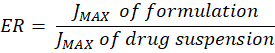
JMAX=amount of drug permeatedtime*Area

ER= JMAX of formulationJMAX of drug suspension
9. Stability Study
The stability of capped flexosomal dispersion is assessed by storing samples at two different temperatures (4–8°C) for three months. Samples are analyzed at 1, 2, and 3-month intervals for changes in physical appearance (color and odor), particle size (PS), PDI, zeta potential (ZP), and entrapment efficiency (%EE).
Applications of Capped Flexosomes12
Capped flexosomes are innovative drug delivery systems that integrate the adaptability of liposomes with controlled drug release capabilities. Their unique properties make them highly valuable in pharmaceutical and biomedical applications. Below are some key uses of capped flexosomes:
1. Targeted Drug Delivery
This enables precise drug delivery to specific bodily locations, including tumor sites. They do this in response to environmental triggers such as pH, temperature or enzymatic activity. Because the caps are only released in the intended area, there are minimal off-target effects with this approach.
2. Controlled Drug Release
Flexosomes have a capping structure, which ensures sustained, controlled, or stimulus-induced drug release. This improves drug efficacy by ensuring therapeutic concentrations of drugs over longer times and also minimizes side effects of high peaks of drug in the circulation.
3. Cancer Treatment
flexosomes, are excellent carriers of anti-cancer agents and are particularly effective in chemotherapy, as they can carry anti-cancer drugs to malignant cells. This specific targeting minimizes the toxicity of chemotherapeutics to normal tissues and increases drug uptake in tumors, leading to improved treatment outcomes.
4. Gene Delivery
These delivery systems are ideal for nucleic acids such as DNA and RNA for mRNA vaccines or gene editing therapies. By necessitating protective measures, the capping structure makes sure that genetic material remains protected from degradation until its delivery to the target cells, thus improving stability and efficacy.
Antigens or adjuvants can be encapsulated in flexosome-based vaccines (VCA), improving stability and efficacy. Vaccine components are protected from degradation, releasing gradually to stimulate immune response at the right tim
Capped flexosomes boost the delivery of active ingredients into the skin in any skincare and cosmetics formulation. They create better efficacy and long-lasting effects by controlling the release of such compounds.
For example, in the case of patients with enzyme deficiencies due to lysosomal storage disorders, capped flexosomes can deliver required enzymes directly to the target cells or tissues. Such enzymes show enhanced stability and can deliver therapeutics more efficiently. These diverse applications highlight the potential of capped flexosomes in modern medicine, offering more effective, safer, and controlled drug delivery solutions.
Future Prospects:
Capped flexosomes are lipid nanoparticles that have multiple powerful applications in targeted drug delivery, including treatment of cancer, autoimmune diseases, infections, and neurodegenerative disorders. Their surface can be altered with targeting ligands to allow for precision dosing of medications to treat disease while minimizing side effects as well as controlling the release and the trigger for the release. Moreover, they have potential applications in gene therapy by facilitating the delivery of genetic material, as well as in vaccine development by delivering mRNA or antigens to boost immune responses. Flexosomes could revolutionise the treatment of neurodegenerative diseases by allowing personalised drug delivery across the blood-brain barrier to the brain. Due to their versatile nature, they can also be applied in environmental fields like slow-release fertilizer delivery in agriculture or bioremediation. Because they could be engineered to deliver any therapeutic based on a person's genetic profile, flexosomes could be pivotal in personalized medicine as the industry evolves. However, optimizing their stability, scalability, and long-term safety is not straightforward. In spite of these obstacles, capped flexosomes are predicted to play a substantial role in advancing gene therapies, precision medicine, and other advanced therapeutics in the coming years.
CONCLUSION:
Capped flexosomes represent an exciting drug delivery breakthrough by integrating the structural versatility of liposomes and their regulated and more targeted drug release. And are suitable for various application such as gene transfer, cancer therapy, vaccine development and dermatological treatments owing to their ability to enhance drug bioavailability, provide stabilizing and reduce toxicity. Indeed, these vesicular carriers offer the advantage of reducing systemic side effects while providing the precise delivery of the therapeutics based on the responsiveness to the physiological stimuli, such as pH, temperature, or enzyme expression. Until further research is done to enhance their long-term safety, scalability, and formulations applicable to the clinical setting, despite the benefits observed. To be widely used, hurdles such as the costs of production and governmental approvals must be crossed. As nanotechnology advances, capped flexosomes may revolutionize personalized medicine and targeted therapy, offering safer and more effective treatment alternatives that will find applications in the biomedical and pharmaceutical sectors.
REFERENCES


Srilakshmi K. T.*, Shalini B., Ganesh Nayak, A. R. Shabaraya, Innovative Drug Delivery with Capped Flexosomes: Applications, and Future Prospects, Int. J. of Pharm. Sci., 2025, Vol 3, Issue 9, 444-451 https://doi.org/10.5281/zenodo.17052841
 10.5281/zenodo.17052841
