Department of Pharmaceutics, Channabasweshwar Pharmacy College (Degree), Latur-413512, Maharashtra, India
Transdermal drug delivery systems (TDDS) offer significant advantages over traditional drug delivery methods but face limitations due to the skin's barrier properties, particularly with conventional liposomal formulations. Ethosomes, a novel lipid-based vesicular carrier system containing high concentrations of ethanol, have emerged as a promising solution. These soft, malleable, and non-invasive vesicles enable deeper skin penetration and systemic drug delivery, overcoming the challenges of the stratum corneum. Ethosomes can effectively deliver both highly lipophilic and cationic drugs such as cannabinoids, testosterone, minoxidil, propranolol, and insulin. Their ease of preparation and scalability make them suitable for commercial applications. This review highlights ethosomes composition, preparation methods, characterization, advantages, limitations, types, and recent research developments, showcasing their potential to enhance drug efficacy, patient compliance, and reduce treatment costs.
Advancement in drug delivery methods are progressing much faster now compared to previous innovations in the field. The skin is a highly accessible organ, making it an ideal route for drug delivery. However, its outermost layer, the stratum corneum, acts as a strong barrier, limiting the passage of most medications, except for lipophilic and low molecular weight drugs. This method, known as transdermal drug delivery use the skin to administer drug systemically. TDD is a method of delivering drugs through intact, healthy skin. The drug penetrates the stratum corneum, passes through the epidermis and dermis, and reaches the dermal microcirculation for systemic absorption without accumulating in the dermis. Transdermal drug delivery systems are effective for drugs with extensive first-pass metabolism, gastrointestinal instability, or severe oral side effects. They offer controlled drug release, reduced dosing frequency, and improved patient compliance.
Skin:
The skin is the largest and most accessible organ of the body, with just a small layer of tissue separating the surface from deeper layers. The skin is a specialized barrier that protects the body and prevents the passage of substances. Transdermal drug delivery is an exciting yet challenging area of research.
Layer of skin:
1.Epidermis:
The epidermis is the outermost skin layer, varying in thickness (0.8 mm on palms and soles), and is composed of multiple layers of epithelial cells. The epidermis consists of four layers: the stratum basale (deepest), stratum spinosum, stratum granulosum, stratum lucidum, and stratum corneum (most superficial).
Stratum basale:
The stratum basale (or stratum germinativum) is the innermost skin layer, connected to the basement membrane by hemidesmosomes. It contains stem cells that generate keratinocytes through mitosis and houses melanocytes
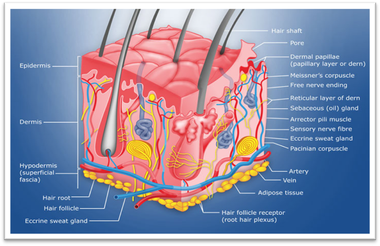
Fig.1: Structure of Skin
Stratum spinosum:
The stratum spinosum, or prickle cell layer, consists of 8–10 layers of irregular, polyhedral cells with "spines" that connect to neighboring cells via desmosomes. It also contains dendritic cells.
Stratum granulosum:
The stratum granulosum, composed of 3-5 layers, contains diamond-shaped cells with keratohyalin and lamellar granules. Keratohyalin granules release keratin precursors that form bundles, while lamellar granules release glycolipids that bind cells together.
Stratum lucidum:
The thicker skin of the palms and soles includes two to three cell layers. The thin, transparent layer is composed of eleidin, a by-product of keratohyalin transformation.
Stratum corneum:
The stratum corneum, the topmost layer, consists of 20-30 layers of anucleate squamous cells (dead keratinocytes). Its thickness varies, especially in callused skin. Dead keratinocytes secrete defensins, part of the body's initial defense.
2.Dermis:
The dermis, 2-3 mm thick, is composed of 70% collagen and elastin fibers, providing strength and elasticity. It contains blood vessels that supply nutrient to both the dermis and epidermis, along with nerves, macrophages, and lymphatic vessels.
3.Hypodermis:
The hypodermis, the deepest skin layer, consists of fat cells and acts as a cushion between the skin and underlying tissues like muscles and bones. It provides protection, heat insulation, support, and conducts vascular and neural signals. Fat cells in the hypodermis account for about 50% of body fat, with fibroblasts and macrophages also present.
Function of Skin(6):
The skin serves several vital functions, including:
Ethosomes
Ethanolic liposomes, are called as Ethosomes, are non-invasive, flexible, and pliable vesicular delivery systems. They enhance the distribution of active ingredients by facilitating the penetration of medication into the deep skin layer. Ethosomes are lipid vesicles containing phospholipids, alcohol (ethanol or isopropyl alcohol), and water, developed by Touitou. They enhance skin drug delivery, with penetration improved by additives like propylene glycol. Age-activator agents, such as ethanol and sodium cholate, boost carrier penetration through the stratum corneum, enabling effective local and systemic delivery of both hydrophobic and hydrophilic drugs. Ethosomes are widely recognized for their efficacy in transdermal drug delivery. Key components include phospholipids, alcohol, cholesterol, polyglycol, dye, and the vehicle. They can encapsulate drugs with hydrophilic, lipophilic, or amphiphilic properties. These soft, malleable vesicles deliver drugs to deep skin layers or systemic circulation. Ethosome sizes range from nanometers to microns.
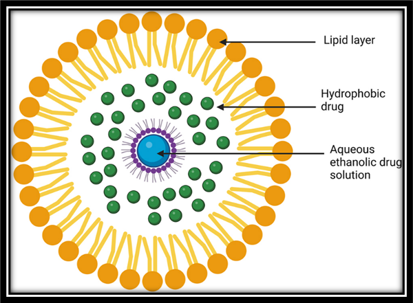
Fig.2: Structure of Ethosome
Advantages of ethosomes (12)
Disadvantages of ethosomes(13)
Types of Ethosomal system(14)(15)
Classical ethosomes are modified liposomes composed of phospholipids, high ethanol concentration (up to 45% w/w), and water. Compared to conventional liposomes, they offer smaller size, negative ζ-potential, enhanced skin permeation, and improved stability, making them more effective for transdermal drug delivery. They can encapsulate drugs with molecular weights ranging from 130.08 Da to 24 kDa.
Binary ethosomes, introduced by Zhou et al., are modified classical ethosomes formulated by incorporating a second alcohol. Propylene glycol (PG) and isopropyl alcohol (IPA) are the most commonly used secondary alcohols in these systems.
Transethosomes, introduced by Song et al. in 2012, are advanced ethosomal systems combining classical components with penetration or edge activator. Designed to merge the benefits of ethosomes and transfersomes, they offer superior skin permeation and can encapsulate drugs ranging from 130.08 Da to 200–325 kDa.
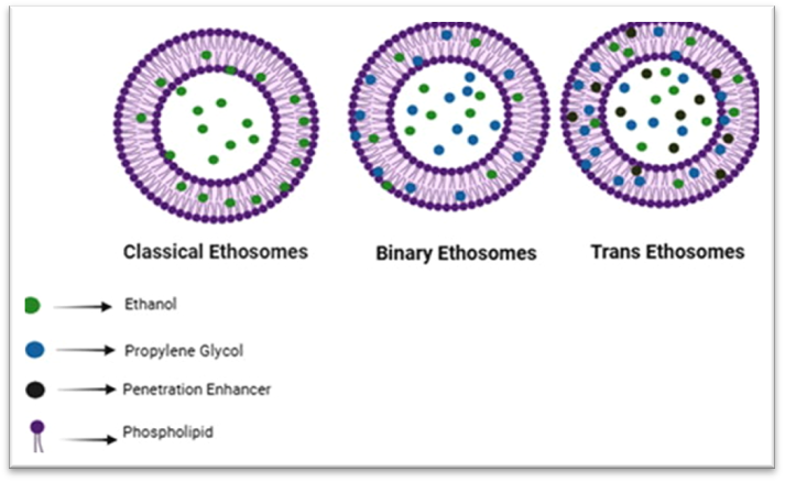
Fig.3: Types of ethosomal system
Composition of Ethosomes(16)(17)
|
Class |
Example |
Uses |
|
Phospholipid |
Soya phosphatidyl choline, Egg phosphatidyl choline, Dipalmitoyl phosphatidyl choline, Distearoyl phosphatidyl choline. |
vesicle forming component |
|
Polyglycol |
Propylene glycol |
Skin penetration enhancer |
|
Alcohol |
Ethanol, Isopropyl alcohol |
For providing the softness for vesicle membrane as a penetration enhancer |
|
cholesterol |
cholesterol |
For providing the stability to vesicle membrane. |
|
Dye |
Rhodamine-123 Rhodamine red Fluorescence Isothiocyanate (FITC) |
For characterization study |
|
Vehicle |
Carbopol934 |
As a gel former |
Method of preparation
1.Cold method:
Phospholipids, drugs, and other lipids are dissolved in ethanol at room temperature with vigorous stirring. Propylene glycol (another polyol) is added during mixing. The mixture is then heated in a water bath to 30°C. Pre-heated water (also at 30°C) is added, followed by stirring for 5 minutes. Vesicle size can be reduced using probe sonication or extrusion. The final formulation is stored under refrigeration.
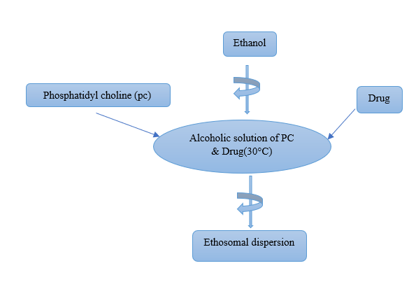
Fig.6: Preparation Of Ethosomes by Cold Method
2.Hot method:
Phospholipid is dispersed in water and heated to 40°C in a water bath until a colloidal solution forms. Separately, ethanol and propylene glycol are mixed and heated to 40°C. Once both phases reach the target temperature, the organic phase is added to the aqueous phase. The drug is dissolved in water or ethanol based on its solubility. Vesicle size can be reduced using probe sonication or extrusion.
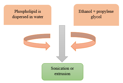
Fig.4: Preparation of Ethosomes by Hot method
3.Classic method:
The phospholipid and drug are dissolved in ethanol and heated to 30°C±1°C in a water bath. Double distilled water is added in a fine stream to the lipid mixture, with constant stirring at 700 rpm, in a closed vessel. The resulting vesicle suspension is homogenized by passing through a polycarbonate membrane using a hand extruder for three cycles.
4.The Ethanol injection and sonication method:
In this method, the phospholipid, previously dissolved in ethanol, is gradually introduced into the aqueous phase using a syringe device, maintaining a controlled flow rate 38?μl/min. this slow injection facilitates the proper mixing of the organic and aqueous phases. After the complete addition of the phospholipid solution, the resulting mixture is subjected to homogenization using an ultrasonic probe for a duration of five minutes to ensure the formation of uniform vesicles and enhance the dispersion of components.
5.Thin-film hydration method:
Lipid are dissolved in an organic solvent within a round-bottom flask. The solvent is then evaporated using a rotary evaporator at a temperature above the lipid phase transition temperature, resulting in the formation of a thin lipid film along the inner walls of the flask. This film is subsequently hydrated with an ethanolic mixture, and the dispersion is sonicated using a probe sonicator to obtain a uniform ethosomal suspension.
Mechanism of Penetration
Ethanol effect: Ethanol function as an effective penetration when applied to the skin. Its mechanism of action in enhancing permeation is well-documented. Ethanol penetrates the intercellular lipid domains of the stratum corneum, where it increases the fluidity of lipid bilayers. This results in a reduction in the structural order and density of the lipid multilayers within the cell membrane. Consequently, the barrier function of the skin is temporarily disrupted, facilitating the enhanced permeation of active compounds through the skin. Effect of ethosomes: Ethosomes enhance cytomembrane lipid fluidity due to the presence of ethanol, which increases skin permeability. As a result, ethosomes can penetrate easily into the deeper layers of the skin. Once inside, they interact with skin lipids and facilitate the release of the encapsulated drug into the deeper skin layers.
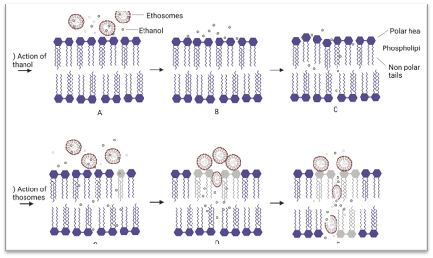
Fig.5: Mechanism of drug Penetration
Characterization and Evaluation of Ethosomes(26)(27)(28)(29)
1.Size and shape:
The size and shape determination of ethosomes intended for skin penetration is commonly carried out using techniques such as scanning electron microscopy (SEM), transmission electron microscopy (TEM), and dynamic light scattering (DLS). SEM is used to observe the surface morphology and overall shape of the ethosomes, providing high-resolution images that help in understanding their structural characteristics. TEM allows for a more detailed visualization at the nanoscale, offering precise measurements of the vesicle size and internal structure. DLS, on the other hand, is used to determine the hydrodynamic diameter and size distribution of ethosomes in suspension, which is essential for evaluating their stability and behavior under physiological conditions. These complementary techniques together provide a thorough analysis of ethosome characteristics, which is crucial for assessing their efficiency in skin penetration.
2.Zeta potential:
Zeta potential is a measure of the overall surface charge of particles in suspension, indicating their colloidal stability. It is determined using a laser-based technique, where a laser beam passes through the sample, and scattered light is detected at an angle (typically ~130°). Upon applying an electric field, particle movement causes fluctuations in the scattered light, which are analyzed to calculate the zeta potential. Values greater than +30 mV or less than −30 mV generally indicate good stability due to sufficient electrostatic repulsion between particles.
3.Entrapment efficiency:
Drug entrapment efficiency refers to the separation of free drug from encapsulated drug using high-speed ultracentrifugation. Ethosomal nano-dispersions are centrifuged at 50,000 rpm for 50 minutes at 4°C using a micro-ultracentrifuge (Thermo Scientific Sorvall MX 150, India). The supernatant is analyzed for free drug using UV, HPLC, or LC-MS.
Entrapment efficiency is calculated using the formula
Entrapment Efficiency = (Encapsulated Drug / Total Drug Used) × 100.
4.Drug content:
A method is developed to determine the drug content in the formulation. Drug content in ethosomes can be quantified using a UV spectrophotometer or a modified high-performance liquid chromatography (HPLC) method.
5.Stability studies:
A stable pharmaceutical dosage form is designed to maintain the physical integrity of the active ingredient throughout its shelf life. For ethosomal formulations, stability studies should assess physical, chemical, and microbial parameters. These studies include product characterization and evaluation of stability during storage. Vesicle stability is monitored by measuring changes in size using DLS and structural integrity using TEM. Ethosomes are stored at 4°C ± 0.5°C, and vesicle size, zeta potential, and entrapment efficiency are evaluated after 180 days using established methods.
6.Invitro dissolution:
"In vitro dissolution studies, in conjunction with Scanning Electron Microscopy (SEM), Transmission Electron Microscopy (TEM), and High-Performance Liquid Chromatography (HPLC), are employed to assess the stability profile and estimate the shelf life of the ethosomal formulation."
7.Skin permeation study:
The skin penetration ability of the ethosomal formulation was evaluated using confocal laser scanning microscopy (CLSM). For diffusion studies, excised animal skin was carefully cleaned of subcutaneous tissue and mounted between the donor and receptor compartments of a Franz diffusion cell (effective area: 1.0 cm²; receptor volume: 10 mL). The receptor chamber was filled with phosphate-buffered saline (PBS, pH 6.5) and maintained at 32?±?1°C. A 1.0 mL ethosomal formulation was applied to the epidermal surface. Samples (0.5 mL) were collected from the receptor compartment at 1, 2, 4, 8, 12, 16, 20, and 24 hours and analyzed using high-performance liquid chromatography (HPLC).
8.Transition temperature:
The transition temperature of the vesicular lipid systems was determined using differential scanning calorimetry (DSC) with a Mettler DSC 60 system (Mettler Toledo, Switzerland) and STAR software. Samples were analyzed in aluminum crucibles at a heating rate of 10?°C/min over a temperature range of 20–300?°C.
9.Degree of deformability and turbidity:
The degree of deformability of the ethosomal formulation can be evaluated using the extrusion method, which involves passing the vesicular suspension through polycarbonate membrane filters of defined pore size under controlled pressure. This technique assesses the flexibility and ability of the vesicles to deform and pass through pores smaller than their diameter, indicating their potential for enhanced skin permeation. The turbidity of the ethosomal preparation, which reflects the clarity and homogeneity of the vesicle suspension, can be quantitatively measured using a nephelometer. This instrument detects the intensity of light scattered by the suspended particles, providing an indirect measure of vesicle size distribution and dispersion stability.
10.Drug uptake studies:
Drug uptake in MT-2 cells (1×10? cells/mL) was studied using 24-well plates. Each well contained 100?µL of RPMI medium and 100?µL of either the drug solution in PBS (pH 7.4), the ethosomal formulation, or a marketed formulation. After incubation, drug uptake was measured using HPLC analysis.
11.HPLC assay:
During in vitro skin permeation studies and experiments conducted using MT-2 cells, the concentration of the drug permeated into the receptor compartment was quantified using a high-performance liquid chromatography (HPLC) assay. The mobile phase employed for the analysis consisted of a mixture of methanol, distilled water, and acetonitrile in a volumetric ratio of 70:20:10 (v/v/v).
12.Statistical Analysis:
The statistical significance of all obtained data was assessed using analysis of variance (ANOVA), followed by a Studentized range (post hoc) test. Data analysis was performed using the PRISM software, with a confidence level set at P < 0.05 for determining statistical significance.
Application of ethosomes(30)(31)
1.Pilosebaceous targeting:
Pilosebaceous units have been effectively targeted in the treatment of follicle-related disorders such as acne and alopecia. Maiden et al. developed a topical minoxidil ethosomal formulation for scalp application, demonstrating improved clinical efficacy.
2.Delivery of Anti-viral drugs:
Jain et al. (2004) demonstrated that ethosomes enhance transdermal flux, sustain drug release, and offer a promising delivery route for zidovudine. Horwitz et al. (1999) formulated an ethosomal gel of acyclovir, achieving faster healing and a higher rate of abortive lesions. Sudhakar et al. (2014) developed ethosomes to improve lamivudine permeability for antiviral therapy. Dubey et al. (2010) reported improved skin deposition and reduced lag time for indinavir using ethosomal delivery.
3.Topical delivery of DNA:
Touitou et al. demonstrated topical delivery of DNA using ethosomes by encapsulating a GFP-CMV construct, achieving gene expression in mouse skin cells. Gupta et al. reported successful immunization with a transfersomal formulation. These studies suggest ethosomes are promising carriers for gene therapy and transdermal immunization.
4.Transdermal delivery of Hormones:
Touitou et al. (2000) investigated the efficacy of ethosomal carriers for the transdermal delivery of testosterone by comparing the skin permeation of an ethosomal testosterone formulation with a marketed transdermal patch (Testoderm) using rabbit pinna skin. The study reported that the ethosomal formulation achieved approximately 30 times higher skin permeation than the marketed product.
5.Delivery of Anti-parkinsonism agent:
Dayan and Touitou (2002) formulated an ethosomal preparation of the psychoactive drug trihexyphenidyl (THP), used in the treatment of Parkinson’s disease, and compared its delivery profile with that of a conventional liposomal formulation. The ethosomal system demonstrated superior transdermal flux, higher skin retention, and greater stability compared to traditional liposomes.
6.Delivery of Anti-biotics:
Ethosomes rapidly penetrate the epidermis, delivering measurable amounts of drugs to deeper skin layers and targeting infections at the source. Ethosomal formulations loaded with erythromycin and bacitracin demonstrated effective dermal and intracellular drug delivery by releasing the entrapped drugs within cells.
7.Delivery of Anti-fungal agent:
Rahul G.S. Maheshwari et al. (2012) developed ethosomes and ultra-deformable liposomes for the transdermal delivery of clotrimazole. Similarly, Sarat Chandan C. et al. (2012) formulated ethosomal carriers containing ketoconazole.
8.Delivery of Anti-Arthritis drug:
Topical delivery of anti-arthritis drugs offers a targeted alternative to oral therapy, minimizing systemic side effects. Ethosomal cannabidiol showed enhanced skin penetration and improved anti-inflammatory effects. Similarly, ethosomal transdermal delivery of piroxicam provides an effective alternative to oral administration, reducing risks such as gastrointestinal bleeding and enzymatic degradation.
9.Used in treatment of herpetic infection:
A 5% acyclovir ethosomal formulation demonstrated significantly improved efficacy in the treatment of herpetic infections compared to the conventional 5% acyclovir cream.
10.Transcellular delivery:
Touitou et al. demonstrated enhanced intracellular uptake of bacitracin, DNA, and erythromycin using CLSM and FACS across various cell lines. Ethosomal delivery of zidovudine and lamivudine showed superior uptake in MT-2 cells compared to marketed formulations, indicating potential for anti-HIV therapy. In 2010, Sheo Datta Maurya developed indinavir sulfate-loaded ethosomes for similar applications.
11.Cosmeceutical Applications of Ethosomes:
Ethosomes have been effectively used in cosmetic formulations due to their enhanced transdermal permeability—especially in elastic forms—and reduced skin irritation from harsh cosmetic ingredients. Key factors for their effectiveness in cosmeceuticals include vesicle size and composition. Ethosomal creams containing Curcuma longa extract have shown promising anti-aging and photoprotective effects in human studies. Osmotic Inc. developed a cellulite cream called Lipoduction, which uses lipid transporters to deliver active ingredients directly to fat cells, reducing cellulite appearance by up to 80% in under 60 days. Additionally, a transethosomal hair dye developed by Yeh et al. demonstrated superior delivery and absorption of black tea extracts compared to a hydroethanolic solution.
REFERENCES


Shripal Kolsure, Dr. Shivappa Nagoba*, Atiya Shaikh, Akshay Babhulkar, Harshada Ghuge, Ethosomes: A Novel Vesicular Carrier for Enhanced Transdermal Drug Delivery, Int. J. of Pharm. Sci., 2025, Vol 3, Issue 5, 5057-5069. https://doi.org/10.5281/zenodo.15558781
 10.5281/zenodo.15558781
