Department of Microbiology, Bhavan’s College, Andheri, University of Mumbai.
Probiotic-based health supplements have gained considerable attention due to their potential benefits in improving gut health, immunity, and overall well-being. This study focuses on the screening, characterization, and formulation of a probiotic health supplement, emphasizing stability assessment to ensure long-term efficacy. Lactic acid bacteria (LAB) were isolated from dairy samples collected from various sources and characterized using biochemical and molecular techniques. Their ability to survive under different pH conditions, bile salt concentrations, NaCl levels, and cholesterol assimilation was evaluated. Acid tolerance and adhesion assays were performed to determine their probiotic potential. A probiotic formulation for skin health was developed incorporating a probiotic blend, hyaluronic acid, grape seed extract, vitamins, and inulin. The formulation was subjected to stability testing at controlled environmental conditions to assess microbial viability and physicochemical properties. This research provides a comprehensive approach to developing a stable probiotic-based health supplement with enhanced functional benefits.
The human gut microbiome plays a crucial role in maintaining overall health by influencing various physiological functions, including digestion, metabolism, immune responses, and neurological pathways 1,2. Comprising trillions of microbial cells, the gut microbiome interacts dynamically with its host, regulating immune homeostasis, synthesizing essential vitamins (such as B-complex and K), and providing protection against pathogenic infections 3,4. An imbalance in the gut microbiome, termed dysbiosis, has been linked to multiple disorders, including inflammatory bowel disease (IBD), metabolic syndrome, obesity, and dermatological conditions such as acne and eczema 5,6. Probiotics, defined by the Food and Agriculture Organization (FAO) and the World Health Organization (WHO) as live microorganisms that confer health benefits when administered in adequate amounts, have garnered significant attention for their potential in improving gut health, enhancing immune modulation, and promoting skin health 7,8. Among various probiotic groups, lactic acid bacteria (LAB) are extensively studied due to their ability to produce antimicrobial compounds (such as bacteriocins), ferment dietary fibers into beneficial short-chain fatty acids (SCFAs), and modulate host immune responses 9,10. LAB strains, particularly from the genera Lactobacillus and Bifidobacterium, have demonstrated significant probiotic potential owing to their strong adhesion to intestinal epithelial cells, high bile salt and acid tolerance, and cholesterol-lowering properties 11,12. The viability and functionality of probiotics are influenced by several factors, including strain selection, storage conditions, pH, temperature, and formulation components 13,14. Stability is a key determinant of probiotic efficacy, as viable microbial cells must survive both processing and gastrointestinal transit to exert beneficial effects 15. This highlights the importance of rigorous screening, characterization, and optimization of probiotic strains to ensure their stability and functional efficacy in health supplements. This study aims to isolate and characterize LAB from dairy sources, evaluate their probiotic properties, and develop a stable oral probiotic supplement specifically formulated for skin health. In addition, stability testing under different environmental conditions will be performed to ensure the long-term viability of the probiotic strains. By conducting a comprehensive analysis, this research will contribute to the development of scientifically validated probiotic-based health supplements with enhanced therapeutic potential, particularly in dermatological applications.
METHODOLOGY
1. Screening and Characterization of lactic acid bacteria from dairy samples
Collection of samples
The different curd samples which are randomly collected from different local made from households and commercially available at local market of milk parlours. These samples will be collected in clean, sterile, wide-mouthed containers, without disinfectant or detergent residue and tight-fitting leak-proof lids. Immediately after collection, the samples will be transferred to the laboratory for microbiological analysis and stored aseptically in low temperature (4oC) refrigerator to protect from contamination and deterioration. 16
Isolation of probiotic bacteria
In the present study, Bacteria will be isolated from curd samples by using MRS medium. Ten gram of each collected samples will be diluted with sterilized phosphate-buffered saline (PBS) and transferred to 100 ml of MRS broth at pH 6.5 MRS (deMan, Rogosa and Sharpe) medium will be used for primary isolation of probiotic Lactobacillus bacteria by diluting the sample with normal saline solution. These solutions will be added to the MRS broth and streaked on to the MRS agar plates after 6 h of incubation. The plates will be aerobically incubated at 37oC for 18-24 h. Cells will be grown under a cool-white light. After incubation, white colonies will be selected for single colony isolation and to isolate different strains of Lactobacillus species. 16
Identification of Lactic Acid Bacteria
The isolated colony formed on the MRS agar (Hi-media) plates will be identified by phenotypically (gram stain and biochemical tests). The identification will be performed according to Bergey's manual of determinative of bacteriology. The culture will be kept in MRS agar slant and stored at 4oC. For long term storage, glycerol stocks will be maintained and stored at 20oC. 16
Determination of optimal growth at different pH
Determination of optimal growth and pH of Lactobacillus spp., 1% (v/v) fresh overnight culture (a single isolated colony will be sub cultured in MRS broth) of Lactobacillus will be inoculated into MRS broth with varying pH ranging from 2 to 6.5. The pH will be adjusted with concentrated acetic acid (99%) and 5 N NaOH. The inoculated broths will be incubated in anaerobic condition 24 h at 37oC. After 24 h of incubation growth of the bacteria will be measured using a spectrophotometer, reading the optical density at 560 nm (OD560) against the uninoculated broth. 16
Bile salt tolerance
This bile salt tolerance test will be examined for optimum growth by inoculating the various isolates separately into MRS broth tubes containing 0.5%, 1%, 1.5%, 2% and 2.5% bile salts. Bacterial growth will be monitored by measuring absorbance at 600 nm after incubation for 18-24 h at 37 o C. Bile salt-free MRS was used as control for this experiment.17
NaCl tolerance
For determination of NaCl tolerance, all the isolates will be grown in MRS broth supplemented with different concentrations of NaCl (1-6%). The broth will be inoculated with 10 ml overnight culture of the isolates and incubated at 37oC for 18-24 h of incubation, bacterial growth will be monitored by measuring absorbance at 600 nm and NaCl free MRS broth used as control. 18
Cholesterol assimilation
Freshly prepared MRS broth, will be supplemented with 0.3% oxgall, as bile salt and filter sterilized water-soluble cholesterol (100 mcg/ml), will be inoculated with each isolate at 1% level and incubated anaerobically at 37 0C for 24 h. After incubation period, cells will be removed by centrifugation (9000 rpm for 15 min) and the remaining cholesterol in the spent broth will be determined calorimetrically using o-phthalaldehyde. One millilitre of the cell- free broth will be added to 1 mL of KOH (33% wt/vol) and 2 mL of absolute ethanol, vortexed for 1 min, followed by heating at 37oC for 15 min. After cooling, 2 mL of distilled water and 3 mL of hexane will be added and vortexed for 1 min. One millilitre of the hexane layer will be transferred into a glass tube and evaporated in water bath at 65oC. The residue will be immediately dissolved in 2 mL of o-phthalaldehyde reagent. After complete mixing, 0.5 mL concentrated sulphuric acid will be added and the mixture will be vortexed for 1 min. Absorbance will be read at 550 nm (T80 UV/Vis spectrometer), after 10 min. All experiments will be replicated twice. 19
Acid tolerance test
Acid tolerant capabilities of the isolates will be carried out by viable count method. One ml of the isolates grown in the MRS broth for three generations having an optical density of 0.280 at 600 nm will be inoculated in 9 ml of sterile MRS broth whose pH will be adjusted to 3.5 with 0.5N HCl. Samples will be incubated at 37?C for 4 h after inoculation. One ml of sample will be taken out immediately after inoculation and after 4 h and will be serially diluted with sterile saline solution and inoculated on MRS agar plates. The agar plates will be incubated at 37?C overnight and the colonies will be counted. The reduction in log cycle after exposure to low pH for 4 h as compared to control will be considered as the criteria for acid resistance. 20
Adhesion assay: Adhesion assay will be carried out after 60-90 passages for HT-29 . The cell suspension with 1 × 10 cells prepared in 4 ml complete DMEM medium will be transferred to each well of six-well tissue culture plates. The medium will be changed every alternate day. When cells reached 80 per cent confluency, the medium will be replenished each day consecutively for 20 days for both the cell lines. The spent medium will be completely removed 24 h before adhesion assay and cells will be fed with DMEM medium lacking antibiotics. The cells will be then washed twice with 3 ml phosphate-buffered saline (PBS, pH 7.4). An aliquot of two ml of DMEM (without serum and antibiotics) will be added to each well and incubated at 37°C for 30 min. Different Lactobacillus cultures (at 1 × 10 cfu) suspended in 1 ml DMEM medium (without serum and antibiotics) will be added to different wells. The plates will be incubated at 37°C in 5 per cent CO -95 per cent air for 2 h. The monolayers will be washed five times with sterile PBS (pH 7.4). The adhesion score will be measured by enumerating adhered bacteria per 20 different microscopic fields. Per cent adhesion will be determined by plating method.
Adhesion score: Methanol will be added to each well of six-well plate at the rate of 3 ml followed by incubation for 10 min at room temperature. Methanol will be completely removed and fixed cells will be stained with Giemsa stain (0.72% w/v) for 20 min at room temperature. The wells will be washed with ethanol to remove excess stain. The plates will be air dried and examined under oil immersion microscope. The number of bacteria will be counted in 20 random microscopic fields and will be grouped into non adhesive (≤40 bacteria), adhesive (41-100 bacteria) and strongly adhesive (>100 bacteria). 21
Formulation of probiotic based oral health supplement and stability testing
Formulation for skin health will include the ingredients such as, Probiotic Blend (in powder form, NLT 1 Billion cfu), hyaluronic acid, grape seed extract, Vitamin C, vitamin E, Inulin, additive, Flavour, Sweetener. This formulation will be made in association with Vital Neutraceuticals Pvt Ltd company. This formulation will be stored in sealed aluminium packets in the dosage form and further will be tested for stability and shelf life.22
The health supplement will be kept at 450C at 70% Rh(humidity) and will be tested for following parameters at the intervals of 0,15, 30,45,60 days. 23
RESULTS
Identification of Lactic Acid Bacteria
Table 1 Morphological, cultural and biochemical characteristics of isolated Lactobacillus spp. from curd samples
|
Sr no. |
Catalase Test |
Gram Staining |
Indole |
Methyl |
Voges |
Citrate |
Nitrate Reduction |
H2S(TSI slant) |
Oxidase |
Urease |
Nitrate reduction |
|
1. |
+ |
NA |
NA |
NA |
NA |
NA |
NA |
NA |
NA |
NA |
NA |
|
2. |
- |
Gram-positive rods |
- |
- |
- |
- |
- |
- |
- |
- |
- |
|
3. |
+ |
NA |
NA |
NA |
NA |
NA |
NA |
NA |
NA |
NA |
NA |
|
4. |
- |
Gram-positive rods |
- |
- |
- |
- |
- |
- |
- |
- |
- |
|
5. |
- |
Gram-positive rods |
- |
- |
- |
- |
- |
- |
- |
- |
- |
|
6. |
+ |
NA |
NA |
NA |
NA |
NA |
NA |
NA |
NA |
NA |
NA |
|
7. |
+ |
NA |
NA |
NA |
NA |
NA |
NA |
NA |
NA |
NA |
NA |
|
8. |
+ |
NA |
NA |
NA |
NA |
NA |
NA |
NA |
NA |
NA |
NA |
|
9. |
- |
Gram-positive rods |
- |
- |
- |
- |
- |
- |
- |
- |
- |
|
10. |
+ |
NA |
NA |
NA |
NA |
NA |
NA |
NA |
NA |
NA |
NA |
|
11. |
- |
Gram-positive rods |
- |
- |
- |
- |
- |
- |
- |
- |
- |
|
12. |
+ |
NA |
NA |
NA |
NA |
NA |
NA |
NA |
NA |
NA |
NA |
|
13. |
+ |
NA |
NA |
NA |
NA |
NA |
NA |
NA |
NA |
NA |
NA |
|
14. |
- |
Gram-positive rods |
- |
- |
- |
- |
- |
- |
- |
- |
- |
|
15. |
- |
Gram-positive rods |
- |
- |
- |
- |
- |
- |
- |
- |
- |
|
16. |
- |
Gram-positive rods |
- |
- |
- |
- |
- |
- |
- |
- |
- |
|
17. |
- |
Gram-positive rods |
- |
- |
- |
- |
- |
- |
- |
- |
- |
|
18. |
- |
Gram-positive rods |
- |
- |
- |
- |
- |
- |
- |
- |
- |
|
19. |
- |
Gram-positive rods |
- |
- |
- |
- |
- |
- |
- |
- |
- |
|
20. |
- |
Gram-positive rods |
- |
- |
- |
- |
- |
- |
- |
- |
- |
|
21. |
- |
Gram-positive rods |
- |
- |
- |
- |
- |
- |
- |
- |
- |
|
22. |
+ |
NA |
NA |
NA |
NA |
NA |
NA |
NA |
NA |
NA |
NA |
|
23. |
+ |
NA |
NA |
NA |
NA |
NA |
NA |
NA |
NA |
NA |
NA |
|
24. |
+ |
NA |
NA |
NA |
NA |
NA |
NA |
NA |
NA |
NA |
NA |
|
25. |
+ |
NA |
NA |
NA |
NA |
NA |
NA |
NA |
NA |
NA |
NA |
|
26. |
+ |
NA |
NA |
NA |
NA |
NA |
NA |
NA |
NA |
NA |
NA |
|
27. |
+ |
NA |
NA |
NA |
NA |
NA |
NA |
NA |
NA |
NA |
NA |
|
28. |
+ |
NA |
NA |
NA |
NA |
NA |
NA |
NA |
NA |
NA |
NA |
|
29. |
+ |
NA |
NA |
NA |
NA |
NA |
NA |
NA |
NA |
NA |
NA |
|
30. |
+ |
NA |
NA |
NA |
NA |
NA |
NA |
NA |
NA |
NA |
NA |
Legend:
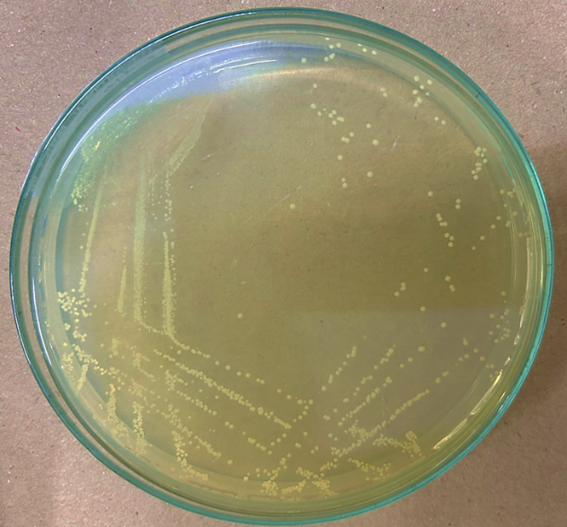
Fig 1. Typical Characteristics of The Isolates Grown on MRS Agar Medium
Identification of Lactic Acid Bacteria
A total of 30 bacterial isolates were obtained from curd samples collected from local dairy and household curd. Among these, 13 isolates were catalase-negative, suggesting a high probability that they belong to the Lactobacillus genus. Catalase-negative bacteria do not produce the enzyme catalase, which is a key characteristic of lactic acid bacteria. Further identification through Gram staining revealed that all 13 catalase-negative isolates were Gram-positive rods. This morphological characteristic is consistent with Lactobacillus species, which are widely recognized as beneficial probiotic bacteria.
Table 2 Sugar fermentation chart of isolated Lactobacillus spp. from curd sample
|
|
Isolate 1 |
Isolate 2 |
Isolate 3 |
Isolate 4 |
Isolate 5 |
Isolate 6 |
Isolate 7 |
Isolate 8 |
Isolate 9 |
Isolate 10 |
Isolate 11 |
Isolate 12 |
Isolate 13 |
|
Glucose |
+ |
+ |
+ |
+ |
+ |
+ |
+ |
+ |
+ |
+ |
+ |
+ |
+ |
|
Lactose |
+ |
+ |
+ |
+ |
+ |
+ |
+ |
+ |
+ |
+ |
+ |
+ |
+ |
|
Maltose |
+ |
+ |
+ |
+ |
+ |
+ |
+ |
+ |
+ |
+ |
+ |
+ |
+ |
|
Sucrose |
+ |
+ |
+ |
+ |
+ |
+ |
+ |
+ |
+ |
+ |
+ |
+ |
+ |
|
Fructose |
+ |
+ |
+ |
+ |
+ |
+ |
+ |
+ |
+ |
+ |
+ |
+ |
+ |
|
Mannitol |
+ |
+ |
– |
– |
+ |
+ |
– |
+ |
– |
– |
+ |
– |
– |
|
Arabinose |
+ |
– |
– |
– |
+ |
– |
+ |
– |
+ |
– |
– |
+ |
– |
|
Xylose |
– |
– |
– |
– |
– |
+ |
– |
+ |
+ |
+ |
– |
+ |
+ |
|
Galactose |
+ |
+ |
+ |
+ |
+ |
+ |
+ |
+ |
+ |
+ |
+ |
+ |
+ |
|
Raffinose |
– |
– |
– |
– |
– |
– |
– |
+ |
+ |
+ |
– |
– |
+ |
|
Sorbitol |
– |
+ |
– |
– |
– |
– |
– |
– |
– |
– |
– |
– |
– |
|
Trehalose |
+ |
+ |
+ |
– |
+ |
+ |
– |
+ |
– |
– |
+ |
+ |
– |
|
Ribose |
+ |
+ |
+ |
+ |
+ |
+ |
+ |
+ |
+ |
+ |
+ |
+ |
+ |
Legend:
The 13 Lactobacillus isolates demonstrate distinct sugar fermentation profiles, reflecting their metabolic diversity. Isolates 1, 2, and 5 show broad capabilities, fermenting most primary sugars like glucose, lactose, maltose, and sucrose, with Isolate 2 uniquely fermenting sorbitol. Isolates 3 and 4 have limited metabolic versatility, focusing on basic sugars and showing no fermentation of secondary sugars like xylose, arabinose, or raffinose. In contrast, Isolates 8, 9, and 10 exhibit broader metabolic potential, fermenting secondary sugars such as arabinose, xylose, and raffinose. Isolates 6 and 7 stand out for their ability to ferment trehalose and xylose, respectively, but show limited utilization of mannitol and raffinose. Isolates 11 and 12 display moderate sugar utilization, focusing on primary sugars but lacking the ability to ferment most secondary sugars. Lastly, Isolate 13 exhibits an extended range, fermenting raffinose and xylose while being unable to utilize trehalose and sorbitol. These varied profiles underscore the strain-specific nature of Lactobacillus isolates, which can be leveraged for targeted applications in nutrition and health.
Bile salt tolerance

Fig. 2 Bile acid tolerance of Lactobacillus isolates from curd samples
Bile salt tolerance is a crucial trait for probiotic bacteria, as it determines their ability to survive and function in the human gastrointestinal tract. The study measured the tolerance of each isolate to different bile salt concentrations (0.5%–2.5%) by recording absorbance at 600 nm. The results indicated that most isolates showed increased absorbance as bile concentration increased. This suggests that these bacteria adapted well to bile salt exposure. Isolate 12 demonstrated the highest absorbance at 2.5% bile salt, suggesting excellent bile resistance, which is essential for probiotic efficacy. In contrast, isolates such as I8 and I11 exhibited lower absorbance at higher bile concentrations, indicating weaker tolerance.
NaCl tolerance
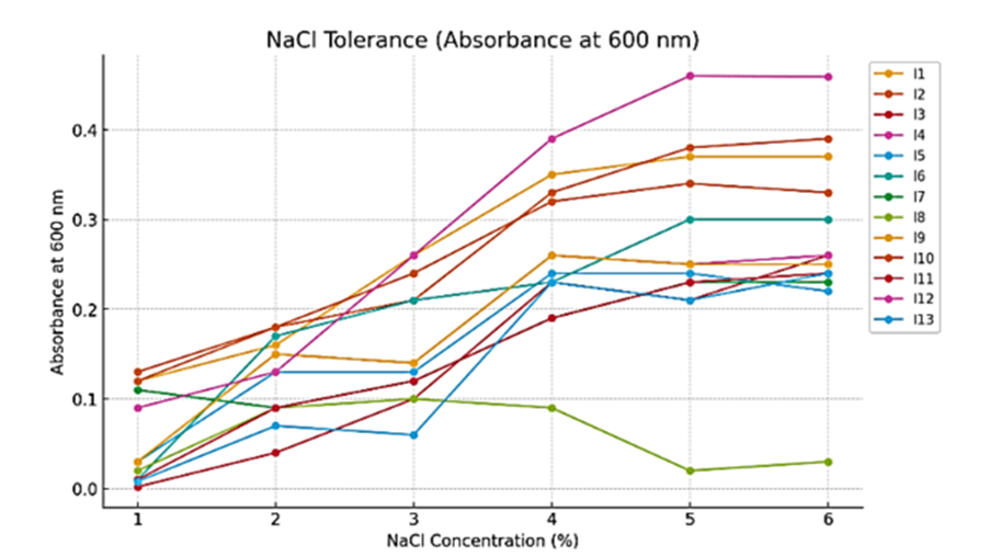
Fig. 3 NaCl tolerance of Lactobacillus isolates from curd samples
The ability of probiotic bacteria to tolerate salt stress is an important characteristic for their survival in food products and the human body. The study evaluated NaCl tolerance at concentrations ranging from 1% to 6%, measuring bacterial growth through absorbance at 600 nm. Many isolates displayed strong growth at 4% NaCl, indicating good salt tolerance. However, at 6% NaCl, only a few isolates, such as Isolate 12, continued to thrive, demonstrating robust resistance to salt stress. This suggests that certain isolates could be used in probiotic formulations where salt tolerance is required.
Determination of optimal growth at different pH

Fig. 4 Optimal growth and pH of isolated Lactobacillus isolates from curd samples.
The optimal growth of an organism at different pH levels by measuring absorbance at 600 nm. The pH values tested include 2.00, 3.00, and 3.50, with growth measurements recorded under different conditions represented by I? to I??. At pH 2.00, the absorbance values range from 0.09 to 0.29, indicating moderate growth. At pH 3.00, the values increase slightly, ranging from 0.11 to 0.32, suggesting improved growth. The highest growth is observed at pH 3.50, with absorbance values between 0.09 and 0.33, indicating that this pH is the most favorable for the organism. Overall, the trend shows that growth improves as pH increases, suggesting that the organism thrives better in slightly less acidic conditions.
Acid tolerance test
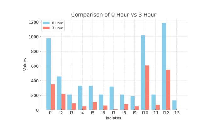
Fig. 5 Acid Tolerance of isolated Lactobacillus isolates from curd samples.
The acid tolerance test evaluated the survival of bacterial isolates under acidic conditions, mimicking the stomach environment. Bacterial counts were taken at 0 hours and 3 hours, and results showed that some isolates maintained better survival over time. Isolate 10 exhibited the highest survival rate, reducing from 102 × 10¹ CFU to 61 × 10¹ CFU, followed by Isolate 12, which reduced from 119 × 10¹ CFU to 55 × 10¹ CFU. These findings indicate that these isolates have strong acid resistance, which is essential for probiotics to survive in the digestive system.
Cholesterol assimilation

Fig. 6 Cholesterol Assimilation of isolated Lactobacillus isolates from curd samples.
Cholesterol assimilation was assessed by testing bacterial growth in a medium containing 0.3% oxgal and 100 mcg/mL cholesterol, measuring absorbance at 550 nm. The results showed that isolates had varying abilities to assimilate cholesterol. Isolate 12 recorded the highest absorbance (0.49), followed by Isolate 10 (0.472) and Isolate 13 (0.467). These results suggest that certain isolates have potential cholesterol-lowering properties, making them suitable for functional food applications aimed at improving heart health.
Adhesion Study of Probiotic Isolates on HT-29 Cell Line
ig 7 Probiotic Adhesion to HT-29 Cells: Experimental Flow and Gram Staining Results of isolates 10,12

Fig. 8 Probiotic Adhesion to HT-29 Cells: Gram Staining Results of Isolates 4,5,6
The adhesion study was conducted to evaluate the ability of Isolate 10, Isolate 12, Isolate 5, Isolate 6, and Isolate 4 to attach to HT-29 human colon carcinoma cell lines. Adhesion ability is crucial for probiotic strains as it determines their ability to colonize the intestinal lining, provide health benefits, and competitively exclude pathogens. The adhesion ability of isolates was evaluated based on the number of bacteria adhering per microscopic field, with classification as strongly adhesive (>100 bacteria per field), moderately adhesive (41–100 bacteria per field), and weakly adhesive (≤40 bacteria per field). The experimental setup included HT-29 cell seeding at 3 × 10? cells/well with a 48-hour incubation period, while probiotic strains were seeded at 6 × 10? – 41 × 10? cells/well, with an adhesion time of 2 hours. These criteria allowed for the accurate assessment of probiotic adhesion potential.
Table 3: Results of Adhesion Study
|
Isolate |
Adhesion Score (Bacteria per Field) |
Adhesion Category |
|
Isolate 10 (I-10) |
41 × 10? CFU/well |
Strongly Adhesive |
|
Isolate 5 (IL-5) |
56 × 10? CFU/well |
Strongly Adhesive |
|
Isolate 4 (IL-4) |
49.1 × 10? CFU/well |
Moderately Adhesive |
|
Isolate 6 (IL-6) |
39.4 × 10? CFU/well |
Moderately Adhesive |
|
Isolate 12 (I-12) |
13.6 × 10? CFU/well |
Weakly Adhesive |
The adhesion study on HT-29 cell lines revealed that Isolate 5 (IL-5) and Isolate 10 (I-10) were the strongest adhesive isolates, with adhesion scores of 56 × 10? CFU/well and 41 × 10? CFU/well, respectively. Their high adhesion is likely due to surface proteins, extracellular polysaccharides (EPS), and bacterial aggregation factors, enhancing their colonization efficiency and ability to exclude pathogens. Moderately adhesive isolates, Isolate 4 (IL-4) and Isolate 6 (IL-6), demonstrated adhesion scores of 49.1 × 10? CFU/well and 39.4 × 10? CFU/well, respectively, indicating decent binding ability but lower colonization potential compared to the strongest isolates. The least adhesive isolate, Isolate 12 (I-12), showed the lowest adhesion at 13.6 × 10? CFU/well, suggesting limited gut persistence despite potential probiotic benefits. Isolate 5 (IL-5) and Isolate 10 (I-10) demonstrated the strongest adhesion, making them ideal probiotic candidates for gut health applications. Isolate 4 (IL-4) and Isolate 6 (IL-6) showed moderate adhesion, indicating some probiotic potential but lower colonization efficiency than the strongest isolates. In contrast, Isolate 12 (I-12) exhibited the weakest adhesion, making it less suitable for gut colonization. These results suggest that Isolate 5 and Isolate 10 could be effectively incorporated into probiotic formulations, while Isolate 12 may require further optimization to enhance its adhesion properties.
2. Formulation of probiotic based oral health supplement and stability testing

Fig. 9 Initial packed preparation of health supplement Sample with potential Lactobacillus isolates I 10 and I 5.

Fig. 10 Reconstituted health supplement Sample with potential Lactobacillus isolates I 10 and I 5.
A probiotic-based oral health supplement was formulated using Isolate 10 and Isolate 5. These isolates were selected due to their strong probiotic characteristics, including acid and bile tolerance, cholesterol assimilation, and adhesion ability. The supplement was developed to prevent acne and improve gut health, aligning with the study’s objectives.
2.2 Stability testing of probiotic based health supplement
Table 4: Results of Stability testing of probiotic based health supplement
|
6 months Accelerated Shelf-life study |
|||||||||
|
Name of Sample: |
Health Supplement |
||||||||
|
No of packets given: |
6 gms x 20 |
||||||||
|
Market storage temperature: |
RT |
||||||||
|
Accelerated Shelf life |
@38 degrees & 90 % RH |
||||||||
|
Time points |
0 day |
1 month |
2 month |
3 month |
4 month |
5 month |
6 month |
LIMITS |
|
|
Parameters |
Test Method |
|
|
|
|
|
|
|
|
|
Sensory |
|||||||||
|
Description |
Visual Examination |
Light pink colour fine powder |
Light pink colour fine powder |
Light pink colour fine powder |
Light pink colour fine powder |
Light pink colour fine powder |
Light pink colour fine powder |
Light pink colour fine powder |
-- |
|
Odour & Taste |
Organoleptic |
Agreeable |
Agreeable |
Agreeable |
Agreeable |
Agreeable |
Agreeable |
Agreeable |
-- |
|
Chemical |
|||||||||
|
pH |
pH meter |
2.97 |
2.99 |
3.03 |
3.06 |
3.07 |
3.09 |
3.97 |
-- |
|
Water activity |
By Water Activity Meter |
0.45 |
0.45 |
0.5 |
0.51 |
0.55 |
0.55 |
0.57 |
NMT 0.9 |
|
Moisture |
FSSAI manual for Milk and Milk powder |
3.38 |
3.4 |
3.46 |
3.4 |
3.48 |
3.5 |
3.59 |
-- |
|
Microbiology |
|||||||||
|
S.aureus |
- |
Absent |
Absent |
Absent |
Absent |
Absent |
Absent |
Absent |
Absent |
|
E.coli |
- |
Absent |
Absent |
Absent |
Absent |
Absent |
Absent |
Absent |
|
|
Salmonella |
- |
Absent |
Absent |
Absent |
Absent |
Absent |
Absent |
Absent |
Absent |
|
Candida albicans |
- |
Absent |
Absent |
Absent |
Absent |
Absent |
Absent |
105Absent |
Absent |
|
Total Bactrerial Count as Probiotic Blend Count |
- |
5.3 X 10⁵ |
5.0 X 10⁵ |
4.48 X 10⁵ |
4.4 X 10⁵ |
3.96 X |
3.8 X 10⁵ |
3.77 X 10⁴ |
-- |
A 6-month accelerated stability study was conducted on the probiotic health supplement at 38°C and 90% relative humidity. The supplement remained stable in terms of color, odor, pH, and microbial viability over six months. However, a slight decrease in probiotic count was observed over time, with the total bacterial count reducing from 5.3 × 10? CFU to 3.77 × 10? CFU by the sixth month. Despite this reduction, the supplement maintained an acceptable shelf-life stability.
DISCUSSION
The study aimed to identify and evaluate Lactobacillus spp. from curd samples, assess their probiotic properties, and formulate a stable probiotic-based oral health supplement. The results provide insights into their potential applications in gut health and disease prevention. Bile tolerance is a key determinant of probiotic efficacy, as bacteria must withstand bile concentrations in the gastrointestinal tract to colonize and exert health benefits 25. The average bile salt concentration in the human intestine ranges from 0.3% to 2.0% 26. The isolates demonstrated significant survival at bile concentrations of 0.5%–2.5%, with Isolate 12 showing the highest tolerance at 2.5%. This suggests that the selected Lactobacillus strains have enhanced bile resistance, a crucial factor for gut persistence27. These results align with previous studies indicating that Lactobacillus spp. can adapt to bile stress by modifying their cell membrane composition 28. Salt tolerance is essential for probiotic strains used in food and pharmaceutical formulations. The study evaluated bacterial growth at NaCl concentrations of 1%–6%, with strong growth observed at 4%. The human intestine generally has an osmolarity equivalent to 0.9% NaCl 29. The ability of some isolates to thrive at 6% NaCl suggests potential for their application in fermented foods and high-salt environments30. Isolate 12 exhibited the highest tolerance, making it suitable for food-based probiotic formulations. The stomach’s acidic environment (pH 1.5–3.5) poses a challenge for probiotics 31. In this study, bacterial growth improved as pH increased from 2.0 to 3.5. The highest growth was observed at pH 3.5, indicating the isolates' ability to survive gastric conditions. Isolate 10 showed the highest survival rate under acidic conditions, making it a strong candidate for probiotic applications 32. These findings align with research demonstrating that Lactobacillus spp. enhance acid resistance through the production of protective proteins and biofilm formation 33. Probiotic strains can lower serum cholesterol by assimilating dietary cholesterol and modifying bile salt metabolism 34. The study demonstrated varying cholesterol assimilation abilities, with Isolate 12 showing the highest absorption (0.49 absorbance), followed by Isolates 10 and 13. This suggests that these strains may contribute to cholesterol reduction, aligning with previous research showing that Lactobacillus strains can enzymatically deconjugate bile acids, reducing cholesterol reabsorption 35. Probiotic adhesion to intestinal cells is crucial for colonization and pathogen exclusion36. The adhesion study categorized isolates into strong, moderate, and weak adhesive groups. Isolates 5 and 10 were the most adhesive, suggesting their ability to persist in the gut and confer probiotic benefits. Strong adhesion is often linked to surface proteins and extracellular polysaccharides, enhancing probiotic efficacy 37. These results are consistent with studies demonstrating that Lactobacillus adhesion improves gut microbiota stability and immune modulation 38. A 6-month accelerated stability study assessed the probiotic supplement’s viability under storage conditions. The total bacterial count decreased from 5.3 × 10? CFU to 3.77 × 10? CFU, indicating a decline in viability over time. However, the formulation-maintained stability in terms of sensory characteristics, pH, and microbial safety. Previous studies suggest that probiotic viability can be improved through encapsulation techniques and optimized storage conditions 39. Despite the decline, the probiotic count remained within acceptable limits, confirming the supplement’s effectiveness 40.
CONCLUSION
The study successfully identified probiotic Lactobacillus strains with strong bile and acid resistance, cholesterol assimilation, and adhesion properties. The formulated supplement demonstrated stability, making it a potential candidate for gut health applications. Future research should explore methods to enhance probiotic viability and optimize formulation strategies for commercial applications.
ACKNOWLEDGMENT
We sincerely thank Vital Nutraceuticals Pvt. Ltd., Ambernath, Maharashtra, for their support in developing the probiotic-based health supplement. Our gratitude also goes to NBHC Lab for conducting the stability tests. Their contributions were invaluable to this project.
REFERENCES


Supriya Yadav*, Dr. S. V. Raut, Development of a Probiotic-Based Health Supplement: Screening, Characterization, and Stability Assessment, Int. J. of Pharm. Sci., 2025, Vol 3, Issue 3, 2658-2677 https://doi.org/10.5281/zenodo.15096989
 10.5281/zenodo.15096989
