View Article

Abstract

Brain tumors are a serious health problem around the world, and their number is increasing every year. India, being a large country, has many patients suffering from brain tumors. The rate of brain tumors in India is different in different regions and among different groups of people. Age, gender, income level, and where a person lives can affect how common brain tumors are in the country. Research and treatment for brain tumors in India are improving. Doctors and scientists are working on new methods like personalized medicine, targeted treatment, immunotherapy, and precision oncology to help patients more effectively. In summary, brain tumors are a big healthcare challenge in India. To deal with them, we need to understand how they occur, how to diagnose them, and how to treat them properly. Working together and using modern techniques can help improve patient care and quality of life.

Keywords

Pediatric brain tumor, adolescents, risk factors, ionizing radiation, genetic predisposition, environmental exposures.

Introduction

Recent health data suggests that the number of brain tumor cases in India is increasing. This rise is adding to the country’s overall burden of brain tumor patients. Several reasons may explain this growth, such as changes in lifestyle, better medical facilities and diagnostic tools, longer life expectancy, and an expanding population. Together, these factors are leading to more people being diagnosed with brain tumors in India. 

Today, many advanced imaging methods are available to help doctors identify and treat brain tumors. Modern MRI scans use special contrast agents, and PET scans use amino-acid-based tracers. Together, these techniques give radiologists clearer and more detailed pictures of tumors, helping them locate and measure tumors.

Brain tumors are the second most common type of cancer in children and young adults. Every year, about 215,000 children aged 0–14 years and around 85,000 teenagers aged 15–19 years are diagnosed with cancer worldwide. According to the International Agency for Research on Cancer, the number of brain tumor cases differs from country to country, ranging from 1.15 to 5.14 cases per 100,000 children. The rates also change depending on the ethnicity of the population. 

Childhood brain tumors include different types of solid growths in the brain and are the leading cause of cancer-related deaths in children under 20 years. They make up nearly 25% of all childhood cancers. Worldwide data shows that both the number of cases and deaths from childhood brain tumors are higher in wealthier countries compared to developing countries

These tumors not only affect physical health but also emotional and mental wellbeing, which can lead to a poor quality of life.(1,6,7,8)

3. LITREATURE  REVIEW 

  1. Onyije F M., Dolatkhah R., Olsson A., et al. Risk factors for childhood brain tumors: A systematic review and meta-analysis of observational studies from 1976 to 2022. Cancer Epidemiol. 2024;88:102510. Synthesises 181 studies (case-control + cohort) on childhood brain tumors, reporting associations for factors like domestic insecticide exposure, maternal consumption of cured meat/coffee, paternal occupational exposures. PMC Useful for summarizing modifiable risk factors and epidemiologic evidence.  
  2. Lutz K., et al. Brain tumors in children and adolescents: incidence trends and risk-factor insights. Children. 2022;9:498. Presents incidence data and discusses that only radiation exposure is a well established environmental risk for paediatric brain tumors. brainlife.org Good for background on incidence and known strong risk factors. 
  3. Vienne-Jumeau A., Tafani C., Ricard D. Environmental risk factors of primary brain tumors: A review. Rev Neurol. 2019;175(10):664-678. Reviews environmental exposures (ionising radiation, pesticides, air-pollution, radio-frequency) for adult CNS tumors, but emphasises childhood radiation exposure.  PubMed Helpful for framing environmental risk exposures in younger populations. 
  4. Diwanji T P., Engelmann A., Snider J W., Mohindra P. Epidemiology, diagnosis, and optimal management of glioma in adolescents and young adults. Adolesc Health Med Their. 2017;8:99-113. Focuses on AYA (adolescents and young adults, ~15-39 yrs.) gliomas: incidence, histology, risk factors like genetic syndromes, radiation.  PMC Valuable to include for ?young? population beyond childhood.  
  5. Rousseau J., Bennett J., Lim-Fat M. Brain Tumors in Adolescents and Young Adults: A Review. Semin Neurol. 2023 Nov 10 (online ahead of print).  Updated review on brain tumors’ in AYA, incorporates molecular classification and emphasises distinct risk/management issues.  brainlife.org Good for discussing how risk and presentation differ in the young adult segment.  
  6. Sheppard J P., Nguyen T., Alkhalid Y., Beckett J S., Salamon N., Yang I. Risk of Brain Tumor Induction from Pediatric Head CT Procedures: A Systematic Literature Review. Brain Tumor Res Treat. 2018;6(1):1?7. Reviews data on head CT (ionising radiation) in children/adolescents and the associated brain tumors risk: excess relative risk ~1.29 (CI 0.66-1.93) after ≥1 scan.  btrt.org Useful for discussing radiation exposure from diagnostic imaging.  
  7. American Cancer Society. Risk Factors for Brain and Spinal Cord Tumors in Children. (Web page) Summarizes well-established vs uncertain risk factors for childhood brain tumors: e.g., head radiation, genetic syndromes; emphasises that most causes are unknown.  Cancer.org Useful for background and framing the ?unknown etiology? challenge.  
  8. MD Anderson Cancer Center. Childhood Brain Tumors – Risk Factors. (Web page)    Highlights risk factors including radiation, inherited genetic conditions (e.g., NF1/2, LiFraumeni, tuberous sclerosis), while noting most tumors have no clear cause.  MD Anderson Cancer Center Useful for genetic predisposition part of risk.  
  9. Environmental Factors and the Risk of Brain Tumors in Young People: A Systematic Review.? (Young people < 25 yrs.) Karger?published article.  Focuses on exposure in younger age (<25 yrs.) to environmental risk factors: cadmium, well-water nitrates, parental smoking, farm exposures etc., albeit with limitations. PubMed Good for covering lessestablished environmental risks.  
  10. Epidemiology and prognostic factors of pediatric brain tumor survival in the US: Evidence from four decades

4. NEED OF THE STUDY 

Brain tumors  in children and young adults cause disproportionate long-term disability and mortality compared with many other illnesses of youth. Yet risk factors, early markers and vulnerable windows of exposure remain poorly consolidated across ages, tumor types and populations. A focused review that synthesizes what is known about environmental, genetic, perinatal and lifestyle risk factors—and highlights gaps and methodological challenges—will help clinicians, researchers and policymakers prioritize prevention, screening and research that targets the young.  

5. AIM  

To evaluate and summarize the potential risk factors, genetic predispositions, and environmental influences contributing to the development of brain tumors among the young population, and to identify preventive and early detection strategies.  

OBJECTIVES 

  1. To review the epidemiological data on the incidence and prevalence of brain tumors in children, adolescents, and young adults.  
  2. To identify genetic, environmental, and lifestyle factors associated with increased brain tumor risk in the young population.  
  3. To analyze current studies highlighting the role of radiation exposure, family history, and environmental toxins in tumor development.  
  4. To assess diagnostic challenges and the importance of early risk assessment in improving prognosis.  
  5. To propose preventive measures and recommendations for early screening and awareness among young individuals  

6. TYPES OF TUMORS 

Brain tumors are growths that develop inside the brain due to cells growing in an abnormal and uncontrolled way. They are a mixed group of tumors, and each type grows and behaves differently. They also have different chances of survival and different treatment options. However, their symptoms, basic tests for diagnosis, and first steps of treatment are mostly similar for all types.  Brain tumors can be classified in different ways, such as by the type of cell they come from, where they are located, how they behave, and their grade (how fast they grow). These classifications help doctors understand the type of tumor and how to treat it. Some important types of brain tumors include:  

1. Gliomas  

These tumors start from glial cells, which support and protect nerve cells in the brain. One very serious and fast-growing type of glioma is called Glioblastoma Multiforme  (GBM). Gliomas can range from low-grade (slow-growing, less harmful) to high-grade (fast-growing, more dangerous) tumors.  

2. Oligodendrogliomas  

These tumors grow from oligodendrocytes, a type of glial cell. Another related tumor, called olingoastrocytoma  develops from ependymal cells found in the brain’s ventricles or the spinal cord canal.  

3. Meningiomas  

These tumors form from the membranes (meninges) that cover and protect the brain and spinal cord. Most meningioma’s  are non-cancerous (benign), but depending on their size and location, they can still cause symptoms by pressing on the brain.  

4. Pituitary Tumors  

These tumors form in the pituitary gland, the part of the brain that controls hormone production. Many pituitary tumors (pituitary adenomas) are benign, but they may affect hormone levels in the body. (the part of the brain that controls balance and coordination). They commonly occur in children. A similar type — previously called Primitive Neuroectodermal  Tumor (PNET) — affects developing nerve cells / PNET Medulloblastomas  are fast-growing tumors usually found in the cerebellum (the part of the brain that controls balance and coordination). They commonly occur in children. A similar type — previously called Primitive Neuroectodermal Tumor (PNET) — affects developing nerve cells(1,3,20)  

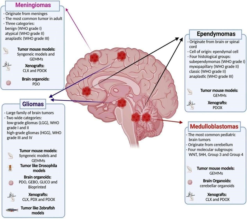
Fig 1: Brain Tumor Types

Location Of Tumor 

  1. Cerebrum  

The largest part of the brain; controls thinking, memory, speech, and movement. Tumors here may cause: Weakness on one side of the body, Speech or language problems, Seizures, Personality or behavior changes, Memory loss.  

  1. Cerebellum  

Located at the back of the brain; controls balance and coordination. Tumors here may cause: Difficulty walking or balancing, Dizziness, Poor coordination, Headache and vomiting  

  1. Brainstem  

Connects the brain to the spinal cord; controls breathing, heart rate, vision, swallowing. Tumors here may cause: Difficulty speaking or swallowing, Double vision, Weakness or paralysis, Breathing or heart rate problems. 

  1. Pituitary Gland (Base of the brain) Regulates hormones.  

Tumors here may cause: Hormonal imbalance, Vision problems (especially peripheral vision loss),Growth abnormalities.  

  1. Pineal Gland  

Deep in the brain; controls sleep cycle Tumors here may cause: Sleep disturbances, Problems with eye movement, Headache.  

  1. Meninges  

Tissue covering the brain (meningiomas). Symptoms include: Headache, Seizures, Vision changes 

  1. Cranial Nerves  

Especially near the ear or face (e.g., acoustic neuroma).  May cause: Hearing loss, Ringing in ears, Facial numbness or weakness(12)  

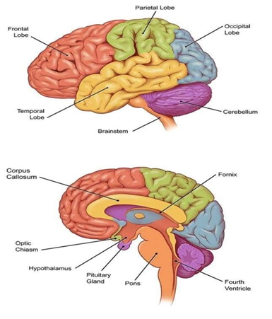
Fig 2: locations of tumor.

7.   MECHANISM OF BRAIN TUMOR FORMATION 

  1. Genetic Mutations  

Normal brain cells acquire DNA mutations (due to heredity or external factors).These mutations affect key genes:Oncogenes get activated (promote cell growth) Tumor suppressor genes get inactivated (e.g., p53, PTEN)  Result: uncontrolled cell division.  

  1. Dysregulated Cell Signaling  

Growth signaling pathways (EGFR, PDGF, PI3K/AKT, RAS/MAPK) become overactive. Cells keep receiving ?grow and divide? signals even when unnecessary.  

  1. Loss of Cell Cycle Control  

Checkpoints that control cell division fail. Tumor cells bypass apoptosis (programmed cell death).They accumulate instead of dying.  

  1. Angiogenesis (New Blood Vessel Formation)  

Tumor cells release VEGF and other factors. New blood vessels grow to supply oxygen and nutrients. Tumor grows rapidly due to enhanced blood supply.  

  1. Invasion and Migration  

Tumor cells secrete enzymes (MMPs) that break down surrounding tissue. They invade nearby brain structures. This makes surgical removal challenging.  

  1. Immune Evasion  

Tumor cells suppress immune cells and produce inhibitory molecules. They avoid being recognized and destroyed by the immune system.      

8. CAUSES OF  BRAIN TUMOR  

I. Genetic (Hereditary) Factors  

Some people inherit abnormal genes from their parents that increase the risk of brain tumors. Example: Neurofibromatosis, Li-Fraumeni syndrome, Tuberous sclerosis                                     

II. Radiation Exposure  

Previous exposure to high-dose radiation, especially to the head, increases the risk.     Example: Radiation treatment for childhood cancers  

III. Age Factor  

Brain tumors can occur at any age but are more common in children and older adults.  

IV. Environmental Factors (less proven but suspected)  

Some studies suggest certain environmental exposures may increase risk.  Possible factors include: Exposure to harmful chemicals (e.g., pesticides, industrial chemicals),  Air pollution, Ionizing radiation in the environment  

V. Weakened Immune System  

People with weak immune systems (e.g., HIV patients or those taking immunosuppressant’s) are at higher risk.  

VI. Family History  

If close family members have brain tumors, your risk may be slightly higher.(8,12)  

Symptoms of Brain Tumor 

Brain tumor symptoms can vary widely because they depend on the tumor’s size, type, growth rate, and location in the brain. As the tumor grows, it may press on nearby nerves, brain tissue, or block the flow of cerebrospinal fluid, leading to increased pressure inside the skull. This results in various physical, mental, and neurological changes.  

General Symptoms  These symptoms can occur regardless of the tumor’s location:  

  1. Headache: Often persistent and worse in the morning or when lying down, Can worsen with coughing, bending, or physical activity, May not respond to usual pain medications over time 
  2. Seizures(Fits): One of the first symptoms in many cases, Can be fullbody convulsions or mild jerking/shaking of a limb Some patients experience a staring spell, confusion, or brief loss of awareness
  3. Nausea And Vomiting: Often due to increased pressure in the skull ,More common in morning and not always related to eating  
  4. Fatigue And Weakness: Extreme tiredness even after rest Reduced physical stamina and energy 
  5. Cognitive& Personality Changes, Tumors in the frontal or temporal lobes often cause: Difficulty thinking, concentrating, or remembering things, Confusion and slow mental processing Mood changes — depression, anxiety, irritability 
  6. Sensory & Motor Symptoms: Tumors affecting movement and sensation can cause: Weakness or paralysis on one side of the body, Difficulty walking or maintaining balance, Clumsiness or frequent falls, Numbness or tingling in arms/legs, Loss of fine motor skills (buttoning clothes, writing) (8,12,20) 

9. DIAGNOSIS OF BRAIN TUMOR 

The diagnosis of brain and spinal cord tumors in adults involves several medical examinations and imaging techniques.

  1. Physical Examination And Medical History: Doctors first examine the patient for general signs of disease, such as noticeable lumps or changes in physical function. They also review habits like smoking and exercise, and collect family medical history to identify risk factors. 
  2. Neurological Examination: A neurological exam is performed to evaluate the function of the brain, spinal cord, and nerves. The doctor may ask questions or request simple tasks to assess: Memory and thinking ability, Speech and facial expressions, Coordination and balance,

Vision and hearing, Walking patterns (gait), Muscle strength and reflexes   

  1. Electrical Activity Test: Tests such as Electroencephalography (EEG) and evoked potential   tests measure electrical activity in the brain and nerves to detect abnormalities.  
  2. Tumor Marker Test: Blood, urine, or tissue samples are analyzed for specific substances called tumor markers, which may indicate the presence of cancer.  
  3. Genetic Test: Tumor tissue or cells are examined to identify genetic or chromosomal changes. These abnormalities help determine cancer risk and behavior.  
  4. Ultrasound: Ultrasound imaging is used for detecting brain abnormalities in newborns and for examining brain blood vessels.  
  5. X-Ray  Imaging: X-rays are used to examine skull bones and detect abnormalities in the brain or nasal sinuses. Traditional X-ray techniques include: Cerebral angiography: Uses contrast dye to visualize blood vessels.
  6. Magnetic Resonance Imaging (MRI): MRI uses magnetic waves to create high-resolution images of brain structures. It can detect tumor location, size, and spread. (1,8,10)

10. PATHOLOGY OF BRAIN TUMOR 

Normal Brain Cells

(Cells start changing due to DNA damage)

Abnormal Cell Growth Begins

Cells divide too fast & don’t die

Formation of Tumor (Mass of abnormal cells)

Tumor grows & presses on brain tissue

↓ ↓ ↓ Effects ↓ ↓ ↓

Headache

Seizures

Weakness

Vision / speech problems

Memory issues

Tumor spreads to nearby brain areas

( Some tumors can be spread through the cerebrospinal fluid)(1)

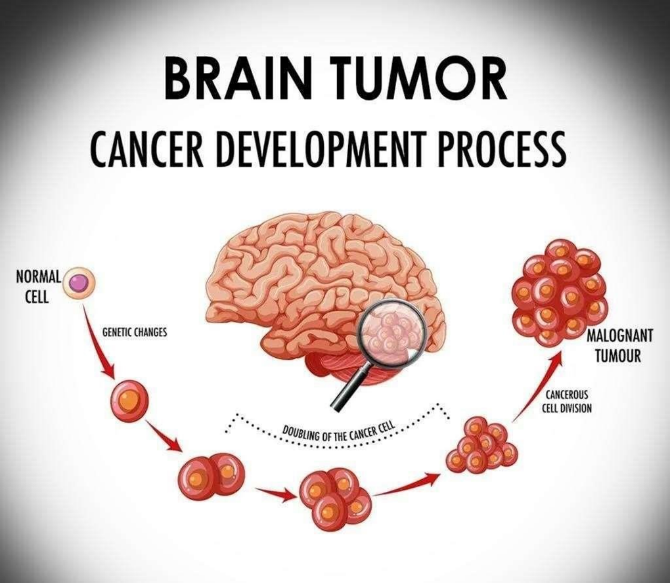
Fig3: Development of Brain Tumor

Pathogenesis Of Brain Tumor 

a) IDH Mutation  

Mutations in the isocitrate dehydrogenase (IDH1/2) genes are common in secondary glioblastomas. These mutations cause abnormal production of 2hydroxyglutarate (2-HG), an oncometabolite that drives tumor formation. Targeted drugs such as AG-120, AG-221, AG881, FT-2102, and IDH305 inhibit mutant IDH enzymes and are under clinical trials. 

b) Notch Pathway  

The Notch signaling pathway regulates cell differentiation and survival. Overactivation contributes to glioblastoma progression and resistance to therapy. Inhibitors of Notch signaling (like γ-secretase inhibitors and CB-103) show promise in reducing tumor growth and improving treatment response.  

  1. Ceramide Signaling  

The enzyme acid ceramidase (ASAH1) converts pro-apoptotic ceramide into sphingosine-1-phosphate (S1P), which promotes cell survival. Overexpression of ASAH1 in glioblastoma reduces apoptosis and enhances tumor spread. ASAH1 inhibitors (like carmofur) can restore apoptosis and are potential therapeutic agents.  

  1. VEGF Signaling  

Vascular endothelial growth factor (VEGF) drives angiogenesis (formation of new blood vessels) to supply tumors with oxygen and nutrients. Blocking VEGF or its receptors (using agents like bevacizumab, sunitinib, or tivozanib) can restrict tumor blood supply. 

Combination therapies are being tested to overcome resistance.  

  1. PDGF signaling  

Platelet-derived growth factor (PDGF) promotes glioblastoma cell growth and survival via its receptor (PDGFR). Tumor cells often activate PDGF signaling through autocrine loops, leading to uncontrolled proliferation. Targeting this pathway can inhibit tumor growth.  

  1. EGFR Pathway in Glioblastoma   

EGFR is a receptor on cell surfaces that normally helps cells grow when growth factors bind to it. In glioblastoma, this receptor often becomes overactive due to gene amplification or mutations (like EGFRvIII). This makes tumor cells grow, divide, and spread uncontrollably.  

  1. PTEN and Glioblastoma   

PTEN is a tumor-suppressor gene that keeps cell growth under control. When PTEN is mutated or lost in glioblastoma, tumor cells grow faster and resist treatment. Research is trying to correct PTEN mutations and restore its function to slow tumor growth.  

  1. SHH (Sonic Hedgehog) Pathway   

The SHH pathway controls cell growth and stem cell behavior. In glioblastoma, mutations keep this pathway permanently ?on,? leading to constant tumor cell growth and survival. Drugs that block this pathway—especially SMO inhibitors like vismodegib—are being tested in clinical trials.(15)  

11. TREATMENT OF BRAIN TUMOR   

For primary brain tumors, the most reliable treatment strategy usually begins with surgical removal of the tumor, followed by chemotherapy and radiotherapy. Surgery offers several benefits, such as maximum tumor excision, reduction of mass effect, decreased tumor load, improved diagnosis, and a better chance of long-term survival. However, brain tumor surgery can lead to a few complications. The most urgent concern after surgery is brain swelling (edema), while issues like infection, bleeding, and anesthesia-related reactions are less common. 

To manage tumor-related edema, corticosteroids such as dexamethasone or prednisone are administered before and after surgery. Patients with larger tumors often present with elevated intracranial pressure and neurological deficits. Neurosurgical patients are also vulnerable to systemic problems such as hemodynamic instability, coagulation abnormalities, and metabolic disturbances. Mortality has been observed to be higher in children younger than 18 years of age.  

1. Radiation Therapy (Re-written Version)  

Advanced radiation techniques such as 3D-Conformal Radiation Therapy (3D-CRT), Intensity-Modulated Radiation Therapy (IMRT), and Proton Beam Therapy (PBT) help deliver radiation precisely to the tumor while protecting surrounding brain tissue.

3D-CRT uses MRI, CT, or PET imaging along with sophisticated computer technology to tailor radiation beams to the tumor’s exact shape. IMRT offers even greater precision by modulating radiation beam intensity with computercontrolled linear accelerators. Compared to these methods, Proton Beam Therapy is more accurate because protons can be focused precisely at the tumor site, sparing nearby healthy tissue; pencil-beam scanning further improves precision. 

Common radiation-related side effects usually appear within 2–3 weeks and may include hair loss, skin irritation, headache, nausea, vomiting, hearing loss, loss of appetite, and cognitive difficulties. PBT patients may additionally experience fatigue, mood changes, depression, insomnia, and nausea.  

2. Targeted Biological Therapy (Precision Medicine)  

Certain targeted therapies work by blocking specific molecules involved in tumor growth. Two commonly used drugs in brain tumors are Bevacizumab (Avastin) and Everolimus (Afinitor). Bevacizumab is a monoclonal antibody that blocks VEGF-A, a key factor in tumor blood-vessel formation. It restricts VEGF binding to endothelial cell receptors, slowing tumor vascular growth. It is frequently used in glioblastoma Multiforme (GBM), especially after radiotherapy and Temozolomide. Avastin can be combined with drugs like irinotecan, carboplatin, and etoposide, though combination therapy does not show much added benefit. Side effects may include proteinuria, hypertension, fatigue, bleeding, headaches, diarrhea, and mouth sores.(1,3,5,8,10,12,17)  

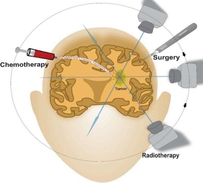
Fig4: treatment of tumor

12.FUTURE PROSPECTS  

Despite major progress in understanding and managing brain tumors, several challenges remain in early detection, precise diagnosis, and effective treatment. Future research and clinical practice are expected to move toward a more personalized, targeted, and technologydriven approach.  

  1. Molecular and Genetic Research:  

Advances in genomics and proteomics will enable identification of novel molecular markers that can predict tumor behavior, treatment response, and prognosis. Integrating molecular profiling into routine diagnostics will help design personalized therapies.  

  1. Artificial Intelligence (AI) and Machine Learning:  

AI-based imaging tools and predictive models are expected to revolutionize tumor detection, segmentation, and classification. Automated radiomics and machine learning algorithms can enhance diagnostic accuracy and treatment planning.  

  1. Targeted and Immunotherapy:  

The future of brain tumor therapy lies in targeted molecular agents and immunebased treatments such as checkpoint inhibitors, CAR-T cell therapy, and oncolytic viruses. These approaches hold promise for improving survival with fewer side effects.  

  1. Nanotechnology and Drug Delivery Systems:  

Overcoming the blood–brain barrier remains a key challenge. Nanoparticle-based drug delivery systems and novel carrier molecules may enhance the delivery of chemotherapeutic and gene-editing agents directly to tumor sites.  

  1. Stem Cell and Gene Therapy:  

Gene-editing tools like CRISPR and the use of neural stem cells for regenerative therapy are emerging areas. These can potentially repair damaged neural tissues and selectively target tumor cells.  

  1. Multimodal and Precision Imaging:  

The combination of MRI, PET, and molecular imaging can provide real-time insights into tumor metabolism and microenvironment, enabling better surgical guidance and radiotherapy planning.  

  1. Public Health and Preventive Strategies:  

In developing countries like India, improved awareness, screening programs, and epidemiological surveillance are essential to detect brain tumors early and reduce mortality.  

  1. Collaborative Research and Big Data:  

Global research collaborations and open-access clinical databases will help integrate large datasets, accelerating discoveries and standardizing treatment protocols. (18) 

OBSERVATION 

The review of existing literature indicates a rising concern regarding the incidence of brain tumors among the young population, including children, adolescents, and young adults. Studies consistently highlight that although brain tumors are relatively rare, they remain one of the leading causes of cancer-related morbidity and mortality in younger age groups

Environmental exposures such as ionizing radiation remain the most strongly supported risk factor across studies. However, increasing attention is directed toward potential associations with non-ionizing radiation (e.g., mobile phone use), although findings remain inconclusive. Genetic predisposition and hereditary syndromes are also observed to play a significant role in a subset of patients.  

Another key observation from the literature is the variability in reported incidence rates across regions, likely influenced by differences in diagnostic capabilities, healthcare access, environmental conditions, and population genetics. Improvements in neuroimaging technologies over recent decades may also contribute to the apparent rise in detected cases. Despite extensive research, significant knowledge gaps persist regarding modifiable risk factors, lifestyle influences, and long-term environmental exposures.  

Overall, the reviewed data suggest that while brain tumor risk in the young pulation remains relatively low, the potential influence of environmental, genetic, and technological factors warrants continued surveillance, targeted research, and improved public health strategies.  

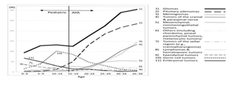
Fig 5: Age specific no. Of patiensts for each brain tumor at the brain tumor registry 2005-08 Japan

CONCLUSION 

In India and across the world, brain tumors are becoming a major health issue because their cases are increasing over time. This growing problem is affected by factors such as a rising population, more elderly people, lifestyle changes, and differences in access to healthcare. A brain tumor is an abnormal growth in the brain that harms normal brain functions. In medical image processing, the goal is to find accurate and useful information with very few errors using computer algorithms. For detecting and classifying brain tumors in MRI scans, four main steps are used: preprocessing the images, segmenting the tumor area, extracting important features, and classifying the tumor type.In the future, tumor imaging will focus on better ways to combine and analyze images from different scans, along with new PET tracers. These improvements will be useful in advanced treatments like intensity-modulated radiotherapy and will help doctors plan radiotherapy more accurately for patients with brain tumors. In recent years, major advances in understanding the molecular features of pediatric brain tumors have improved how we diagnose and treat them. These developments support precision medicine, helping create more targeted therapies. As research grows, future WHO classifications will rely more on molecular markers than traditional histology. Next-generation sequencing continues to provide valuable insights into tumor types, patient characteristics, and treatment outcomes.

REFERENCES

  1. Jaiswal NK, Singh KP, Kumar D. Assessing the Risk of Brain Tumors in Young Populations: A Review. Indian Journal of Pharmacy Practice. 2025;18(3):243–248. Available from: https://ijopp.org/article/8214 
  2. Sheppard JP, Nguyen T, Alkhalid Y, Beckett JS, Salamon N, Yang I. Risk of Brain Tumor Induction from Pediatric Head CT Procedures: A Systematic Literature Review. Brain Tumor Res Treat. 2018;6(1):1–7. Available from: https://doi.org/10.14791/btrt.2018.6.e4 
  3. Kaifi R. A Review of Recent Advances in Brain Tumor Diagnosis Based on AI-Based Classification. Diagnostics. 2023;13(18):3007. Available from: https://doi.org/10.3390/diagnostics13183007 
  4. Mehndiratta A, Giesel FL. Brain Tumour Imaging. In: Diagnostic Techniques and Surgical Management of Brain Tumors. 2011. Available from: https://doi.org/10.5772/23507 
  5. Wagner MW, Jabehdar Maralani P, Bennett J, Nobre L, Lim-Fat MJ, Dirks P, Laughlin S, Tabori U, Ramaswamy V, Hawkins C, Ertl-Wagner BB. Brain Tumor Imaging in Adolescents and Young Adults: 2021 WHO Updates for Molecular-based Tumor Types. Radiology. 2024;310(2):e230777. Available from: https://doi.org/10.1148/radiol.230777 
  6. Zumel-Marne A, Castaño-Vinyals G, Kundi M, Alguacil J, Cardis E. Environmental Factors and the Risk of Brain Tumours in Young People: A Systematic Review. Neuroepidemiology.           2019;53(3-4):121–141.      Available        from: https://doi.org/10.1159/000500601 
  7. Onyije FM, Dolatkhah R, Olsson A, Bouaoun L, Deltour I, Erdmann F, Bonaventure A, Scheurer ME, Clavel J, Schüz J. Risk factors for childhood brain tumours: a systematic review and meta-analysis of observational studies from 1976 to 2022. Cancer. Epidemiology.  2024;88:102510. Available from: https://doi.org/10.1016/j.canep.2023.102510 
  8. Elshaikh BG, Omer H, Garelnabi MEM, Sulieman A, Abdella N, Algadi S, Toufig H. Incidence, Diagnosis and Treatment of Brain Tumours. J Res Med Dent Sci. 2021;9(6):340– 347. Available from: https://www.jrmds.in/articles/incidence-diagnosisand-treatment-ofbrain-tumours.pdf 
  9. AlRayahi J, Alwalid O, Mubarak W, Maaz AUR, Mifsud W. Pediatric Brain Tumors in the  Molecular Era: Updates for the Radiologist. Seminars in Roentgenology. 2023;58(1):47–66. Available from: https://doi.org/10.1053/j.ro.2022.09.004 
  10. Rasheed S, Rehman K, Akash MSH. An insight into the risk factors of brain tumors and their therapeutic interventions. Biomedicine & Pharmacotherapy. 2021;143:112119. Available from: https://doi.org/10.1016/j.biopha.2021.112119 
  11. Yamasaki F. Adolescent and young adult brain tumors: current topics and review. Int J Clin Oncol.2022;27(3):457–464.Availablefrom: https://pmc.ncbi.nlm.nih.gov/articles/PMC8782686/ 
  12. Kabitha KK, Moses Samuel Rajan, Karunakar Hegde, Sunil Koshy, Ashok Shenoy. A COMPREHENSIVE REVIEW ON BRAIN TUMOR. International Journal of Pharmaceutical, Chemical and Biological Sciences (IJPCBS). 2013;3(4):1165–1171. Available from: https://www.ijpcbs.com/articles/a-comprehensive-review-on-braintumor.pdf 
  13. Delaidelli A, Moiraghi A. Recent Advances in the Diagnosis and Treatment of Brain Tumors. Brain Sci. 2024;14(3):224. Available from: https://doi.org/10.3390/brainsci14030224 
  14. Nadeem MW, Al Ghamdi MAA, Hussain M, Khan MA, Khan KM, Almotiri SH, Butt SA.  Brain Tumor Analysis Empowered with Deep Learning: A Review, Taxonomy, and Future Challenges. Brain  Sci. 2020;10(2):118. Available from: https://doi.org/10.3390/brainsci10020118 
  15. Rajaratnam V, Islam MM, Yang M, Slaby R, Ramirez HM, Mirza SP. Glioblastoma: Pathogenesis and Current Status of Chemotherapy and Other Novel Treatments. Cancers. 2020;12(4):937. Available from: https://doi.org/10.3390/cancers12040937 
  16. Khalighi S, Reddy K, Midya A, Pandav KB. Artificial intelligence in neuro-oncology: advances and challenges in brain tumor diagnosis, prognosis, and precision treatment. NPJ Precision Oncology. 2024;8(1):80. Available from: https://doi.org/10.1038/s41698-024- 00575-0 
  17. Dorfner FJ, Patel JB, Kalpathy-Cramer J, Gerstner ER, Bridge CP. A review of deep learning for brain tumor analysis in MRI. npj Precision Oncology. 2025;9(1):2. doi:10.1038/s41698024-00789-2. 
  18. Zhang R, Wang D-M, Liu Y-L, Tian M-L, Zhu L, Chen J, Zhang J. Symptom management in adult brain tumours: A literature review. Nurs Open. 2023;10(8):4892–4906. doi:10.1002/nop2.1795.  Available  from: https://pmc.ncbi.nlm.nih.gov/articles/PMC10333888/ 
  19. Burström G, Amini M, El-Hajj VG, Arfan A, Gharios M, Buwaider A, Losch MS, Manni F, Edström E, Elmi-Terander A. Optical Methods for Brain Tumor Detection: A Systematic Review. J Clin Med. 2024;13(9):2676. doi:10.3390/jcm13092676. Available from: https://pmc.ncbi.nlm.nih.gov/articles/PMC11084501/ 
  20. Smith N. Brain Tumour: A Comprehensive Analysis. Journal Of Multiple Sclerosis. 2024; 11(4):001-002. doi:10.35248/2376-0389.24.11.04.001-002. Available from: https://www.iomcworld.org/open-access/brain-tumour-a-comprehensive-analysis.pdf 21. Herholz K, Langen K-J, Schiepers C, Mountz JM. Brain tumors. Semin Nucl Med. 2012;42(6):356–370. doi:10.1053/j.semnuclmed.2012.06.001. Available from: https://pmc.ncbi.nlm.nih.gov/articles/PMC3925448/ 
  21. Abdusalomov AB, Mukhiddinov M, Whangbo TK. Brain Tumor Detection Based on Deep Learning Approaches and Magnetic Resonance Imaging. Cancers. 2023; 15(16):4172. https://doi.org/10.3390/cancers15164172 
  22. Eslaminejad T, Nematollahi-Mahani SN, Ershad Sarabi R, Ohadi M, Eslaminejad T, Ansari M, Mirzaie V. Brain Cancer Treatment; A Systematic Review. Int J Cancer Manag. 2022; 15(6):e121473. https://doi.org/10.5812/ijcm-121473 
  23. Pagar KR, Mahale MR. A Review on Brain Tumour, Etiology and Treatment. Asian Journal of Pharmaceutical Research. 2023; 13(1):51–54. doi:10.52711/22315691.2023.00010. Available from: https://asianjpr.com/AbstractView.aspx?PID=2023-13-1-10 

Reference

  1. Jaiswal NK, Singh KP, Kumar D. Assessing the Risk of Brain Tumors in Young Populations: A Review. Indian Journal of Pharmacy Practice. 2025;18(3):243–248. Available from: https://ijopp.org/article/8214 
  2. Sheppard JP, Nguyen T, Alkhalid Y, Beckett JS, Salamon N, Yang I. Risk of Brain Tumor Induction from Pediatric Head CT Procedures: A Systematic Literature Review. Brain Tumor Res Treat. 2018;6(1):1–7. Available from: https://doi.org/10.14791/btrt.2018.6.e4 
  3. Kaifi R. A Review of Recent Advances in Brain Tumor Diagnosis Based on AI-Based Classification. Diagnostics. 2023;13(18):3007. Available from: https://doi.org/10.3390/diagnostics13183007 
  4. Mehndiratta A, Giesel FL. Brain Tumour Imaging. In: Diagnostic Techniques and Surgical Management of Brain Tumors. 2011. Available from: https://doi.org/10.5772/23507 
  5. Wagner MW, Jabehdar Maralani P, Bennett J, Nobre L, Lim-Fat MJ, Dirks P, Laughlin S, Tabori U, Ramaswamy V, Hawkins C, Ertl-Wagner BB. Brain Tumor Imaging in Adolescents and Young Adults: 2021 WHO Updates for Molecular-based Tumor Types. Radiology. 2024;310(2):e230777. Available from: https://doi.org/10.1148/radiol.230777 
  6. Zumel-Marne A, Castaño-Vinyals G, Kundi M, Alguacil J, Cardis E. Environmental Factors and the Risk of Brain Tumours in Young People: A Systematic Review. Neuroepidemiology.           2019;53(3-4):121–141.      Available        from: https://doi.org/10.1159/000500601 
  7. Onyije FM, Dolatkhah R, Olsson A, Bouaoun L, Deltour I, Erdmann F, Bonaventure A, Scheurer ME, Clavel J, Schüz J. Risk factors for childhood brain tumours: a systematic review and meta-analysis of observational studies from 1976 to 2022. Cancer. Epidemiology.  2024;88:102510. Available from: https://doi.org/10.1016/j.canep.2023.102510 
  8. Elshaikh BG, Omer H, Garelnabi MEM, Sulieman A, Abdella N, Algadi S, Toufig H. Incidence, Diagnosis and Treatment of Brain Tumours. J Res Med Dent Sci. 2021;9(6):340– 347. Available from: https://www.jrmds.in/articles/incidence-diagnosisand-treatment-ofbrain-tumours.pdf 
  9. AlRayahi J, Alwalid O, Mubarak W, Maaz AUR, Mifsud W. Pediatric Brain Tumors in the  Molecular Era: Updates for the Radiologist. Seminars in Roentgenology. 2023;58(1):47–66. Available from: https://doi.org/10.1053/j.ro.2022.09.004 
  10. Rasheed S, Rehman K, Akash MSH. An insight into the risk factors of brain tumors and their therapeutic interventions. Biomedicine & Pharmacotherapy. 2021;143:112119. Available from: https://doi.org/10.1016/j.biopha.2021.112119 
  11. Yamasaki F. Adolescent and young adult brain tumors: current topics and review. Int J Clin Oncol.2022;27(3):457–464.Availablefrom: https://pmc.ncbi.nlm.nih.gov/articles/PMC8782686/ 
  12. Kabitha KK, Moses Samuel Rajan, Karunakar Hegde, Sunil Koshy, Ashok Shenoy. A COMPREHENSIVE REVIEW ON BRAIN TUMOR. International Journal of Pharmaceutical, Chemical and Biological Sciences (IJPCBS). 2013;3(4):1165–1171. Available from: https://www.ijpcbs.com/articles/a-comprehensive-review-on-braintumor.pdf 
  13. Delaidelli A, Moiraghi A. Recent Advances in the Diagnosis and Treatment of Brain Tumors. Brain Sci. 2024;14(3):224. Available from: https://doi.org/10.3390/brainsci14030224 
  14. Nadeem MW, Al Ghamdi MAA, Hussain M, Khan MA, Khan KM, Almotiri SH, Butt SA.  Brain Tumor Analysis Empowered with Deep Learning: A Review, Taxonomy, and Future Challenges. Brain  Sci. 2020;10(2):118. Available from: https://doi.org/10.3390/brainsci10020118 
  15. Rajaratnam V, Islam MM, Yang M, Slaby R, Ramirez HM, Mirza SP. Glioblastoma: Pathogenesis and Current Status of Chemotherapy and Other Novel Treatments. Cancers. 2020;12(4):937. Available from: https://doi.org/10.3390/cancers12040937 
  16. Khalighi S, Reddy K, Midya A, Pandav KB. Artificial intelligence in neuro-oncology: advances and challenges in brain tumor diagnosis, prognosis, and precision treatment. NPJ Precision Oncology. 2024;8(1):80. Available from: https://doi.org/10.1038/s41698-024- 00575-0 
  17. Dorfner FJ, Patel JB, Kalpathy-Cramer J, Gerstner ER, Bridge CP. A review of deep learning for brain tumor analysis in MRI. npj Precision Oncology. 2025;9(1):2. doi:10.1038/s41698024-00789-2. 
  18. Zhang R, Wang D-M, Liu Y-L, Tian M-L, Zhu L, Chen J, Zhang J. Symptom management in adult brain tumours: A literature review. Nurs Open. 2023;10(8):4892–4906. doi:10.1002/nop2.1795.  Available  from: https://pmc.ncbi.nlm.nih.gov/articles/PMC10333888/ 
  19. Burström G, Amini M, El-Hajj VG, Arfan A, Gharios M, Buwaider A, Losch MS, Manni F, Edström E, Elmi-Terander A. Optical Methods for Brain Tumor Detection: A Systematic Review. J Clin Med. 2024;13(9):2676. doi:10.3390/jcm13092676. Available from: https://pmc.ncbi.nlm.nih.gov/articles/PMC11084501/ 
  20. Smith N. Brain Tumour: A Comprehensive Analysis. Journal Of Multiple Sclerosis. 2024; 11(4):001-002. doi:10.35248/2376-0389.24.11.04.001-002. Available from: https://www.iomcworld.org/open-access/brain-tumour-a-comprehensive-analysis.pdf 21. Herholz K, Langen K-J, Schiepers C, Mountz JM. Brain tumors. Semin Nucl Med. 2012;42(6):356–370. doi:10.1053/j.semnuclmed.2012.06.001. Available from: https://pmc.ncbi.nlm.nih.gov/articles/PMC3925448/ 
  21. Abdusalomov AB, Mukhiddinov M, Whangbo TK. Brain Tumor Detection Based on Deep Learning Approaches and Magnetic Resonance Imaging. Cancers. 2023; 15(16):4172. https://doi.org/10.3390/cancers15164172 
  22. Eslaminejad T, Nematollahi-Mahani SN, Ershad Sarabi R, Ohadi M, Eslaminejad T, Ansari M, Mirzaie V. Brain Cancer Treatment; A Systematic Review. Int J Cancer Manag. 2022; 15(6):e121473. https://doi.org/10.5812/ijcm-121473 
  23. Pagar KR, Mahale MR. A Review on Brain Tumour, Etiology and Treatment. Asian Journal of Pharmaceutical Research. 2023; 13(1):51–54. doi:10.52711/22315691.2023.00010. Available from: https://asianjpr.com/AbstractView.aspx?PID=2023-13-1-10 

Photo
Nikita Achintalwar
Corresponding author

Swami Vivekanand College of Pharmacy, Udgir.

Photo
Dr.Ganesh S. Tolsarwad
Co-author

Swami Vivekanand College of Pharmacy, Udgir.

Photo
Maheshwar S Mukta
Co-author

Swami Vivekanand College of Pharmacy, Udgir.

Photo
Shaikh Sharmin
Co-author

Swami Vivekanand College of Pharmacy, Udgir.

Photo
Vishwajeet Belkone
Co-author

Swami Vivekanand College of Pharmacy, Udgir.

Photo
Supriya Bansode
Co-author

Swami Vivekanand College of Pharmacy, Udgir.

Photo
Sheyash Bawage
Co-author

Swami Vivekanand College of Pharmacy, Udgir.

Nikita Achintalwar, Dr. Ganesh Tolsarwad, Maheshwar S Mukta, Shaikh Sharmin, Vishwajeet Belkone, Supriya Bansode, Sheyash Bawage, Assessing the Risk of Brain Tumor in Young Population, Int. J. of Pharm. Sci., 2026, Vol 4, Issue 2, 3906-3919. https://doi.org/10.5281/zenodo.18761408

More related articles
Retrospective Study Of Adverse Reaction In Paediat...
Shreyas Bururte , S. M. Pore, I. B. Deshmukh, A. S. , F. M. , ...
Formulation and Evaluation of Multi-Unit Particula...
Shashikant Saini, Sunita Rani, Rohit Saini, ...
A Detailed Pythopharmacological And Biochemical Review On Mimosa Pudica (Laajvan...
Syed Mohammad Abdullah, Anuska Shivani, Raushan Kumar, Aditya Kumar, Raunak Kumar Singh, ...
Scope of Artificial Intelligence (AI) in Data Privacy and Security Concerns in H...
Sonu Deshmukh, Shivani Shahu, Thorvi Kubde, Kunal Darote, Payal Deshmukh, ...
A Comprehensive Review on Nanosponge: A Novel Class of Versatile Drug Delivery S...
P. Sreeja, Dr. J. Jaslin Edward, T. Jaghatha, Arshith H. M., Felin Varsha V. J., Keerthika A., Nawaz...
Related Articles
An Overview of Synergistic Effect of Anti-Inflammatory Activity Shown by Moringa...
Rupali Joshi, Shruti Dhere, Abhijeet Dhage, Sarthak Dhumal, Komal Dhakane, ...
Role Of Steroids Regulation in Development of Uterine Fibroids...
Dr. Gunjan, Muskan Kumari, Dr. Preeti Singh, ...
Retrospective Study Of Adverse Reaction In Paediatric Patients...
Shreyas Bururte , S. M. Pore, I. B. Deshmukh, A. S. , F. M. , ...