1Department of Pharmaceutics, Mewar University. Gangrar, Chittorgarh, Rajasthan-312901
2Assistant Professor, Faculty of Pharmaceutical Sciences, Mewar University. Gangrar, Chittorgarh, Rajasthan-312901.
Herein, the advantages of mesoporous silica nanoparticles (MSNs) over various different types of vectors of drug delivery will be discussed, focusing on MSNs in targeted cancer therapy. Different methods of synthesis such as solvothermal and evaporative-induced self- assembly (EISA) are covered for their suitability to make MSNs with required properties. The solvothermal method allows a more homogeneous preparation of MSNs, whereas particle sizes are generally around 50 nm in diameter, making MSNs ideal candidates for drug delivery applications due to their significant surface area and inherent stability. This EISA process is particularly aimed at improving loading efficiency of therapeutic agents, including miRNA and anticancer drugs, via surface functionalization with polyethyleneimine (PEI) and hyaluronic acid (HA). These changes allow for the preferential targeting of cancer cells, thereby increasing treatment efficacy and minimizing side effects. Also presented are novel enabling methods for controlled drug release, such as catalytic surface functionalization of nanoparticles with coumarin moieties. This alteration facilitates pore accessibility in a light-gated manner, thereby improving the resistance against the premature release of the drug and limiting off-target effects. Moreover, incorporation of biodegradable polymers, such as poly (lactic-co-glycolic acid) (PLGA), allows a more prolonged release of the drug, in addition to compatibility with in vivo conditions. All in all, this research reflects the flexibility and capacity of the MSNs for selective tumor therapy. Future research will need to take the necessary steps in optimizing synthesis and stimuli-responsive systems to ultimately improve therapeutics and clinical relevant (grams).
DDS has been used in clinical and preclinical practices to deliver therapeutic agents for the treatment of disease [1]. Conventional DDS is delivered through oral administration or injection. Benefits of DDS including all the advantages of conventional DDS like easy administration and patient acceptance; it has some major limitation and disadvantages also.
Controlled drug delivery systems can address many of the limitations that conventional drug delivery systems have. For example, chemotherapeutic drugs are used in the treatment of cancer, but they are always dispersed non-specifically, causing damage to both normal and cancer cells, leading to low efficacy and high toxicities [2]. Controlled DDSs will be perfect carriers for chemotherapeutic agents, directing the chemotherapeutic agents to the tumor, which increases the drug concentration in cancer cells and avoids toxicity in normal cells [3,4]. Furthermore, controlled DDSs shield the drugs from degradation and clearance, which is beneficial for the delivery of proteins and novel therapeutic agents including gene therapy and RNA interference. They can provide the evasion against the uptake by reticuloendothelial or other tissues and enzymatic degradation [5]. Nanotechnology is an emerging field and nanoparticles are considered an ideal for controlled drug delivery systems. Basically, nanoparticles are defined as having a Ps sizes typically in the range of 10-1000 nm in diameter. As a DDS, nanoparticles can enhance drug efficacy by extending drug half-life, enhancing the solubility of some hydrophobic drugs and delivering the drugs in a controlled/sustained manner [84]. In addition, stimuli- responsive nanoparticles can assist in reducing the toxicity and actually regulate the bio distribution of the drugs. The first nanoparticles DDS to be discovered were liposomes, which were employed as carriers for proteins and drugs in the 60s [6]. Since then, the number of materials for the development as DDSs (Fig. 1). As reviewed by Bobo et al. In 2016, FDA has approved 51 nanoparticles and 77 products are in clinical trials. [7] A large fraction of this approved nanoparticle space consists of polymeric and liposomal materials. However, such simple materials are unable to deliver many drugs and only nanomaterial’s, such as micelle, metallic and protein-based materials are able to form nanoparticles DDSs. This review will be cantered on three types of nanoparticles DDS which belong to three different origins of materials used for the fabrication of nanoparticles namely, chitosan nanoparticles a member of natural polymeric material group; silica nanoparticles, formed from inorganic materials; and Plolactide-co-glycolic acid (PLGA) nanoparticles, formed from synthetic polymeric material. Then their fabrication approach and the application in drug delivery for the treatment of cancer will be discussed. Current issues that nanoparticles are likely to confront for clinical applicability and prospects for the development of nanoparticles DDSs will also be discussed.
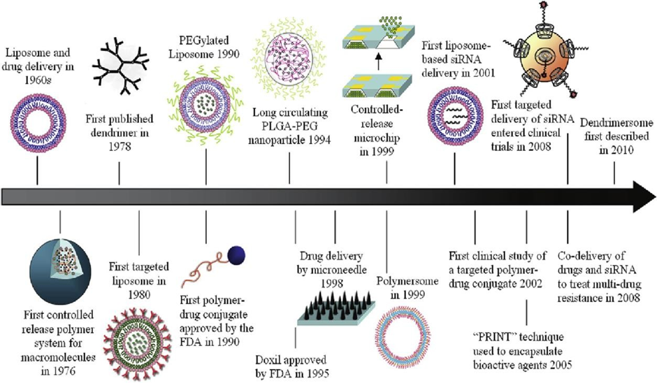
Figure 1: Timeline for the development of nanoparticle drug delivery systems. Reprinted with permission from [5]. Copyright (2010) American Chemical Society.
2.Chitosan Nanoparticles
Chitosan is a natural carbohydrate polymer derived from the de acetylation of chitin, the principal component of crab, lobster, and shrimp shells. Chitosan is regarded as suitable for pharmaceutical applications because of its low price, good biocompatibility, low toxicity and degradability in the body by chattiness. Since chitosan dissolves in acidic aqueous solutions at room temperature, the preparation of nanoparticles may be performed under mild conditions without the need for toxic organic solvents or heating. Chitosan DDS can accommodate a wide range of drugs, such as small molecules, proteins and poly- nucleotides [8]. This allows chitosan to carry the drug and release it in a controlled manner. Chitosan possesses the ability to ionically crosslink due to the free amine groups available on the chitosan polymer [9].
2.1. Methods of Fabrication of Chitosan Nanoparticles
2.1.1. Ionotropic Gelation Method
Calvo et al. reported the manufacturing of chitosan-PEO nanoparticles using the ionotropic gelation method [10]. Chitosan has positively charged amine groups, which can lead to a sol- gel transition when interacts with a negatively charged polyanion to produce nanoparticles under favorable conditions [11]. Tripolyphosphate (TPP) is a frequently-used macro-anion [12,13]. Calvo et al. prepared different concentrations of chitosan in acetic acid aqueous solutions and TPP in purified water at the same concentrations as the chitosan solutions (0.05 wt%, 0.1 wt%, 0.5 wt%, and 1 wt%). By mixing different volumes of TPP with the chi- tosan solutions, the optimal condition for producing chitosan nanoparticles was found. The preparation of chitosan/PEO and chitosan/PEO-PPO nanoparticles was carried out by drop wise adding TPP aqueous solution to chitosan solution containing PEO and PEO-PPO individually under continuous magnetic stirring. The optimal chitosan/TPP ratio was 5/1, with a minimum particle size of 260 nm for the chitosan nanoparticles obtained. The particles of chitosan/PEO and chitosan/PEO-PPO were about 300–1000 nm in size based on various concentrations of PEO and PEO-PPO. As the zeta potential of chitosan/PEO and chitosan/PEO-PPO nanoparticles was lower than that of the chitosan nanoparticles, it proved that chitosan/PEO and chitosan/PEO-PPO nanoparticles were more stable than chitosan nanoparticles.
2.1.2. Emulgation Solvent Diffusion Method
The emulsification solvent diffusion method was initially described for the preparation of poly D, L-lactide/glycolide (PLGA) nanoparticles [14], and then modified for the production of chitosan nanoparticles by El-Shabouri [15]. Cy-A and lecithin were initially dissolved in methylene chloride and subsequently, mixed into acetone. Aqueous chitosan solution was added to the mixed solution with magnetic stirring and high-pressure homogeni- zation for 5 min. This suspension was then subjected to low-pressure evaporation, filtration, centrifuge, resuspension in water and re-centrifugation to give particle size of approximately 150 nm.
2.1.3. Method with polyelectrolyte complex (PEC)
Chitosan can also form nanosized particles with DNA according to the poly- electrolyte complex (PEC) method. DNA and cationic chitosan can self- assemble for nanoparticle formation due to the charge neutralization between them, resulting in their hydrophilicity decrease [8]. Erbacher et al. [16] presented chitosan/DNA complexes for gene delivery. Complexes were prepared by incubating varying polymer:DNA phosphate equivalents. The reaction was performed and completed over a 15 min time period under room temperature with continuous stirring. Dynamic light scattering evidenced size distribution for DNA/chitosan ratio 0.5-10 ranging from 80-500 nm. But at near zero zeta potential the particle size was about 1–5 μm while transmission electron microscopy (TEM) indicated complexes sizes about 50–100 nm and their shapes like donut and rod. Out of the three described methods for the fabrication of chitosan nanoparticles, the ionotropic gelation method has the least number of steps, and all steps were conducted in mild and non-toxic aqueous conditions. It is the most simple and applicable method. This process is particularly beneficial when the drug used is hydrophobic, but has certain disadvantages such as requiring high amounts of pressure and toxic solvent. Chitosan nano- particles could be utilized in gene delivery by virtue of PEC method.
2.2.Chitosan Nanoparticles for Drug Delivery
Chitosan nanoparticles are widely used in the application of cancer therapies. Chitosan nanoparticles mainly use passive targeting (known as enhanced permeability and retention (EPR) effect [17]), active targeting, and physical targeting of stimuli-sensitive targeting, using their characteristics to target tumors on specific organs.
2.2.1. Tumor passive and active targeting using chitosan nanoparticles
Hyaluronic acid (HA) conjugated chitosan nanoparticles were synthesized by Jain and Jain [18] via ionotropic gelation. Colon cancer drug 5-fluorouracil (5FU) Because there are many ha receptors around the tumor tissues, ha was added into the chitosan nanoparticles DDS. Thus, nanoparticles are not only able to target colon tumors by means of EPR effect but also by means of the interaction between HA and HA receptors. The size of synthesized chitosan nanoparticles incorporating 5FU in that study was approximately 135 nm and after conjugation of HA with free amino groups with chitosan through a condensation reaction, particle size increased to approximately 150 nm. Hyaluronic acid coupled nanoparticles displayed inhibited drug release in 24 h compared to uncoupled chitosan nanoparticles, partly as a result of a double barrier arising from an additional HA layer placed on the nanoparticles. The HA conjugated nanoparticles also have higher uptake rate in cancer cells during 4 h of incubation time at 37 ?C, which makes HA-conjugated chitosan nanoparticles promising carriers for targeted drug delivery to colon tumor.
2.2.2. Chitosan Nanoparticles Targets Tumors Stimuli-Sensitively
Chitosan nanoparticles sensitive to stimuli are also extensively used in can- cer therapies. pH and temperature are typically selected as physiologi- cal signals due to the presence of acidosis or hyper- thermia in inflamed or neoplastic tissues. Zhang et al. [19] described a pH- mediated chito- san-based microgel for cancer therapeutic drug release during inflamma- tory response. Initially, chitosan powder was dissolved in water at 85 ?C and then was reacted with Glycidyltrimethylammonium chloride to form N-[(2-hydroxy-3- trimethylammonium)propyl]chitosan chloride (HTCC). HTCC nanoparticles were prepared by TPP based ionotropic gelation method. At pH decrease from 7.4 to 5.0, the chitosan nanoparticles size goes from average 200 nm to 400 nm or more (2.2 times increase), relative volume of the nanoparticles increase 11 times After the microgels are internalized into the cells, they will swell dramatically and stay at the diseased site to release the drug. The characteristics of loading efficiency, release kinetics and cell viability(mortality) were identified after loading with methotrexate disodium (MTX). Microgels released the drug at a higher rate at pH ¼ 5.0 (93% on day 1), while only ∼30% drug was retained in microgel after 5 days at pH ¼ 7.4. The MTX-loaded HTCC exerted the most significant mortality on the HeLa cells in the cell viability experiments as compared to the pure drug group and MTX-chitosan nanoparticles that were non-conjugated.
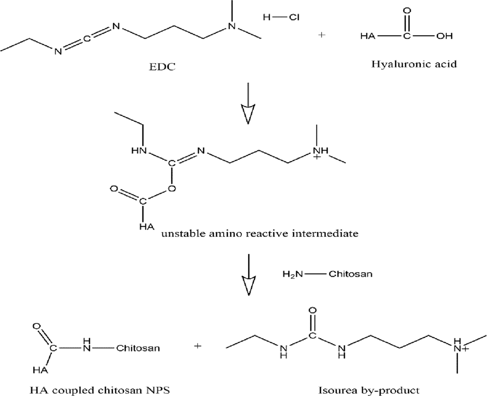
Scheme 1: Coupling of hyaluronic acid with chitosan nanoparticles.
3. Silica Nanoparticles
Silica xerogel has been predominantly used as inorganic carriers for drug delivery [65]. It is biocompatible, highly porous and amenable to functionalization. In 1992, Kresge et al. The fabrication of mesoporous silica nanoparticles (MSNs) was first reported in [20], and MSNs have many advantages over silica xero- gels in their application as drug delivery systems: i) when materials are nano-sized, they have a larger surface area (usually more than 1000 m2/g) and pore volume, thus they are superior for adsorption and drug loading in pores [21]; ii) by controlling nanoparticle size, the loading and release kinetics of the drugs in nanoparticles can be easily modulated [22]; iii) since surface modification of MSNs is easily and early achievable, it improves the targeting ability of nanoparticles, increasing drug delivery efficiency and decreasing systemic toxicity [23,24]; iv)MSNs can be used as cellular drug delivery systems and as bioimaging probes when incorporated with magnetic materials or luminescent compounds [25,26].
3.1. Fabrication of MSNs
3.1.1. Solvothermal approach to MSNs construction
Hexagonal mesopores MCM-41 is among the most famed type of MSNs. When Kresge et al. In the paper [20], from which the Liu group derived firstly the synthesis of MSNs via a liquid-crystal template mechanism, the particle sizes are submicron. Many researchers, however, had diversified their way of having the synthesis of MCM-41 and the particle size of MCM-41 can be upto 50 nm now. Meng et al. [27] reported synthesizing 50 nm mesoporous silica core-based MSNs. A solution of pluronic F127, n- Cetyltrimethylammonium bromide (CTAB), and water were heated up to 80 ?C for 0.5 h. tetraethyl orthosilicate (TEOS) was mixed with the N-(2-Aminoethyl)- 3- aminopropyltrimethoxysilane (NAPTS) with a volume and mixed in a ratio of 5:1 (V:V) and then added into F127 and CTAB combined solution. After 20 min, trihydroxysilypropyl methylphosphonate was added. The solution was filtered by 220 nm filter, combined with methanol and NH4NO3 to remove CTAB 70 ?C 30 min. The MSNs were subsequently centrifuged and washed with methanol. As for the TEM tests, it indicated that the particle size of synthesized MSNs is about 50 nm, and the hydrodynamic size of MSNs is about 70 nm after coated by PEG. This method uses the alkylammonium salt (CTAB for instance) as a liquid crystal template, which can under− take self−assembly into micelles when its concentration exceeds the critical micelle concentration (CMC) and form liquid crystal mesophases when its concentration becomes even higher [28]. Silica precursors can thus condense to give rise to electrostatically stabilized, hydrogen-bonded, amorphous silica mesophases.
3.1.2. Evaporative-induced self-assembly (EISA) process
The evaporation-induced self-assembly (EISA) method was first invented by Lu et al. to prepare mesoporous silica films [29]. TEOS was first mixed with ethanol and hydrochloric acid (HCl) and refluxed at 60C for 90 min. A, and adding more water and HCl to achieve a higher concentration of HCl. The two-step method was established primarily to reduce the siloxane condensation rate in the solution. The solution was stirred for 15 min and aged for 15 min, then diluted 1 : 3 with ethanol. Surfactant CTAB was introduced into the solution at concentration much lower than CMC. The mesoporous film was prepared through dip- coating at 7.6 cm/min. In the dip-coating process, the evaporation of ethanol increases the concentration of surfactants to more than CMC, and self-assembly of silica in the micelle began. The film stabilized only several seconds later. After two years, Lu et al. adapted the EISA technique for making silica mesoporous nanoparticles with hexagonal or cubic stable pore mesostructures [30]. Transmission electron microscopy (TEM) was performed and showed the successful synthesis of additional nanoparticles with a diameter of approximately 200–300 nm. The process started with a solution of silica, ethanol, water, and surfactant initial concentration below CMC. (6) An aerosol dispersion was produced in a tubular reactor, and during 6 s alcohol evaporated and promoted micelles formation leading to solid silica-surfactant self-- assembled MSNs (Fig. 2). Comparing the particle size of MSNs synthesized by the liquid- crystal template method and the EISA method can find that the former can synthesize MSNs with a smaller size that is more regular shape and more stable hexagonal or cubic pore meso- structures (Fig. 2). Another possible benefit of EISA could be that nonvolatile drugs could have their molecular states readily encapsulated within the silica-surfactant mesophase. Keep in mind that in EISA process CTAB is not removed, this could lead to toxicity problems in animal experiments. It was discovered that rats consuming the CTAB dose (45 mg/kg/day per 1 year) CTAB with drinking water resulted in slower growth rate (Isomaa et al., 1999) [31].
3.2. Silica Nanoparticles Applications in Drug Delivery
3.2.1. MSNs based gene delivery for cancer therapy
However, MSNs have been investigated as a potential carrier for gene delivery 810. The mesoporous structure of MSNs provides a more homogeneous distribution of drug through the matrix system compared to the more common polymer or lipid based delivery systems, furthermore the pore size of MSNs can be readily tuned to accommodate different molecular sizes of drugs (Scheme 2) [32]. MSNs also has a higher drug loading capacity in comparison with chi- tosan and other polymers[33]. When applied in gene delivery, gene molecules are buried deep inside the mesopores and consequently can escape from the attack of nuclease until they arrive at the target site [34]. The surface of positively-charged MSNs is often decorated in order to enhance the binding and loading capacity of negatively-charged nucleic acids. Xia et al. Coating of MSNs outside using a cationic polymer: the impact of poly(ethyleneimine) (PEI) [PMID: 25770361]. [49]Their results indicated that the surface coating of PEI enhance the binding avidity of DNA and SiRNA, and cell uptake rate of the nanoparticles. SiRNA attached to the MSN-PEI surface allows for the knockdown of GFP from HEPA-1 cells. MSN-PEI system is comparable to the commercial drug delivery system for the plasmid DNA delivery. Nevertheless, an issue for the PEI coated MSNs is that toxic PEI coating may boost the cytotoxicity of MSNs delivery system [35]. Yang et al. Hence, [36] first demonstrated that the combined delivery of tumor suppressor miRNA, miRNA-204-5p, with the anti-cancer drug Oxaliplatin (OXL) in PEI/hyaluronic acid-assembled mesoporous silica nanoparticles (Oxmi-HSMN) exhibited synergistic anticancer effect in treating colon cancer. According to this, PEI was used to modify MSN by electrostatic interaction to enhance the loading of miRNA, and the HA was covalently conjugated on the MSN surface to achieve selective targeting of CD44 receptors on the colon cancer cells. The size of Oxmi-HMSN was approximately 138 nm and narrow distributed (PDI ¼ 0.165) for passive delivery to the cancer site based on the EPR effect. OXL-MSN did display a more rapid release of drug in an in vitro study compared with the OXmi-HMSN formulation, likely because the surface assembly of PEI polymer on Oxmi- HMSN inhibited the diffusion of drug into the release medium. The Cellulate uptake study demonstrates that OXmi-HMSN shows greater cell uptake compared to OXmi-MSN (non- targeted). OXmi-HMSN group had the strongest inhibition of tumor growth compared to OXL and OXL-MSN group (P 80%) under treatment with the nanoparticles at a concentration of 100 μg/ml.
3.2.2. MSNs based zero premature release in anti-cancer therapy
A number of drugs used in the treatment of tumors are toxic not only for tumors but also for other tissues or organs. Hence, achieving ?zero premature release? (i.e., drug distribution in non-targeted sites approaching 0) is highly desirable [33]. Mal et al. [37] then prepared the coumarin group-modified MSNs that could dynamically photo-regulate the pore accessibility (opening and closing) under UV irradiation. The coumarin substituents are viewed as ?double hinged doors?. The boules are coated with MSNs whose surface is modified with coumarin moieties such that upon irradiation with 310 nm light the coumarin groups photopolymerize to form cyclobutane coumarin dimmers and the doors are shut preventing any drug from being absorbed into the pores. By irradiating the MSNs with 250 nm wavelength UV light, photocleavage of the dimers can occur which leads to opening of the doors and gives accessibility to the inner part of the MSNs. Controlled release study showed that 28 wt% cholestane was absorbed in irradiated coumarin modified MSNs after washing with n-hexane (its small molecular size allows cholestane to be stored in the pores of the MSNs) while no cholestane remained in the coumarin modified MSNs without irradiation after washing. Other stimuli such as pH [38], enzymes [39], and magnetic fields [40] have been studied to achieve zero premature release in MSNs despite photo-stimulating.
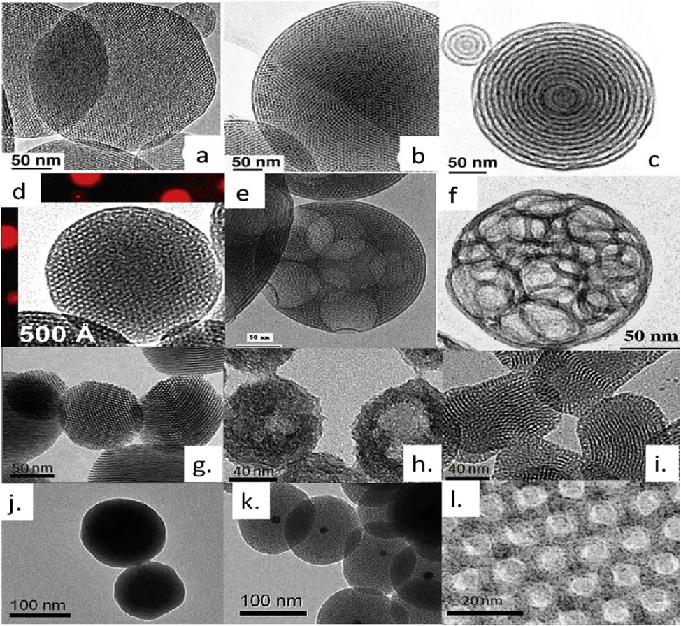
Fig. 2. TEM images of MSNs prepared by EISA (a.-d.) and solution-based methods (e.-l.). Reprinted with permission from [28]. Copyright (2013) American Chemical Society.
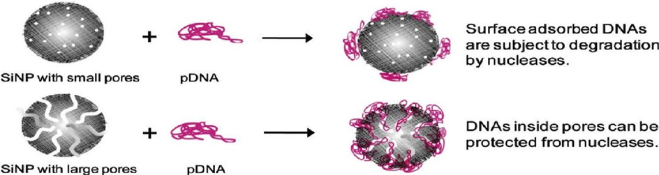
Scheme 2. Monodispersed MSN (MMSN) can be tailored to have a large pore size (>15 nm) for increased plasmid DNA loading capacity and better protection from nucleases. Reprinted with permission from [32]. Copyright (2011) American Chemical Society.
4.PLGA : Polylactide-co-glycolic acid (PLGA) nanoparticles
PLGA (polylactide-co-glycolic acid) is a copolymer of polylactic acid (PLA) and polyglycolic acid (PGA) prepared by the ring-opening polymerization of lactide (LA) and glycolide (GA). PLGA has several advantages as biomaterials for drug delivery systems. Adamantane-2: First, its application in the human body has already been approved by the U.S. Food and Drug Administration (FDA) and the European Medicine Agency (EMA) [41]. Second, because it is a synthetic polymer, it has high purity, tail- orable molecular weight, and high reproducibility compared with many natural polymers. Third, in terms of biodegradability, the obtained PLGA peptide copolymers will be degraded into lactic acid and glycolic acid through hydrolytic scission in vivo and be further degraded through the Krebs circle to form CO_2 and water [42]. Fourthly, the copolymer is less prone to hydrolysis than PLA and PGA and therefore shows promise for sustained drug release over a period of days, weeks or months.
4.1. Preparation of PLGA nanoparticles
4.1.1. Emulsion evaporation method
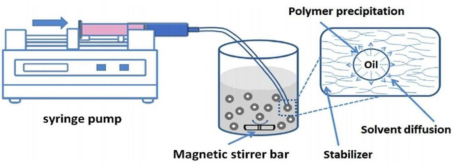
Emulsion evaporation method [43] is one of the most common tech- niques for the fabrication of PLGA nanoparticles. Two types of emulsion processes can be used, depending on the solubility of the drug in water. Hydrophilic drugs are encapsulated in double-emulsion (water-in-oil-in-water, w/o/w) while hydrophobic drugs are encapsulated in single-emulsion (oil-in-water, o/w). In the single-emulsion technique, PLGA polymer and hydrophobic drug are dissolved in an organic solvent before emulsion formation by adding water and surfactant into the organic phase. The control of droplet size is often performed by sonication or applying high shear stress such as rapid stirring to prevent the coalescence of emulsion droplets [41,43]. Last, the organic phase is removed by evaporation or extraction, the synthesized nanoparticles can then be collected by centrifugation (Scheme 3) [44,45]. The above method is modified to form a double emulsion for encapsulating hydrophilic drugs. The first emulsion can then be created by injecting the organic solvent into an aqueous solution, which is followed by injection into the aqueous solution with stabilizer. While the emulsion evaporation method is good for laboratory fabrication of PLGA nanoparticles, it is not good for large-scale fabrication. Especially due to the energy- demanding nature of high-speed homogenization and sonication [45]. Dropwise evaporation from a double emulsion is also a promising method, but it has the disadvantages of larger size of nanoparticles and low encapsulation efficiency.
Scheme 3. Fabrication of nanoparticles by emulsion evaporation method. Reprinted with permission from [46].
4.1.2.Salting out method
In the salting out method to form PLGA nanoparticles, PLGA is initially dissolved in a water-miscible organic solvent like acetone. Through high-speed stirring, the polymer solution is then injected into the aqueous phase comprising of emulsifier and high concentration of salts (e.g. calcium chloride). Dilution of the salt concentration induces the formation of PLGA nanoparticles, allowing higher diffusion rates of the organic phase into the aqueous phase. The organic solvent and salts were removed by centrifugation or cross- flow filtration (Scheme 4). Salting out approach is suitable for encapsulation of peptide or protein as it reduces tension [47]. All of this could happen at room temperature; therefore, it is suitable for heat-sensitive drug encapsulation [48]. This method has disadvantages, such as it can only embed hydrophobic drugs, and the extra salt removal washing steps are carried out [49,50].
4.1.3. Nanoprecipitation method
Nanoprecipitation method is also referred as solvent diffusion or sol- vent displacement method [41]. In this one-step method, PLGA polymer is first dissolved in water-miscible and polar solvent, for example, acetone or THF, and subsequently, the solution is added dropwise into an aqueous solution containing sta- bilizer under constant rapid stirring. Due to high diffusion of organic solvent into the aqueous phase, nanoparticles precipitate out. The solvent not needed can then be blown off with nitrogen or air, or reduced-pressure evaporation can be used (Scheme 5). The nanoprecipitation method is used for encapsulation of both hy- drophobia and hydrophilia drugs. However, loading efficiency of hydrophilic drugs is very low. Various factors, including polymer solution concentration [51], polymer molecular weight [41], stabilizer type [14], and stirring speed [52], can influence the size of nanoparticles formed from the nanoprecipitation method.
Scheme 4. Fabrication of nanoparticles by emulsion evaporation method. Reprinted with permission from [46].
Scheme 5. Fabrication of nanoparticles by emulsion evaporation method. Reprinted with permission from [46].
4.2. PLGA nanoparticles schematic illustration in cancer therapy
Since the conventional EPR effect-based passive targeting scenario leads to poor tumor deposition of nanodrugs, the majority of PLGA nanoparticles finds explanation through the use of active targeting method for the delivery of chemotherapeutic agents towards cancer therapy. PLGA nanoparticles are frequently coated or grafted with targeting ligands on their surface. The selected ligands offer a selective binding towards receptors that tend to be overexpressed on cancer cells or tumoral endothelial cells. Hyaluronic acid (HA) is a naturally occurring polysaccharide itself and an apt targeting ligand for PLGA NP DDS in cancer therapy as it binds to CD44 receptors which are known to be over-expressed on certain tumor types. In addition, HA as targeting ligands has many superiority comparing with other targeting ligands, such as nontoxicity, biocompatibility, biodegradability and non-immunogenici. Cerqueira et al. synthesized HA-decorated PLGA nano- particles for targeted delivery of paclitax- el (PTX) in the setting of triple-negative breast cancer. Single emulsion method was used to fabricate cetrimide added positively charged PLGA nano- particles (2). The surface charge was then altered by adding negatively-charged sodium hyaluronate following solvent evaporation (dichloromethane) of the pre-formed dodecanethiol layer. Various formulation parameters like the PVA concentration, the sonication time, the polymer concentration, and the polymer type have been explored for its effect on the nanoparticles. The authors note that increasing the PVA concentration from 0.5% to 2% does reduce the size and polydispersity index (PDI) of the nanoparticles. Varying the sonication time from 1 min to 4 min caused a reduction in the particle size and improved the encapsulation efficiency (EE%) and drug loading (DL%) of PTX. The formulation that achieved the highest EE% (90.8 1.5) and was discovered was 100 mg of 50:50 PLGA and 2% w/v PVA. The nanoparticle size for 0.05% w/v is nearly 250 nm and zeta potential after sodium hyaluronate modification is approximately —37.5 mv. In the in vitro release study, PTX was released office from HA-coated PLGA nanoparticles vs bare PLGA nanoparticles. The ppH hands over in large part because the HA layer provides a sparse surface of the PLGA nanoparticles, augmenting the hydrolysis of the PLGA by promoting the volume of pearly penetrability. The results demonstrated that the nano- particle of HA-PTX- PLGA has a lower IC50. This further portrays that the HA-PTX-PLGA nano- particles illustrate more cellular uptake than the PTX-PLGA nano- particles which could be due to the interaction of HA with the over expressed CD44 receptors in the MDA- MB-231 cells. An alternative way in active targeting is using an external prompt example as a magnetic field to attract the nanoparticles to the location of tumor [53, 54]. Cui et al. [55] that utilized dual-targeting magnetic PLGA nano-par- ticles against brain tumors. Due to the restriction of the blood-brain barrier (BBB), the choice of chemo drug for glioma treatment has been extremely limited. To overcome this challenging sit- uation for glioma treatment, the authors mediated dual-functional PLGA nanoparticles DDS (MNP/T7-PLGA NPs). For ligand- mediated active targeting, human transferrin receptor-binding peptide T7 was selected, and magnetic nanoparticles (MNP) were encapsulated into the poly(lactic-co-glycolic acid) (PLGA) NPs using a single-emulsion solvent evaporation method for magnetic-guided targeted delivery. The anticancer agent selected for tumor treatment was paclitaxel (PTX) and Curcumin (CUR). Human malignant glioma U 87 cells and mouse brain endothelial cell line bEnd were used in the in vitro cell uptake experiment. 3. Confocal image results indicated that the dominant mechanism of intracellular delivery was attributed to T7-mediate intracellular delivery. To investigate the brain targeting ability of the MNP/T7-PLGA NPs in vivo, mice with orthotopic glioma were treated with Cy5-labeled NPs. IVIS imaging at 4 h after tail vein injection indicated that the group with MNP/T7-PLGA NPs + MAG (with magnetic field) exhibited the highest fluorescence in the brain. MNP/T7-PLGA NPs + MAG group exhibited the highest inhibition of glioma growth and 100% survival, compared with groups without magnetic field, or with free CUR+PTX in a 35-day experimental course.
5.Nanoparticles: Challenges to Clinical Utilization
Nanoparticles, as drug delivery systems have many merits, however, only a limited number of nanoparticle drugs available so far (Table 1.) the market for cancer treatment. Their concerns still have many limitations and disadvantages of nano- particles DDS.
5.1.Scaling up problems
Nanoparticles have in general only been synthesized at the lab-scale, as many current fabrication methods are not applicable at a scale up. Methods involving less energy input must be developed to produce large amounts of nanoparticles. Moreover, dry forms of nanoparticles readily aggregate due to their small size and high specific surface area, making them difficult to process [4]. Chitosan is a naturally occurring polymer obtained from many different sources there- fore there are variations in molecular weight, molecular weight distri- bution, and purity levels of chitosan materials. Chitosan is also very sensitive to environmental conditions: high relative humidity (>60%) can cause an obvious increase in the water content of chitosan, which reduces the mechanical properties of chitosan [59,60]; high temperature (>40 ?C) can accelerate the degradation of chitosan [61,62].
5.2. Toxicity and biodistribution
Inferior to bulk materials, nanoparticles have many unique properties. Consequently, the toxicological profile of the nanoparticles differs from that of the bulk-compounds [63]. NPs are not necessarily compatible with our existing procedure and tests, to evaluate toxicity of drugs. There are also concerns about bio- distribution of the nanoparticles. It has been found that nanoparticles in the range 50–100 nm show greater accumula- tion in liver, spleen, and lung [64,65], resulting in nonspecific accumulation of nanoparticles. It can also be toxic to those organs if the drug included in the nanoparticles is harmful to normal cells. Currently, very little is known about the biological behavior of nanoparticles [63]. Concerns on the cytotoxicity and in vivo toxicity have limited the application of the silica nanoparticles as DDSs. Silica can also be toxic due to the presence of silanol groups, which can lead to membranolysis. Radicals on the silica surface, which can react with water to generate reactive oxygen species (ROS), are also involved in cell death. However, repeated studies regarding the toxicity of MSNs reported conflicting conclusions [66–68]. So, it cannot be concluded that MSNs are nontoxic as DDS at this moment. Although MSNs exhibit acute toxicity, long-term toxicity studies also need to be performed prior to clinical application.
Table 1
List of FDA-approved nanomedicines for cancer treatment [7,56,57,58].
|
Tradename |
Material |
Drug |
Company |
Indication |
Year(s) approved |
|
Doxil® |
Liposome- PEG |
doxorubicin |
Janssen |
Metastatic breast cancer, metastatic ovarian cancer |
1995 |
|
Eligard® |
PLGA |
Leuprolide acetate |
Tolmar |
Prostate Cancer |
2002 |
|
Abraxane® |
Albumin |
Paclitaxel |
Celgene |
Metastatic breast cancer |
2005 |
|
|
|
|
|
Pancreatic cancer |
2013 |
|
Genexol PM® |
mPEG- PLA |
Paclitaxel |
Samyang Corporation |
Metastatic breast cancer |
2007 |
|
Onivyde® |
Liposome |
Irinotecan |
Merrimack |
Pancreatic cancer |
2015 |
5.3. Low control of loading and unloading of drugs
A low rate of drug loading is one of the major pitfalls for many polymeric nanoparticles [43,69]. Several studies defined the loading rate as less than 10% [70–72]. Burst release is another major issue for nanoparticles as drug delivery systems. This process occurs usually within a few minutes after the nanoparticles interact with the surroundings with a high likelihood if oriented to the environment [73]. This phenomenon occurs on nanoparticles made from varied materials. For example, Janes et al. [12] published burst release of dextran sulfate entrapped in chitosan nanoparticles 17% during 2 h whereas Suh et al. in the first three days, resulting in 40% burst release of an antiproliferative drug in polyethylene oxide- PLGA nanoparticles. Due to burst release, which can mainly be linked to the adsorption of the drugs attached to surface, drug may not reach the targeted site, thus resulting in loss of drug efficiency.
5.4. Physiological barriers
Physiological barriers between nanoparticles and tumor cells are another obstacle for nanoparticles DDSs in cancer treatment. After sys- temic administration, nanoparticles cross the microvessel wall, the extracellular matrix, and the plasma membrane of cells to deliver the drug; each of the three barriers obstructs the delivery of the nanoparticle in a unique manner, through disruptions in transvascular, interstitial and transmembrane trans- port, respectively [74]. While the EPR effect is common in tu- mors, there are also areas with permeability microvessels absent or low, creating heterogeneity of the nanoparticles distribution [75]. After arriving at the interstitial space, the nanoparticles still face transport difficulty because of the low convective transport driving force in solid tumors. As a result, the systemically given nanoparticles are typically limited to the perivascular and peripheral areas in solid tumors [74].
Fig. 3. Schematic diagram for future multifunctional nanoparticles. Republished with permission of Annual Reviews, Inc, from [83]; permission conveyed through Copyright Clearance Center, Inc. Abbreviations: GNP, gold nanoparticles; MNP, magnetic nanoparticles; QD, quantum dot; HfO, hafnium oxide nanoparticles; UCNP, upconversion nanoparticles.
6. Nanoparticle drug delivery prospects
Nanoparticles have several merits versus the traditional drug delivery approaches. To broaden the scope of application of nanoparticles in the pharmaceutical market, it is necessary to perform additional in vivo studies and clinical trials in order to demonstrate toxicity and long-term biological behavior of nanoparticles [126]. Recently, there have been many studies are concentrating on the surface modi- fication of nanoparticles in order to prolong the retention time of the nanoparticles. Nanoparticle surface modification, for example, polyethylene glycol. In addition, when grafted on the surfaces of nanoparticles [76], the formation of a hydrating layer on the nanoparticles can enhance the circulation lifetimes after intravascular administration. An additional top-down biomimetic strategy, nanoparticles coated with cell membranes has also been extensively investigated [77–82]. The isolated cell membranes still maintain the original membrane proteins and lipid bilayer structure, so they can still perform the functions of natural cell membranes. Nanoparticles cloaked with cell membrane could not only potentially avoid the drawbacks of naked nanoparticles but also possess the advantages of reduced immune response and improved biodistribution. Another emerging area of research is the development of multi-functional nanoparticles (Fig. 4). 3). The increasing medical needs drive this field. In cancer treatment, the nanoparticles need to act not only as a drug vector but also as diagnostic and drug monitoring agents. For instance, dynamic imaging describes the concept of image-guided drug delivery that combines magnetic resonance imaging (MRI) with drug delivery nanoparticles, allowing non-invasive monitoring of the biodistribution, circulation, and targeting behavior of nanoparticles.
CONCLUSION
Most of MSNs are mainly synthesized and applied in cancer therapeutic. The results highlight the potential of MSNs as efficient nanocarriers for targeted drug delivery, as several synthesis methods have their own unique advantages in use for therapy. By this approach, MSNs with uniform particle size, ~50 nm in width can be prepared, which benefits drug delivery performance as they combine high surface area with controlled morphology. In addition, evaporative-induced self-assembly (EISA) process is promising in improving drug loading capacity of delivery carriers, especially sensitive delivery carriers like miRNA, which enable sustained-release, thus improving the effect of targeted cancer therapy. The study further underlines the importance of surface modifications to improve the drug delivery properties of MSNs. Functionalization with polyethyleneimine (PEI), hyaluronic acid (HA), and other compounds not only enhances drug loading capability but also warrants selective targeting towards cancerous tissues, thus personalizing the drug regimen. Novel approaches for controlled drug release were also explored. The incorporation of coumarin segments in MSNs allows for controlled access of drug molecules to the porous shell by switching off the epoxy groups under UV irradiation, preventing drug precocitation. Advancements like these are important for reducing off-target effects, increasing drug stability and improving the therapeutic output of cancer therapies. In addition, the use of biodegradable polymers such as PLGA (poly(lactic-co-glycolic acid) provide an excellent means of sustained and controlled release of drugs without adverse effects. For the most monograph MSN structures, as a result of these polymers possess high biocompatibility, biocompatibility, and reproducibility, they can be used with in vivo applications and ultimately enhance the clinical potential of this MSN can carry drug delivery systems (293). Our perspective of future research trends includes optimization of synthesis techniques as well as the exploration of multi-functional modifications to adapt the targeting specificity and release profiles of MSNs. The conjugation of stimuli-responsive components, such as pH, temperature, or enzyme-triggered systems, would greatly enhance cancer treatment accuracy and effectiveness. And definitive clinical studies will be important to confirm the real-world medical safety, biodegradability, and therapeutic efficacy of MSNs. Concerning, MSNs serve as versatile and promising connector for next-generation cancer treatments that aim to revolutionize targeted medications delivery and improve patient collection. In this regard, further research and loss of innovation in this field will ensure the future generation of more competent, tailored, and safer cancer care.
Consent for Publication
Not Applicable
Conflicts of Interest
The authors declare that there are no conflicts of interest, whether financial or otherwise.
ACKNOWLEDGEMENTS
The author is sincerely grateful to MD. Zulphikar Ali, Assistants Professor, Department of pharmacy, Mewar University, Chittorgarh, Rajasthan for constant support and guidance during the review. He offered such thoughtful commentary and support, without which this work would not be possible. Also shall the author thank Mewar University to provide its opportunity and resources which greatly helped in completing this project.
REFERENCES


Rifisha Basumatary*, Md. Zulphikar Ali, Advances in Nanoparticle-Driven Drug Delivery for Targeted Cancer Therapy, Int. J. of Pharm. Sci., 2025, Vol 3, Issue 3, 2491-2511 https://doi.org/10.5281/zenodo.15084887
 10.5281/zenodo.15084887
