Matoshree Miratai Aher College of Pharmacy.
The analysis of blood and urine specimens is the primary emphasis of this paper, which explores the complexities of pathology. Blood and urine are vital tools for evaluating physiological and pathological states, and pathology is essential to the diagnosis and comprehension of many diseases. With measures including total blood count, blood chemistry, and coagulation studies helping to identify problems ranging from anemia to metabolic diseases, blood testing offers a thorough understanding of the body's health. Furthermore, the identification of genetic markers and circulating tumor cells is made possible by developments in molecular pathology, which transforms cancer surveillance and diagnosis. Informing the lab and clinical physician about the clinicopathologic relevance of human disease is the aim of pathology. The collection of samples for analytical testing in a clinical laboratory is known as clinical pathology, and it encompasses clinical chemistry, hematology, and microbiology. This review covers basic blood tests, the introduction of a light microscope, a semi-auto analyzer, anemia, a WBC disorder, and hematologic value. In order to ensure that the study can be carried out using the available tissue, that the processes for acquiring tissue do not adversely influence patient care, and that all local, state, federal, and hospital rules are followed, pathology reviews are undertaken.
Module1:
Clinical Chemistry of Blood Blood:
It is a fluid form of connective tissue.
1. The color of blood is crimson.
2. The typical pH of blood is 7.4.
3. A typical adult's blood volume is 5 L.
Blood Composition:
1. Blood contains a liquid component known as plasm and blood cells known as formed elements.
• Blood cells come in three different varieties. • The most prevalent cells in the blood are red blood cells, or erythrocytes.
•Leukocytes, or white blood cells, make up only around 1% of the blood.
• Thrombocytes (platelets)
Specific Gravity of Blood:
A gravimetric analysis was performed to estimate the specific gravity, or relative density, of human whole blood and plasma from 25 healthy participants. It was determined to be 1.0621 (95% CI: 1.0652-1.0590) for whole blood at 4 °C and 1.0506 (95% CI: 1.0537-1.0475) at 37 °C.
BLOOD VISCOSITY: Blood has thixotropic and viscoelastic characteristics and is a non-Newtonian, shear-thinning fluid. The usual range for blood viscosity is 3.5 to 5.5 cP, according to many cardiovascular handbooks. The biochemical examination of bodily fluids to aid in disease diagnosis and therapy is known as clinical chemistry. Chemical reactions are used in this specialty's testing to detect or measure the concentrations of chemical substances in body fluids.[2]
2) Abnormal Cells and the Importance of Erythrocyte
• Red blood cells, also known as erythroid cells, erythrocytes, heamatids, red blood corpuscles, or red cells.
• Your body's tissues receive oxygen from red blood cells, or erythrocytes. The presence of these cells in
1. Megaloblastic anemia
2. Reduce blood osmotic pressure
3. Anisocytes: In pernicious anemia, these cells are found.
4. Sickle cell anemia: This condition is caused by aberrant hemoglobin molecules that leave the body stiff and bent. Fifth, poikilocytosis:
RBC LIFE SPAN: RBCs have an average lifespan of 120 days.
-How Red Blood Cells Work -RBCs' hemoglobin molecules.
-Hemoglobin releases oxygen while absorbing CO2. -The release of CO2 when hemoglobin forms a connection with oxygen.
-The enzyme carbonic anhydrase is kept in red blood cells. In a reversible reaction, carbonic anhydrase catalyzes the conversion of CO2 to HCO3. -.
Abnormal Cells and Their Significance
The size of RBC's is venous blood is a slightly larger than those in arterial blood.
1. Microcytes [ Smaller cell]
2. Macrocytes [Larger cell]
3. Anisocytes [cell with different Size]
4. Sickle Cell
5. Poikilocytosis.
1. Microcytosis: - This cell present in
• Iron-deficiency anemia
• prolonged forced breathing
• Increase osmotic pressure in blood[3]
3) Anemia
Anemia is defined as a low number of red blood cells.
Anemia is a blood disorder.
Abnormal decrease in :-
• Red blood
• Haemoglobin
• Packed cell volume cause anemia
Types Of Anemia
1.Iron deficiency anemia: Inadequate Iron Anemia is a disorder where the body has insufficient amounts of iron. Anaemia, or low levels of healthy red blood cells in the body, is frequently caused by iron deficiency.
2.Aplastic anemia: Another name for aplastic anemia is bone marrow aplasia. The body stops making enough new blood cells in this uncommon illness.
3.Sickle Cell Anemia: A collection of illnesses that result in the breakdown of red blood cells. Red blood cells that have sickle cell disease, a hereditary group of illnesses, twist into a sickle shape.
4. Hemolytic Anemia: This condition occurs when the production of red blood cells is outpaced by the destruction of existing ones. Hemolysis is the breakdown of red blood cells. Every component of your body receives oxygen from red blood cells.
4) WBC lymphoma disorder: This type of cancer affects the lymphatic system. White blood cells called lymphocytes are where it grows. These cells are vital to the body's immune system and aid in the battle against illness.[4]
• Lymphocytes are responsible for immune responses.
There are two main types of lymphocyte
1. B Cell :-The B cells make antibodies that attack bacteria and toxins.
2. T Cell: When bodily cells become malignant or have been overtaken by viruses, T cells target the cells themselves. One kind of white blood cell is a lymphocyte. They support the immune system's defense against germs, viruses, and cancer. At your doctor's office, your lymphocyte count can be determined via a routine blood test. The progenitors produced from bone marrow are the source of T, B, and NK cells as well as their corresponding subsets (Table I). The T-cell lineage is committed by progenitors that move to the thymus and receive signals via the Notch receptor.
One In humans, IL-7 for T cells2 and IL-15 for NK cells are essential for lineage formation.
3. The development of T-cell receptors (TCR) or B-cell receptors (BCR), which are important processes in the adaptive immune system, results in the acquisition of lymphocyte specificity and diversity. Your lifestyle, altitude, sex, age, and race all affect your lymphocyte levels. Blood and lymph tissue contain this type of immune cell, which is produced in the bone marrow. B and T lymphocytes are the two primary subtypes of lymphocytes. T lymphocytes aid in the destruction of tumor cells and the regulation of immunological responses, while B lymphocytes produce antibodies. One kind of white blood cell is a lymphocyte.[5]
Little blood cells called platelets aid in the formation of clots that stop bleeding. Your blood arteries transmit signals to the platelets if they are damaged. After that, the platelets rush to the damaged area and create a clot, or plug, to repair the damage. Adhesion is the process of spreading over a damaged blood vessel's surface to halt bleeding. This is because platelets develop sticky tentacles that aid in their adhesion to one another once they reach the site of the lesion. To draw in more platelets, they also emit chemical cues.
Aggregation is the mechanism by which the extra platelets heap onto the clot. A platelet appears like a tiny plate when viewed under a microscope. To determine if your bone marrow is producing the appropriate quantity of platelets, your doctor may do a blood test known as a complete blood count. Between 150,000 to 450,000 platelets per microliter of blood is considered normal.
•If your platelet count drops below 10,000 to 20,000, you run the risk of bleeding. If you are cut or injured, the bleeding is probably going to be more severe when the platelet count is below 50,000. [6]
6)Abnormal constituent of urine
Urinalysis (UA) simply means analysis of urine, it is a laboratory test done to detect problems withyour body that canappear in your urine.
The abnormal constituents found in urine are as followsAbnormal Constituents:
•Proteins
•Sugar(Glucose & others)
•Ketone bodies
•Bile pigments
•Blood
Normal protein levels are up to 150 mg per day or 10 mg per 100 ml in a single sample. Qualitative tests Acetic acid and heat coagulation test: The test relies on the idea that acetic acid precipitates proteins and causes heat coagulation. Test for sulphosalicylic acid: Sulphosalicylic acid causes precipitation by neutralizing protein cations. A chemical test called Heller's Nitric Acid Ring test demonstrates that precipitated proteins become desaturated when exposed to strong acids. A protein solution is mixed with concentrated nitric acid from the test tube's side to create two layers. If the test is positive, a white ring will show up between the two layers. Urine protein levels are frequently checked using Heller's test. [7]
Module 2:
1) practice in injecting drug by intramuscular: -
An intramuscular (IM) injection is a drug that is injected into a muscle. Certain drugs can be absorbed quickly using the intramuscular method. Selecting a muscle depends on the patient's age or size as well as the dosage amount. Adverse events, suboptimal drug absorption, and site responses might result from poor technique and inaccurate injection site landmarking. By stretching the muscle fibers for retention, a slow injection (10 seconds per milliliter) reduces the possibility of leakage along the needle track. To give the medication time to permeate the muscular mass, wait ten seconds. Withdraw the needle steadily and smoothly.

Figure:2
2)Practice in injecting drug by subcutaneous: -
Generally speaking, oral delivery is incompatible with biopharmaceuticals such vaccines, heparin, insulin, growth hormone, hematological growth factors, interferons, monoclonal antibodies, etc. As a result, they must be given parenterally via subcutaneous (SC) injection, intramuscular (IM), or intravenous (IV) injection. Local anesthetics and medications used in palliative care, such morphine and fentanyl, are also administered via the SC route. Parenteral routes might be regarded as nearly perfect methods of administration from a pharmacokinetic perspective because of their great bioavailability and typically quick beginning of action. While IM and SC administrations involve an absorption process from the injection site, which results in a delayed response because drug molecules must diffuse in the interstitial space in order to reach the capillaries (i.e., to be absorbed), IV administration allows the entire dose to enter the systemic circulation and an immediate physiological response to be achieved. Numerous physicochemical (such as molecule size, electrostatic charge, and hydrophilicity) or physiological factors can affect this absorption process. Monoclonal antibodies and other molecules bigger than 16 kDa are primarily absorbed into lymphatic capillaries, while molecules smaller than 1 kDa are preferentially absorbed into blood capillaries. Compared to IV and IM administrations, SC injections have several notable advantages over other injection types, including the fact that skilled personnel are not needed, the injections are less painful, the risk of infection is lower with SC than with IV injections, and if an infection does occur, it is typically confined to a local infection rather than a systemic infection. Additionally, for individuals who need several doses, SC injections provide a wider variety of potential locations than IM injections [8].
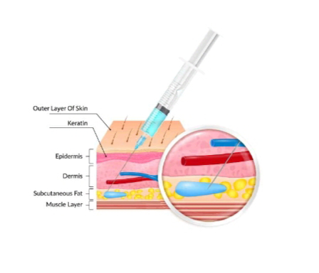
Figure:3
3) Practice in injecting drug by intravenous: -
Intravenous drug injection is not recommended or safe for non-medical people, even if it requires sterile equipment, proper technique, and knowledge of medicine administration. The biggest danger of contracting infections like HIV, Hepatitis B, and Hepatitis C occurs when using shared or non-sterile equipment. Intravenous drug administration offers several advantages over other ways of administration.
–intravenous (and intra-arterial) drug administration provides the most complete drug availability with a minimal delay
–by control of the administration rate, constant plasma concentrations can be obtained at a required level.
–unexpected side effects observed during the administration period can be halted by stopping the infusion (pleading for an extended infusion time)
–compounds that are poorly absorbed by the gastrointestinal tract may be advantageously administered intravenously
–compounds that are unacceptably painful when administered intramuscularly or subcutaneously may present no difficulties by the intravenous route.[9]

Figure:4
# Withdrawing of blood samples:
MATERIALS:
• Safety Needles, 22g or less
• Butterfly needles. 21g or less
• Syringes
• Vacutainer tube holder
• Transfer Device
• Blood Collection Tubes.
* The vacuum tubes are designed to draw a predetermined volume of blood.
* Tubes with different additives are used for collecting blood specimens for specific types of tests.
* The color of cap is used to identify these additives.
• Tourniquets. Single use, disposable, latex-free tourniquets
• Antiseptic. Individually packaged 70% isopropyl alcohol wipes.
• 2x2 Gauze
• Sharps Disposal Container. An OSHA acceptable, puncture proof container marked “Biohazardous”
• Bandages or tape [10]
Procedure:
1. Determine who the patient is; two active kinds of identification are needed. Request that the patient provide their name and birthdate, which must correspond with the requisition.
2. Assure the patient that only the bare minimum of blood will be extracted for the test.
3. Confirm that any time or diet constraints have been fulfilled.
4. Draw Order "The following order of draw is the approved order as established by CLSI." Whenever more than one tube is drawn during a single venipuncture, this order of draw should be adhered to.
1. Blood Culture
2. Light Blue Top (plasma): 3.2% sodium citrate. These tubes are used for coagulation tests and need to be completely filled to ensure the proper ratio of blood to anticoagulant.
3. Red Top (serum): Plain and gel. Used for chemistry and reference tests.
4. Green Top (plasma): With and without gel, contains lithium heparin. These tubes are used primarily for chemistry tests. 5. Lavender or Pink Top (plasma): Contains EDTA. Used primarily for hematology and blood bank testing.
5. Gray Top (plasma): Contains sodium fluoride/potassium oxalate. Used by chemistry for glucose testing.[11]
Module 3
# Introduction to light microscope
Despite being an essential component of contemporary biomedical sciences, light microscopy is frequently considered an outdated method. Despite being more than 400 years old, light microscopy is still developing, and its full potential in the biological sciences may not yet be fully realized.
Providing magnified images of objects that are lighted or emit light in the visible spectrum or the nearby ultraviolet or near-infrared regions is the aim of light microscopy. To achieve optical magnification, light must be passed through lenses. A digital photomicrograph can be readily enlarged using contemporary digital photography technologies. After a while, the image gets extremely fragmented and magnification stops revealing any further details. This demonstrates that the resolution has not risen despite the magnification. This is a basic example of how, in the majority of situations, resolution matters more than magnification. A light microscope is a piece of equipment used in biology labs that detects, magnifies, and enlarges extremely small objects using visible light. [12]
Parts of microscope:
• Illuminator -This is the light source located below the specimen.
• Condenser-Focuses the ray of light through the specimen.
• Stage -The fixed stage is a horizontal platform that holds the specimen.
• Objective -The lens that is directly above the stage.
• Nosepiece-The portion of the body that holds the objectives over the Stage.
• Diaphragm – Regulates the amount of light into the condenser.
• Base- Base supports the microscope which is horseshoe shaped.
• Coarse focusing knob – Used to make relatively wide focusing adjustments To the microscope.
• Fine focusing knob – Used to make relatively small adjustments to the microscope. Body- The microscope body.
• Ocular eyepiece- Lens on the top of the body tube. It has a magnification of 10x normal vision.
#Types of Light microscope:
1. Bright-field Microscopy
2. Dark-field Microscopy
3. Phase contrast Microscopy [13]
4. Fluorescence microscopy
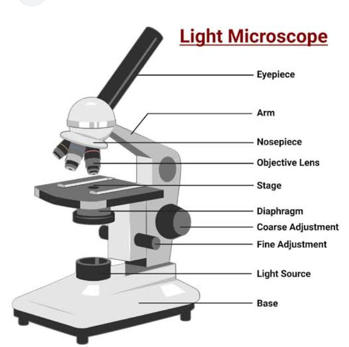
Figure:5
Module 4:
Experimental
#Introduction to semi auto analyser:
A semi-auto analyzer is a hybrid lab tool that requires manual procedures for biological sample analysis, such as loading samples and preparing reagents, but automates other aspects of testing, including measurement. These analyzers are perfect for tiny labs, clinics, and emergency situations when efficiency and adaptability are essential because they are affordable, versatile, and yield faster results than manual methods. [14]

Figure:6
Procedure:
1. Pre-Analysis Preparation & Calibration
Power On: Switch on the main power and the instrument's power button.
Environment Check: Ensure the instrument is on a smooth, dust-free, and dry working platform at a stable room temperature (15-30°C).
Instrument Setup: Power on the instrument and select the desired test from the menu.
Daily Checks: Perform a daily appearance check and clean the instrument's pipelines and colorimetric pool with distilled water.
Baseline/Zero Calibration: Aspirate distilled water to set a baseline reading, effectively zeroing the instrument for the photometric measurement.
2. Sample & Reagent Handling
Reagent Addition: Reagents are manually added to the colorimetric pool or reaction cup.
Sample Aspiration: Manually insert the sample ID, then introduce the patient sample into the instrument. The instrument's peristaltic pump will then aspirate the correct volume of the sample.
Sample Mixing: The aspirated sample is then mixed within the colorimetric pool with the reagents.
3. Measurement & Calculation
Light Measurement: A light beam is passed through the mixture in the flow cell.
Colorimetric Reading: The sample and reagents will absorb a portion of the light. The remaining light is passed through a filter to isolate specific wavelengths and then measured by a photodetector.
Microprocessor Action: The photodetector converts the light energy into an electrical signal, which is sent to the microprocessor.
Result Generation: The microprocessor calculates the light absorbed and uses Beer-Lambert's Law to determine the concentration of the analyte in the patient sample.
4. Post-Analysis & Maintenance
Result Display: The final result is displayed and can be printed.
Daily Cleaning: After each measurement, aspirate distilled water to thoroughly clean the colorimetric pool and pipelines to prevent cross-contamination.
Long-Term Maintenance: To extend the life of the pump tubes and other components, remove the pump tubes when the analyzer is not in use for extended periods.[15]
ADVANTAGES:
DISADVANTAGES:
#Various test performed semi auto analyser
a) Estimation Of Urea by Urease Method (Semi-Auto Analyzer)
1. The urease enzyme and water were used to hydrolyze urea, producing carbon dioxide and ammonia: NH2 + CO + NH2 + H2O 2NH3 + CO2 Urease
2. In the presence of GLDH, the ammonia reacted with α-ketoglutaric acid and reduced NADH to produce nicotinamide adenine dinucleotide (NAD) and glutamic acid: NH3+ α Keto glutarate + NADH+H+ GLDH glutamate +NAD + H2O Using a semiauto analyzer, the rate at which NADH was converted to NAD at 340 nm was related to the content of urea.
b) Estimation of Urea by Fully Automatic Analyzer (Kinetic Urease and Glutamine Dehydrogenase Method)
A fully automatic analyzer was utilized to estimate the urea levels in the serum sample. Urease hydrolyzes urea in the process, producing carbon dioxide and ammonia. In addition to oxidizing reduced β-NADH to β-NAD, GLDH catalyzes the condensation of ammonia and α-ketoglutarate to glutamate. The urea levels were directly correlated with changes in absorbance.
C) Estimation of Total Cholesterol by Semiauto Analyzer (Cholesterol Oxidase and Peroxidase Method):
The esterified cholesterol was hydrolyzed to liberate cholesterol by cholesterol esterase (CHE). Hydrogen peroxide (H2O2) was produced by oxidizing free cholesterol, and peroxidase then combined it with phenol and 4-aminopyrine to create a red quinonimine dye complex. The amount of cholesterol in the serum sample was directly correlated with the color's intensity. [17]
Module:5
Analysis Of Constituents of Blood and Urine
Examination of blood and urine's normal and aberrant components Blood, sugar, proteins, bile salts, bile pigments, and ketone bodies are all abnormal components of urine. It is seen in Addison's disease, diabetes mellitus, diabetes insipidus, chronic progressive renal failure, excessive water intake, and the use of diuretics like alcohol and caffeine, among other conditions. An aqueous solution that contains more than 95% water is called urine.[18]
Normal Constituents of Urine
Normal urine has various organic and inorganic constituents.
The normal inorganic constituents are chloride, phosphates, sulphates, calcium, etc. while organic constituents are urea, uric acid, and creatinine.
Abnormal Constituents of Urine: -
The abnormal constituents of urine are blood cells, albumin, and glucose. Presence of albumin, glucose, and blood cells in the urine causes the pathological conditions called albuminuria, glycosuria, and haematuria respectively.
Hematological Value.
The branch of medicine that deals with the diagnosis and treatment of diseases of the blood and bone marrow.
Blood Cells
Values Units Variable (Common Abbreviations) M = 14–18 F = 12–15 g/dL blood Hemoglobin (Hb, Hgb)
Hematological Values of Blood
Red blood cell: Male: 4.35-5.65 trillion count cells/L* (4.35-5.65 million cells/mcL**)
Female: 3.92-5.13 trillion cells/L (3.92-5.13 million cells/mcL)
Hemoglobin:Male: 13.2-16.6 grams/dL*** (132-166 grams/L)
Female: 11.6-15 grams/dL (116-150 grams/L)
Hematocrit:Male: 38.3-48.6 percent
Female: 35.5-44.9 percent White blood cell 3.4-9.6 billion cells/L count (3,400 to 9,600 cells/mcL)
Platelet count :Male: 135-317 billion/L (135,000 to 317,000/mcL)
Female: 157-371 billion/L (157,000 to 371,000/mcL)
???? Complete Blood Count (CBC) Values
Thyroid function test:
A thyroid function test (TFT) is a blood test that assesses the health of your thyroid gland, which regulates metabolism, by measuring hormones. Hyperthyroidism (overactive thyroid) and hypothyroidism (underactive thyroid) are among the conditions it screens for. TSH (thyroid-stimulating hormone) and the primary thyroid hormones, T3 and T4, are usually measured by the assays.
Glucose test:
In order to detect and track diseases including diabetes and gestational diabetes, a glucose test monitors blood glucose levels. The oral glucose tolerance test (OGTT), which measures glucose before and after consuming a glucose solution, the fasting blood glucose test (which measures glucose after fasting for at least eight hours), and the glucose challenge test (a condensed form of the OGTT that is frequently used during pregnancy) are examples of common tests. Tests for gestational diabetes, or diabetes that develops during pregnancy, include the glucose challenge test.
Urea test:
In order to evaluate kidney and liver function, a urea test, also called the Blood Urea Nitrogen (BUN) test, analyzes the quantity of urea nitrogen in your blood. The breakdown of proteins produces urea, a waste product that the kidneys typically filter from the blood and eliminate in urine. Conditions including renal illness, dehydration, liver problems, or problems with protein metabolism can all be indicated by abnormal urea levels. The test is a straightforward diagnostic technique for tracking general health and the effectiveness of waste removal from the kidneys, and it includes a simple blood draw.
Creatine test:
A creatine test is a medical procedure used to assess kidney function by measuring creatinine, a waste product from muscle activity, in your blood and/or urine. Because it is filtered out of the blood by healthy kidneys, elevated creatinine levels can be a sign of kidney disease. The test is used to identify kidney illness, track its progression, and look for adverse drug reactions.
Creatinine test:
A creatinine test evaluates kidney function by measuring the amount of creatinine, a waste product from muscles, in your blood or urine. High blood creatinine is a sign of inadequate renal filtration since healthy kidneys filter this waste product. This test aids in kidney disease diagnosis, renal disease monitoring, and therapy efficacy assessment.
Cholesterol test:
In order to determine your risk of heart disease, a cholesterol test, also known as a lipid panel, evaluates the amounts of various fats in your blood, such as triglycerides, high-density lipoprotein (HDL), and low-density lipoprotein (LDL). A tiny blood sample is taken from your arm by a medical specialist in order to perform the test. For a fasting lipid panel, which precisely analyzes all lipid components, you might need to fast for up to 12 hours prior to the test. The findings are used to assess your cardiovascular health and identify any lifestyle or treatment modifications that may be required.
Bilirubin test:
Total Serum Bilirubin (Bilirubin Total, Serum) - MRLDC... In order to assess liver function and identify diseases such as jaundice, liver disease, anemia, and bile duct abnormalities, a bilirubin test quantifies the amount of the yellow pigment bilirubin in your blood. In order to determine your bilirubin levels, a medical professional will take a blood sample from a vein in your arm. The test aids in determining the origin of symptoms such as black urine, yellow skin and eyes, and unexplained exhaustion.
Diastase test:
The diastatic activity of honey, which reveals the quantity and existence of diastase (a kind of amylase) in the sample, is measured by a diastase test (also known as diastase number, or DN). The test uses a starch substrate that, when broken down by the enzyme, releases a colored material. Since heat lowers enzyme levels, a high diastatic activity indicates that the honey has not been exposed to extreme heat or unfavorable storage conditions.
Serum glutamic pyruvic transaminase (SGPT):
The Alanine Aminotransferase (ALT) enzyme, which is mostly present in liver cells, is measured by a blood test called an SGPT test, also referred to as an ALT test. It is used by physicians to evaluate liver function, identify liver illness or damage, such as fatty liver disease or hepatitis, and track the effectiveness of liver disease treatment. A high SGPT test result frequently indicates a liver issue since elevated blood levels of the enzyme can show that liver cells are damaged or irritated. [19]
Module :6
Report analysis
Using techniques including microscopy, chemical analysis, and genetic testing, laboratory diagnosis in pathology verifies illnesses by examining body fluids, tissues, and cells to find anomalous alterations, infectious agents, or molecular abnormalities. In order to identify diseases including infections, malignancies, and metabolic problems and to guide treatment decisions, pathologists rely on tests like blood testing, tissue biopsies, and microbiology assays. [20]
How it Works
1. Sample Collection: A patient's biological samples, such as blood, urine, sputum, or tissue biopsies, are collected.
2. Sample Processing: Lab technicians and pathologists process these samples. For tissue, this involves embedding it in wax, sectioning it into thin slices, and placing it on a microscope slide.
3. Microscopic Examination: Pathologists examine the prepared slides under a microscope to identify cellular changes, abnormalities, and other disease indicators.
4. Laboratory Tests: Various tests are performed depending on the suspected condition:
Hematology: Analyzes blood cells to diagnose anemia, infections, and blood cancers.
Clinical Chemistry: Measures levels of chemicals, enzymes, and hormones to check organ function and detect toxins.
Microbiology: Identifies bacteria, viruses, and fungi to diagnose infections and determine effective antibiotic or antiviral treatments.
Molecular Pathology: Detects genetic abnormalities, crucial for diagnosing genetic disorders and certain cancers, and guiding precision medicine.
5. Pathologist's Report: Based on the findings, a pathologist generates a report with a diagnosis or helps clarify the nature of the illness.[20]
Module :7
Collection and completion of data
In pathology, information is gathered by taking patient samples (such as blood, tissue, or urine), which are then painstakingly processed and examined in a lab to produce a diagnosis. A number of meticulous procedures are followed in order to complete this data, including slide preparation, staining, pathologist microscopic inspection, and the production of an extensive diagnostic report. In order to ensure correct tracking from the patient to the final report, data collecting also entails appropriate labeling and recording.
1. Sample Collection
Patient Data: Healthcare professionals collect samples like blood, urine, faeces, or tissue from patients.
Labeling: Each sample is crucialy labeled with a unique patient identifier, ensuring it is associated with the correct patient and procedure.
Request Form: A request form, either paper or electronic, accompanies the sample and contains vital patient details and clinical history, guiding the diagnostic process.
2. Sample Processing and Analysis
Pre-analysis: The sample is placed in a container and sent to the lab with its request form.
Sample Preparation: Samples are processed through steps like embedding in paraffin wax to allow for precise sectioning.
Slide Preparation: The embedded sample is then cut into thin slices (sections) and placed on a glass slide.
Staining: Various stains are applied to the slide to make specific cell structures, tissues, or disease markers visible for microscopic examination.
Data Compilation for Research:
Dataset Diversity: Test datasets in computational pathology aim to be representative and diverse, covering various forms of biological and technical variability to minimize biases and improve AI model generalizability.
Consistent Protocols: To avoid selection bias, data should be collected prospectively following a Systematic Data Extraction: Datasets from pathology reports, such as those from TCGA, are pre-processed to remove missing or 13]. Lymphocytes By, David F. La Rosa M D Jordan S. Orange MD [21]
CONCLUSION:
While pathophysiology aims to explain the functional changes occurring within an individual as a result of a disease or pathologic state, pathophysiology describes the abnormal or undesirable position. The results for displaying pathology information are included in this report. Consequently, the structure of the results might offer more than just a lab output; it might also offer a useful tool that can be shared. The ultimate goal is to better understand and manage their sickness in order to enhance their health and other physiological anomalies that affect people.
REFERENCES


Naikwadi Komal*, Gagare Rutuja, Sathe Poonam, Sah Ritu, Mundekar Yashraj, Dalvi Rushikesh, Musale Yogesh, A Review of Pathology, Int. J. of Pharm. Sci., 2025, Vol 3, Issue 10, 2881-2896 https://doi.org/10.5281/zenodo.17455526
 10.5281/zenodo.17455526
